. 2017 Oct 5:6:1804.
doi: 10.12688/f1000research.12252.2. eCollection 2017.

Endoplasmic reticulum-to-Golgi transitions upon herpes virus infection

Affiliations

Endoplasmic reticulum-to-Golgi transitions upon herpes virus infection

Peter Wild et al. F1000Res. .

Abstract

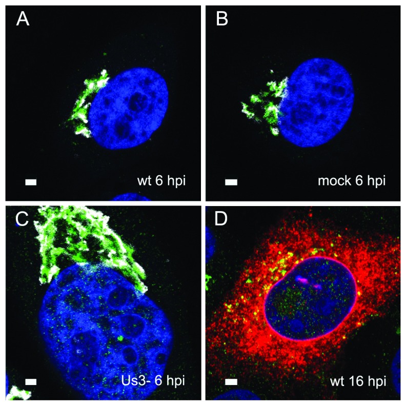
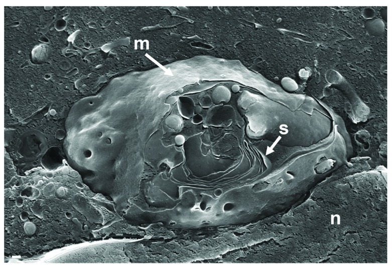
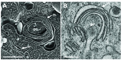
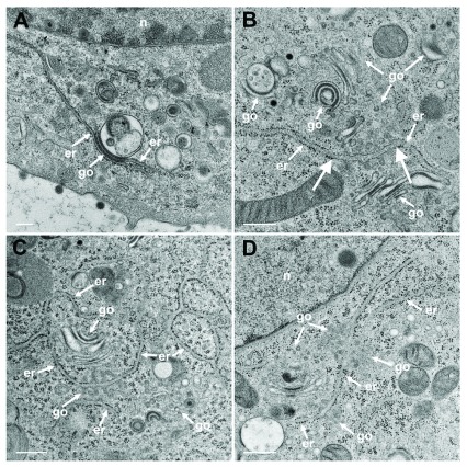
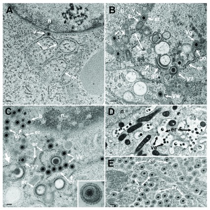
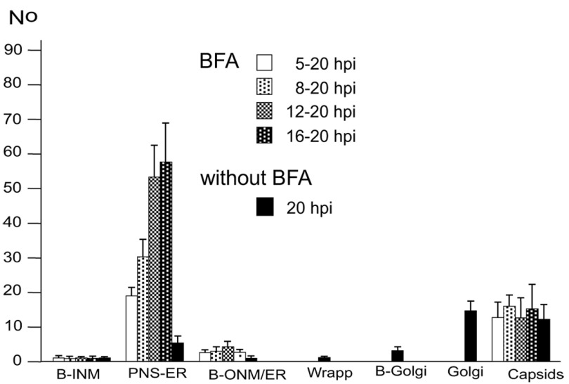
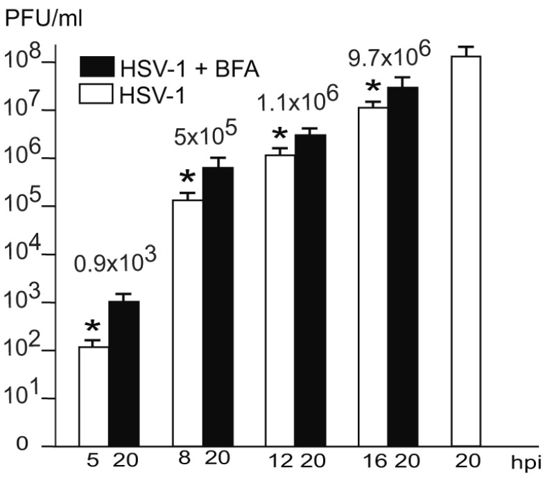
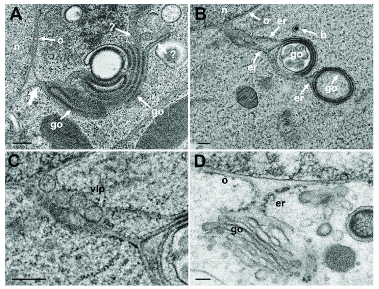
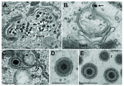
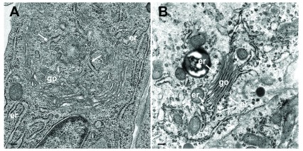
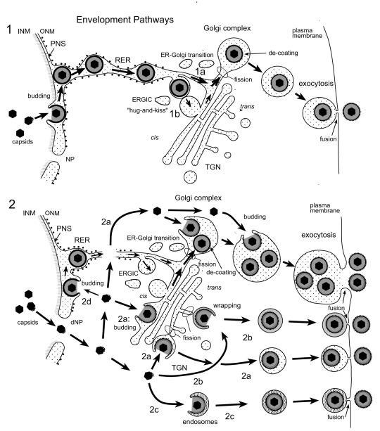
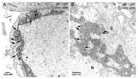
Background: Herpesvirus capsids are assembled in the nucleus, translocated to the perinuclear space by budding, acquiring tegument and envelope, or released to the cytoplasm via impaired nuclear envelope. One model proposes that envelopment, "de-envelopment" and "re-envelopment" is essential for production of infectious virus. Glycoproteins gB/gH were reported to be essential for de-envelopment, by fusion of the "primary" envelope with the outer nuclear membrane. Yet, a high proportion of enveloped virions generated from genomes with deleted gB/gH were found in the cytoplasm and extracellular space, suggesting the existence of alternative exit routes. Methods: We investigated the relatedness between the nuclear envelope and membranes of the endoplasmic reticulum and Golgi complex, in cells infected with either herpes simplex virus 1 (HSV-1) or a Us3 deletion mutant thereof, or with bovine herpesvirus 1 (BoHV-1) by transmission and scanning electron microscopy, employing freezing technique protocols. Results: The Golgi complex is a compact entity in a juxtanuclear position covered by a membrane on the cis face. Golgi membranes merge with membranes of the endoplasmic reticulum forming an entity with the perinuclear space. All compartments contained enveloped virions. After treatment with brefeldin A, HSV-1 virions aggregated in the perinuclear space and endoplasmic reticulum, while infectious progeny virus was still produced. Conclusions: The data suggest that virions derived by budding at nuclear membranes are intraluminally transported from the perinuclear space via Golgi -endoplasmic reticulum transitions into Golgi cisternae for packaging. Virions derived by budding at nuclear membranes are infective like Us3 deletion mutants, which accumulate in the perinuclear space. Therefore, i) de-envelopment followed by re-envelopment is not essential for production of infective progeny virus, ii) the process taking place at the outer nuclear membrane is budding not fusion, and iii) naked capsids gain access to the cytoplasmic matrix via impaired nuclear envelope as reported earlier.

Keywords: Golgi complex; brefeldin A; egress pathway; endoplasmic reticulum; envelopment; herpes virus; intraluminal transport.


Conflict of interest statement

No competing interests were disclosed.

Figures

Figure 1.
Figure 1.. Immunolabelling of the Golgi complex.
Confocal microscopy of the trans-face (green) and the cis-face (white) of Vero cells, immunolabeled with anti TGN46 antibodies (green) and anti GM130 antibodies (white) at 6 h after inoculation with HSV-1 ( A), R7041(ΔUs3) ( C) after mock infection ( B), as well a at 16 hpi with wt HSV-1 ( D) together with immunolabeling of the viral glycoprotein gB (red). The Golgi complex was always in a juxtanuclear position and enormously enlarged after R7041(ΔUs3). Note the enlargement of the nucleus after infection with R7041(ΔUs3). Bars 1 µm.
Figure 2.
Figure 2.. Cryo-FESEM of a Golgi complex in close vicinity to the nucleus (n) in a Vero cell, at 10 hpi with wt HSV-1.
The entire visible surface is covered by an intact membrane (m) except at the part it is broken away giving view to Golgi stacks (s). Bars: 200 nm.
Figure 3.
Figure 3.. Golgi fields not involved in virus maturation and transport.
The Golgi complex, as revealed in freeze-fracture planes ( A) and in thin sections ( B), is entirely covered at the cis-face by the membrane (m) of the outermost cisterna. Bars: 100 nm.
Figure 4.
Figure 4.. TEM of R7041(ΔUs3) infected Vero cells at 12 hpi.
( A) The ER (er) runs from the nuclear (n) periphery towards a Golgi field (go), continuing into the membrane of the outermost stack and further into the cytoplasmic matrix. The membranes of the second stack continue also into ER membranes. ( B) An ER cisterna runs through multiple small Golgi fields, whereby the ER membranes turn into Golgi membranes (thick arrows). ( C) ER membranes forming a network continue into Golgi membranes. ( D) ER membranes run through two Golgi fields, turning each time into Golgi membranes. Bars: 500 nm.
Figure 5.
Figure 5.. The effect of BFA.
TEM of Vero cells at 9 hpi with R7041(ΔUs3) ( A), at 16 hpi ( B) and at 20 hpi ( C) with wt HSV-1, and at 15 or 17 hpi with wt HSV-1 and BFA exposure ( D and E). ( A) The ER (er) runs from the nucleus (n) towards the cell periphery forming an entity with the PNS that contains a virion (v). ( B) The ER contains virions. One capsid is in the stage of budding (b) into the ER. The ER continues into Golgi (go) membranes at two sites. One Golgi cisterna contains a virion (Vg), one virion has been derived by wrapping (Vw). Close to Golgi stacks, there is probably a virion (V?) of abnormal size. ( C) One capsid buds (b) at the nuclear (n) periphery. The ER is dilated and filled with virions (Ve) and dense material: An ER membrane turns into a Golgi membrane (thick arrow). ( D) After exposure to BFA from 8 to 15 hpi with wt HSV-1, the ER was dilated and contained some virions. ( E) The ER was almost filled with virions after exposure to BFA from 8 to 17 hpi with wt HSV-1. Note that virions in the PNS and ER are covered by a dense coat hiding spikes whereas spikes are clearly apparent on virions in the extracellular space ( C inset). Bars: 200 nm.
Figure 6.
Figure 6.. Means and standard deviations of the phenotype of HSV-1 infected Vero cells.
BFA was added to monolayers at 5, 8, 12 or 16 hpi (MOI of 5) and incubated until 20 hpi. For control, inoculated cells were incubated for 20 h without addition of BFA. Cells were rapidly frozen at 20 hpi and processed for electron microscopy. The phenotypes of envelopment were counted in 10 cellular profiles of 5 independent experiments: Capsids budding at the INM (B-INM), at the ONM and ER membranes (B-ONM/ER) and at the Golgi complex (B-Golgi); virions in the PNS-ER compartment (PNS-ER); virions derived by wrapping (Wrapp); virions in Golgi cisternae or large vacuoles (Golgi); capsids in the cytoplasmic matrix (capsids).
Figure 7.
Figure 7.. Virus yields at the time of BFA administration or controls (white) and at 20 hpi (black).
The difference between virus yields (indicated with numbers) at the time of BFA addition and harvesting at 20 hpi is considered to be due to virus production after Golgi disintegration. These infectious virions correspond to the virions accumulating in the PNS-ER compartment. n = 4, p<0.01.
Figure 8.
Figure 8.. TEM of Vero cells at 12 hpi with R7041(ΔUs3) and of BoHV-1 infected MDBK cells, showing Golgi fields close to the nucleus (n).
( A) Golgi (go) membranes continue (thick arrow) into the ONM (o) as well as towards the cytoplasm indicated by (?) because the destination is unknown. ( B) Golgi membranes continue via ER membranes (er) into the ONM. The ER contains 4 virus-like particles. ( C) Details of panel B. ( D) PNS, ER and Golgi complex form an entity in a BoHV-1 infected MDBK cell ( D: This figure has been reproduced with permission of P. Wild et al., Micron 33, 2002, Elsevier). Bars 200 nm.
Figure 9.
Figure 9.. Virions in Golgi cisternae versus budding of capsids.
( A) Golgi cisternae engulfing BoHV-1 virions at 20 hpi. Many of them are tangentially (t) sectioned. ( B) Budding BoHV-1 capsid at a Golgi membrane of the cis-face (arrow). ( C) HSV-1 virion in a Golgi cisterna that connects to the ER (arrow). Note the dense content within the ER and Golgi cisterna indicating little loss of material during processing. ( D) Concentric vacuole derived by wrapping containing a single BoHV-1 virion. The space between viral envelope and vacuolar membrane is always filled in well preserved cells. ( E) Virions in a large vacuole or cisterna exhibiting clearly spikes even in tangentially (t) sectioned virions. Bars: 200 nm.
Figure 10.
Figure 10.
( A) TEM image of a cultured epithelial cell in which the Golgi complex is embedded in the ER. The ER membranes (arrows) run into the Golgi complex, close to a structure that probably represents a tangential section of the Golgi organizing center. ( B) TEM image of a parathyroid cell prepared according to conventional protocols showing Golgi membranes (go) continuing into ER membranes (er). Bars: 200 nm.
Figure 11.
Figure 11.. Schematic representation of proposed herpes virus envelopment pathways.
(1) Capsids bud at the INM into the PNS acquiring tegument and an envelope covered with a dense coat. These perinuclear virions are transported into the RER and further via Golgi transitions (1a) or the ERGIC (“hug-and-kiss”, 1b) into Golgi cisternae where they are packaged into transport vacuoles, which are detached from Golgi membranes by fission. The dense coat is shed off while vacuoles are transported to the cell periphery for exocytotic release of uncoated virions into the extracellular space. (2) Capsids gain direct access to the cytoplasmic matrix via dilated nuclear pores (dNP), and are transported to any site of the Golgi complex. They either bud into Golgi cisternae and vacuoles, respectively (2a) or are enveloped by a process designated wrapping (2b) that involves budding and concomitant formation of a small transport vacuole engulfing a single virion. Capsids can also be enveloped by endosomal membranes (2c). Occasionally, capsids may bud at the OM or RER (2d), and the resulting virions are intraluminally transported as in pathway 1. Finally, vacuoles derived by fission from Golgi membranes or from membranes of vacuoles or endosomes transport virions to the cell periphery and release them into the extracellular space via exocytosis. The dense coat, which derived during the budding process at the INM and ONM and probably protects the viral envelope from fusion with membranes the virions are transported along, is shed of (de-coating) in transport vacuoles at latest when virions are released into the extracellular space. During budding at Golgi cisternae and vacuoles, a dense rim of tegument is closely attached to the inner layer of the viral envelope. However, no dense coat is formed so that spikes (glycoproteins) are readily seen in high resolution micrographs.
Figure 12.
Figure 12.. Virus transport across the ONM in the absence of the fusion proteins gB/gH.
gB/gH null virions accumulate in the PNS-ER compartment. The arrows (inserted by P. Wild) point to interactions of virus particles with the ONM and ER membranes showing the characteristics of budding. The fusion proteins gB/gH are missing. Therefore, these virus membrane interactions represent various stages of budding – not of fusion. Reproduced from Farnsworth et al., 2007, Figure 2), copyright (2007) National Academy of Sciences, U.S.A.

Similar articles

Cited by

References

    1. Albecka A, Laine RF, Janssen AF, et al. : HSV-1 Glycoproteins Are Delivered to Virus Assembly Sites Through Dynamin-Dependent Endocytosis. Traffic. 2016;17(1):21–39. 10.1111/tra.12340 - DOI - PMC - PubMed
    1. Ben-Tekaya H, Miura K, Pepperkok R, et al. : Live imaging of bidirectional traffic from the ERGIC. J Cell Sci. 2005;118(Pt 2):357–367. 10.1242/jcs.01615 - DOI - PubMed
    1. Bigalke JM, Heldwein EE: Structural basis of membrane budding by the nuclear egress complex of herpesviruses. EMBO J. 2015;34(23):2921–2936. 10.15252/embj.201592359 - DOI - PMC - PubMed
    1. Bigalke JM, Heldwein EE: Nuclear Exodus: Herpesviruses Lead the Way. Annu Rev Virol. 2016;3(1):387–409. 10.1146/annurev-virology-110615-042215 - DOI - PMC - PubMed
    1. Bigalke JM, Heldwein EE: Have NEC Coat, Will Travel: Structural Basis of Membrane Budding During Nuclear Egress in Herpesviruses. Adv Virus Res. 2017;97:107–141. 10.1016/bs.aivir.2016.07.002 - DOI - PMC - PubMed

Grants and funding

This study was supported by the Foundation for Scientific Research at the University of Zürich, Switzerland.

LinkOut - more resources