. 2018 Aug 23;3(16):e99007.
doi: 10.1172/jci.insight.99007.

BDNF inhibits neurodegenerative disease-associated asparaginyl endopeptidase activity via phosphorylation by AKT

Affiliations

BDNF inhibits neurodegenerative disease-associated asparaginyl endopeptidase activity via phosphorylation by AKT

Zhi-Hao Wang et al. JCI Insight. .

Abstract

AEP is an age-dependent lysosomal asparaginyl endopeptidase that cleaves numerous substrates including tau and α-synuclein and mediates their pathological roles in neurodegenerative diseases. However, the molecular mechanism regulating this critical protease remains incompletely understood. Here, we show that Akt phosphorylates AEP on residue T322 upon brain-derived neurotrophic factor (BDNF) treatment and triggers its lysosomal translocation and inactivation. When BDNF levels are reduced in neurodegenerative diseases, AEP T322 phosphorylation is attenuated. Consequently, AEP is activated and translocates into the cytoplasm, where it cleaves both tau and α-synuclein. Remarkably, the unphosphorylated T322A mutant increases tau or α-synuclein cleavage by AEP and augments cell death, whereas phosphorylation mimetic T322E mutant represses these effects. Interestingly, viral injection of T322E into Tau P301S mice antagonizes tau N368 cleavage and tau pathologies, rescuing synaptic dysfunction and cognitive deficits. By contrast, viral administration of T322A into young α-SNCA mice elicits α-synuclein N103 cleavage and promotes dopaminergic neuronal loss, facilitating motor defects. Therefore, our findings support the notion that BDNF contributes to the pathogenesis of neurodegenerative diseases by suppressing AEP via Akt phosphorylation.

Keywords: Cell Biology; Molecular pathology; Neurodegeneration; Neuroscience.


Conflict of interest statement

Conflict of interest: The authors have declared that no conflict of interest exists.

Figures

Figure 1
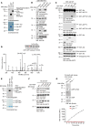
Figure 1. Akt phosphorylates AEP on residue T322.
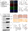
(A) In vitro kinase assay. His-AEP was incubated with GST-Akt in the presence of [γ-32P]-ATP. The reaction products were separated in SDS-PAGE and analyzed by autoradiography. GST and GST-tau were used as negative and positive control, respectively. The protein inputs are shown in the lower panel. (B) Akt phosphorylates AEP in intact cells. Mammalian GST-AEP was cotransfected with HA-Akt WT, KD, or CA into HEK293 cells. At 48 hours, GST-AEP was pulled down and analyzed with anti–p-Akt substrate antibody (first panel) and anti-GST antibody (second panel). Expression of GST-AEP (third panel) and HA-tagged p-/total Akt (fourth and fifth panel) in cell lysates are also shown. (C) Western blot showing phosphorylation of full-length and fragmented AEP by Akt in HEK293 cells. (D) MS/MS spectrum showing the phosphorylation of AEP on residue T322 in GST-AEP purified from HEK293 cells that were cotransfected with GST-AEP and HA-Akt. (E) T322 in AEP is the major phosphorylation residue. In vitro kinase assay showing phosphorylation of WT and mutated AEP (T to A, unphosphorylated mutant) by Akt (upper panel). The inputs are shown in the lower panel. (F) Western blot analysis of phosphorylation level of WT and various mutated AEP constructs after cotransfection with Akt. (G) Knockout of AEP abrogated p-T322 antibody immunoblotting signals on AEP. AFU, arbitrary fluorescence units.
Figure 2
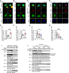
Figure 2. BDNF triggers Akt-phosphorylated AEP lysosomal translocation.
(A and B) Colocalization between AEP pT322/Akt (A) and Akt pS473/AEP (B) in rat primary cortical neurons (DIV 13) treated with or without 100 ng/ml BDNF for 30 minutes. (C and D) Colocalization between AEP pT322/Akt (C) and Akt pS473/AEP (D) in rat primary cortical neurons (DIV 13) treated with 2 or 20 μM preaggregated Aβ for 16 hours. p-AEP T322 was biotinylated. Scale bars: 5 μm. (EH) Quantification of colocalization in AD. Data shown as the mean ± SEM (n = 10 cells per group). *P < 0.05; **P < 0.01 by 2-tailed t test for E and F, 1-way ANOVA with Tukey’s multiple-comparisons test for G and H. (I) p-T322 mediates AEP lysosomal translocation. Cells were treated with 100 ng/ml BDNF for 30 minutes, and then were subjected to subcellular fractionation. LAMP1 was used as specific marker for the lysosomal fraction. Tubulin was used as a loading control for the cytoplasmic fraction. (J) Akt phosphorylation translocates cytoplasmic AEP into the lysosomes. BR6 cells were treated with control or Akt inhibitor (10 μM), followed by BDNF (100 ng/ml) treatment at different time points. Western blot data in I and J are representative of 3 independent experiments.
Figure 3
Figure 3. AEP phosphorylation by Akt is inhibited in neurodegenerative diseases.
(AD) Immunofluorescent staining of Akt/AEP pT322 and Akt pS473/AEP in human AD hippocampal CA1 (A and B) and PD substantia nigra (C and D) compared with age-matched healthy control. Confocal imaging shows colocalization. Scale bars: 10 μm. (EH) Quantification of colocalization in AD. Data shown as the mean ± SEM (n = 5 cases per group). *P < 0.05, **P < 0.01 by 2-tailed t test.
Figure 4
Figure 4. BDNF reduction in neurodegenerative diseases elicits AEP cytosolic residency and activation.
(AE) Western blot analysis of phosphorylated AEP and Akt in AD (AC) or DLB brains (D and E) compared with age-matched healthy controls. Western blot data are representative of 3 independent experiments. Data in C and E shown as the mean ± SEM (n = 3–5 per group). *P < 0.05, **P < 0.01 by 2-tailed t test.
Figure 5
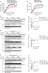
Figure 5. Akt phosphorylation of AEP inhibits its autocleavage and enzymatic activation.
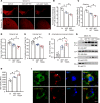
(A and B) AEP activity assay of HEK293 cell lysates. HEK293 cells were cotransfected with AEP and Akt WT/KD/CA (A) or WT and mutated AEP (B). Data shown as the mean ± SEM of 3 independent experiments. *P < 0.05, **P < 0.01 by 2-way ANOVA and Bonferroni’s post hoc test. (CE) Western blot analysis of autocatalytic process of WT/mutated AEP (C) and cleavage of Tau (D) and α-synuclein (E) by WT/mutated AEP after incubation in pH 5.5 reaction buffer at 37°C. Truncated bands were quantified using densitometry. After being normalized with total AEP, tau, or α-synuclein levels, the relative levels were plotted against reaction times. Each curve (right) was fitted with GraphPad Prism 5 using the Boltzmann sigmoidal function. Data are representatives of 4 independent experiments. AFU, arbitrary fluorescence units.
Figure 6
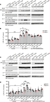
Figure 6. Akt-phosphorylated AEP blocks tau P301S or α-synuclein–induced cell death.
(A) AEP T322A increases truncated tau and T322E mutant blocks the cleavage. Western blot was conducted using BR6 cells cotransfected with AEP and tau P301S. Cells were treated with 100 ng/ml BDNF 1.5 hours before harvesting. (B) LDH assay showing that AEP mutants regulate cell death induced by tau P301S. Data shown as the mean ± SEM (n = 3). (C) AEP T322A increases truncated α-synuclein and T322E mutant blocks the cleavage. Western blot was conducted using BR6 cells cotransfected with AEP and α-synuclein. Cells were treated with 100 ng/ml BDNF 1.5 hours before harvesting. (D) LDH assay showing that AEP mutants regulate cell death induced by cleaved α-synuclein. Data shown as the mean ± SEM (n = 3). *P < 0.05, **P < 0.01 by 2-way ANOVA with Bonferroni’s post hoc test.
Figure 7
Figure 7. Akt phosphorylation mimetic T322E mutant inhibits tau pathologies in Tau P301S mice.
(A) Western blot analysis of phosphorylated AEP and Akt in 9-month-old Tau P301S mouse brains compared with age-matched WT littermates. (BE) Immunofluorescent staining of Akt pS473 and AEP pT322 in 9-month-old Tau P301S mouse hippocampal CA1 compared with age-matched WT littermates. Scale bar: 50 μm. Quantification of intensity (C and D) and colocalization (E) are shown. Data shown as the mean ± SEM (n = 5 per group). (F) Relative BDNF level by ELISA in P301S mouse hippocampus. Data shown as the mean ± SEM (n = 5 per group). (G) T322E mutation reduces AEP and tau fragments. Hippocampal CA1 tissues from AAV-injected Tau P301S mice were analyzed by immunoblotting (n = 4 mice per group). Molecular weight of untagged exogenous AEP is similar to endogenous AEP. (H) T322E mutation reduces AEP enzymatic activity. Data represent tyhe mean ± SEM (n = 3 mice per group). (I and J) Tau pathology is attenuated by AEP T322E. IHC staining with anti–p-Tau AT8 antibody. The brain sections were immunostained with AEP (upper) and AT8 (middle). Scale bar: 25 μm. AT-8 immunoreactivity quantification (lower, mean ± SEM; 9 sections from 3 mice). Western blot data in A and G are representative of 3 independent experiments. *P < 0.05; **P < 0.01 by 2-tailed t test (CF) or 1-way ANOVA with Tukey’s multiple-comparisons test (H and J).
Figure 8
Figure 8. Akt phosphorylation mimetic T322E mutant ameliorates cognitive dysfunctions in Tau P301S mice.
(A) T322E mutant increases the spine density. Scale bar: 5 μm. Golgi staining was conducted on brain sections from CA1 regions of Tau P301S mice (mean ± SEM; n = 6). *P < 0.05 by 1-way ANOVA with Tukey’s multiple-comparisons test. (B) Electrophysiological analysis. Shown traces are the representative fEPSPs recorded before (black) and 60 minutes after (red) 3-theta-burst stimulation. T322E mutant increased the ratio of paired pulses (lower left) and rescued the LTP defects in Tau P301S mice (lower right) (mean ± SEM; n = 6 in each group). *P < 0.05 P301S-T322E versus P301S-Ct; #P < 0.05 P301S-T322E versus P301S-AEP WT; 1-way ANOVA with Tukey’s multiple-comparisons test. (CE) Morris water maze analysis of cognitive functions. T322E mutant in CA1 rescues the learning (C and D) and memory (E) impairments in Tau P301S mice (mean ± SEM; n = 8 mice per group). *P < 0.05 by 2-way ANOVA with Bonferroni’s post hoc test for C or 1-way ANOVA with Tukey’s multiple-comparisons test for D and E.
Figure 9
Figure 9. Unphosphorylated AEP T322A mutant facilitates α-synuclein pathologies and stimulates motor deficits in young SNCA-Tg mice.
(AF) AEP T322A overexpression induces more TH+ dopaminergic cell loss and motor dysfunction in SNCA mice, compared with control or AEP-overexpressing group. AAV-control (Ct), AAV-AEP, or AAV-AEP T322A was injected into right substantial nigra (SN) of SNCA-Tg mice. (A) TH expression in SN and striatum of the above animals was analyzed by immunofluorescent staining. Scale bar: 200 μm. (B and C) Quantification of TH+ fluorescent signals in SN (B) and striatum (C). Data are shown as the mean ± SEM (n = 3 per group). Motor behavioral assays, Rotarod (D), cylinder test (E), and amphetamine-induced rotation (F) were conducted by a blinded observer 2 months after the virus injection. AAV-AEP T322A infection induced more severe motor dysfunction than AAV-AEP WT. Data are shown as the mean ± SEM (n = 7–9 per group). (G) The T322A mutation enhances AEP’s protease activity and cleavage of α-synuclein. SN lysates were probed with various indicated antibodies. (H) AEP enzymatic assay. AEP activity in AEP T322A–injected mice was increased more than in the AEP WT–injected group. Data are shown as mean ± SEM (n = 3 per group). (I) α-Synuclein localizes in the Lewy body in the SN of AEP T322A–injected brains. Immunofluorescent signals of anti–α-synuclein (green) and anti–14-3-3 (red) or anti–α-synuclein pS129 (green) and anti-ubiquitin (red) as Lewy body marker were detected in AEP T322A–injected brain sections. The nuclei were stained with DAPI. Scale bar: 20 μm. *P < 0.05 by 1-way ANOVA.

Similar articles

Cited by

References

    1. Lin MT, Beal MF. Mitochondrial dysfunction and oxidative stress in neurodegenerative diseases. Nature. 2006;443(7113):787–795. doi: 10.1038/nature05292. - DOI - PubMed
    1. Zhang Z, et al. Delta-secretase cleaves amyloid precursor protein and regulates the pathogenesis in Alzheimer’s disease. Nat Commun. 2015;6:8762. - PMC - PubMed
    1. Zhang Z, et al. Cleavage of tau by asparagine endopeptidase mediates the neurofibrillary pathology in Alzheimer’s disease. Nat Med. 2014;20(11):1254–1262. doi: 10.1038/nm.3700. - DOI - PMC - PubMed
    1. Zhang Z, et al. Asparagine endopeptidase cleaves α-synuclein and mediates pathologic activities in Parkinson’s disease. Nat Struct Mol Biol. 2017;24(8):632–642. doi: 10.1038/nsmb.3433. - DOI - PMC - PubMed
    1. Zhang Z, et al. Inhibition of delta-secretase improves cognitive functions in mouse models of Alzheimer’s disease. Nat Commun. 2017;8:14740. - PMC - PubMed

Publication types

MeSH terms