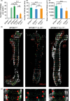
Constitutive overexpression of TNF in BPSM1 mice causes iBALT and bone marrow nodular lymphocytic hyperplasia
- PMID: 30107066
- PMCID: PMC6378607
- DOI: 10.1111/imcb.12197
Constitutive overexpression of TNF in BPSM1 mice causes iBALT and bone marrow nodular lymphocytic hyperplasia
Abstract
BPSM1 (Bone phenotype spontaneous mutant 1) mice develop severe polyarthritis and heart valve disease as a result of a spontaneous mutation in the Tnf gene. In these mice, the insertion of a retrotransposon in the 3' untranslated region of Tnf causes a large increase in the expression of the cytokine. We have found that these mice also develop inducible bronchus-associated lymphoid tissue (iBALT), as well as nodular lymphoid hyperplasia (NLH) in the bone marrow. Loss of TNFR1 prevents the development of both types of follicles, but deficiency of TNFR1 in the hematopoietic compartment only prevents the iBALT and not the NLH phenotype. We show that the development of arthritis and heart valve disease does not depend on the presence of the tertiary lymphoid tissues. Interestingly, while loss of IL-17 or IL-23 limits iBALT and NLH development to some extent, it has no effect on polyarthritis or heart valve disease in BPSM1 mice.
Keywords: Arthritis; BPSM1; IL-17; IL-23; NLH; TNF; heart valve disease; iBALT; nodular lymphoid hyperplasia; tertiary lymphoid organs, bronchus-associated.
© 2018 The Authors Immunology & Cell Biology published by John Wiley & Sons Australia, Ltd on behalf of Australasian Society for Immunology Inc.
Figures


Similar articles
-
Spontaneous retrotransposon insertion into TNF 3'UTR causes heart valve disease and chronic polyarthritis.Proc Natl Acad Sci U S A. 2015 Aug 4;112(31):9698-703. doi: 10.1073/pnas.1508399112. Epub 2015 Jul 20. Proc Natl Acad Sci U S A. 2015. PMID: 26195802 Free PMC article.
-
Pneumocystis-Driven Inducible Bronchus-Associated Lymphoid Tissue Formation Requires Th2 and Th17 Immunity.Cell Rep. 2017 Mar 28;18(13):3078-3090. doi: 10.1016/j.celrep.2017.03.016. Cell Rep. 2017. PMID: 28355561 Free PMC article.
-
Inducible bronchus-associated lymphoid tissue (iBALT) in patients with pulmonary complications of rheumatoid arthritis.J Clin Invest. 2006 Dec;116(12):3183-94. doi: 10.1172/JCI28756. J Clin Invest. 2006. PMID: 17143328 Free PMC article.
-
Friend or Foe: The Protective and Pathological Roles of Inducible Bronchus-Associated Lymphoid Tissue in Pulmonary Diseases.J Immunol. 2019 May 1;202(9):2519-2526. doi: 10.4049/jimmunol.1801135. J Immunol. 2019. PMID: 31010841 Free PMC article. Review.
-
Inducible Bronchus-Associated Lymphoid Tissue: Taming Inflammation in the Lung.Front Immunol. 2016 Jun 30;7:258. doi: 10.3389/fimmu.2016.00258. eCollection 2016. Front Immunol. 2016. PMID: 27446088 Free PMC article. Review.
References
-
- Foo SY, Phipps S. Regulation of inducible BALT formation and contribution to immunity and pathology. Mucosal Immunol 2010; 3: 537–544. - PubMed
-
- Engels K, Oeschger S, Hansmann ML, et al. Bone marrow trephines containing lymphoid aggregates from patients with rheumatoid and other autoimmune disorders frequently show clonal B‐cell infiltrates. Hum Pathol 2007; 38: 1402–1411. - PubMed
Publication types
MeSH terms
Substances
LinkOut - more resources
Full Text Sources
Other Literature Sources
Molecular Biology Databases

