Unexpected contribution of lymphatic vessels to promotion of distant metastatic tumor spread
- PMID: 30101193
- PMCID: PMC6082649
- DOI: 10.1126/sciadv.aat4758
Unexpected contribution of lymphatic vessels to promotion of distant metastatic tumor spread
Abstract
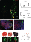
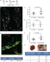
Tumor lymphangiogenesis is accompanied by a higher incidence of sentinel lymph node metastasis and shorter overall survival in several types of cancer. We asked whether tumor lymphangiogenesis might also occur in distant organs with established metastases and whether it might promote further metastatic spread of those metastases to other organs. Using mouse metastasis models, we found that lymphangiogenesis occurred in distant lung metastases and that some metastatic tumor cells were located in lymphatic vessels and draining lymph nodes. In metastasis-bearing lungs of melanoma patients, a higher lymphatic density within and around metastases and lymphatic invasion correlated with poor outcome. Using a transgenic mouse model with inducible expression of vascular endothelial growth factor C (VEGF-C) in the lung, we found greater growth of lung metastases, with more abundant dissemination to other organs. Our findings reveal unexpected contributions of lymphatics in distant organs to the promotion of growth of metastases and their further spread to other organs, with potential clinical implications for adjuvant therapies in patients with metastatic cancer.
Figures


Similar articles
-
Molecular control of lymphatic metastasis.Ann N Y Acad Sci. 2008;1131:225-34. doi: 10.1196/annals.1413.020. Ann N Y Acad Sci. 2008. PMID: 18519975 Review.
-
Tumor necrosis factor superfamily 15 promotes lymphatic metastasis via upregulation of vascular endothelial growth factor-C in a mouse model of lung cancer.Cancer Sci. 2018 Aug;109(8):2469-2478. doi: 10.1111/cas.13665. Epub 2018 Jul 20. Cancer Sci. 2018. PMID: 29890027 Free PMC article.
-
Blockade of FLT4 suppresses metastasis of melanoma cells by impaired lymphatic vessels.Biochem Biophys Res Commun. 2016 Sep 16;478(2):733-8. doi: 10.1016/j.bbrc.2016.08.017. Epub 2016 Aug 6. Biochem Biophys Res Commun. 2016. PMID: 27507214
-
Vascular endothelial growth factor C disrupts the endothelial lymphatic barrier to promote colorectal cancer invasion.Gastroenterology. 2015 Jun;148(7):1438-51.e8. doi: 10.1053/j.gastro.2015.03.005. Epub 2015 Mar 6. Gastroenterology. 2015. PMID: 25754161
-
Tumor lymphangiogenesis and melanoma metastasis.J Cell Physiol. 2008 Aug;216(2):347-54. doi: 10.1002/jcp.21494. J Cell Physiol. 2008. PMID: 18481261 Review.
Cited by
-
Loss of CYLD accelerates melanoma development and progression in the Tg(Grm1) melanoma mouse model.Oncogenesis. 2019 Oct 7;8(10):56. doi: 10.1038/s41389-019-0169-4. Oncogenesis. 2019. PMID: 31591386 Free PMC article.
-
Biomechanical control of lymphatic vessel physiology and functions.Cell Mol Immunol. 2023 Sep;20(9):1051-1062. doi: 10.1038/s41423-023-01042-9. Epub 2023 Jun 2. Cell Mol Immunol. 2023. PMID: 37264249 Free PMC article. Review.
-
ZNF468-mediated epigenetic upregulation of VEGF-C facilitates lymphangiogenesis and lymphatic metastasis in ESCC via PI3K/Akt and ERK1/2 signaling pathways.Cell Oncol (Dordr). 2024 Oct;47(5):1927-1942. doi: 10.1007/s13402-024-00976-0. Epub 2024 Aug 14. Cell Oncol (Dordr). 2024. PMID: 39141315
-
Lymph Node Stromal Cells: Mapmakers of T Cell Immunity.Int J Mol Sci. 2020 Oct 21;21(20):7785. doi: 10.3390/ijms21207785. Int J Mol Sci. 2020. PMID: 33096748 Free PMC article. Review.
-
Diagnostic accuracy of de-escalated surgical procedure in axilla for node-positive breast cancer patients treated with neoadjuvant systemic therapy: A systematic review and meta-analysis.Cancer Med. 2022 Nov;11(22):4085-4103. doi: 10.1002/cam4.4769. Epub 2022 May 3. Cancer Med. 2022. PMID: 35502768 Free PMC article. Review.
References
-
- Stacker S. A., Williams S. P., Karnezis T., Shayan R., Fox S. B., Achen M. G., Lymphangiogenesis and lymphatic vessel remodelling in cancer. Nat. Rev. Cancer 14, 159–172 (2014). - PubMed
-
- Dieterich L. C., Detmar M., Tumor lymphangiogenesis and new drug development. Adv. Drug Deliv. Rev. 99, 148–160 (2016). - PubMed
Publication types
MeSH terms
Substances
Grants and funding
LinkOut - more resources
Full Text Sources
Other Literature Sources
Medical
Molecular Biology Databases

