

Fluorescence Probes for ALKBH2 Allow the Measurement of DNA Alkylation Repair and Drug Resistance Responses
- PMID: 30098084
- PMCID: PMC6478024
- DOI: 10.1002/anie.201807593
Fluorescence Probes for ALKBH2 Allow the Measurement of DNA Alkylation Repair and Drug Resistance Responses
Abstract
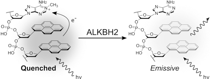
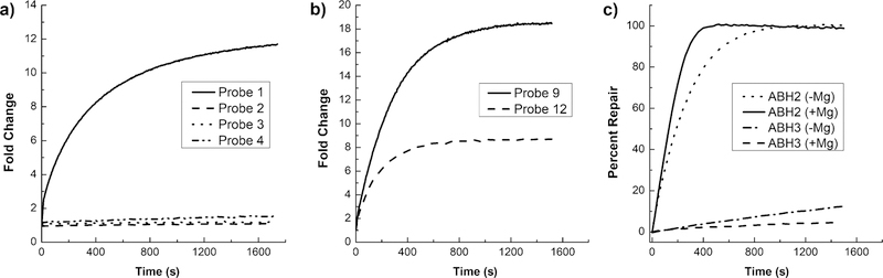
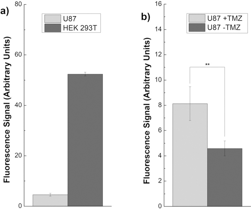
The DNA repair enzyme ALKBH2 is implicated in both tumorigenesis as well as resistance to chemotherapy in certain cancers. It is currently under study as a potential diagnostic marker and has been proposed as a therapeutic target. To date, however, there exist no direct methods for measuring the repair activity of ALKBH2 in vitro or in biological samples. Herein, we report a highly specific, fluorogenic probe design based on an oligonucleotide scaffold that reports directly on ALKBH2 activity both in vitro and in cell lysates. Importantly, the probe enables the monitoring of cellular regulation of ALKBH2 activity in response to treatment with the chemotherapy drug temozolomide through a simple fluorescence assay, which has only previously been observed through indirect means such as qPCR and western blots. Furthermore, the probe provides a viable high-throughput assay for drug discovery.
Keywords: DNA repair; biosensors; cancer; fluorescent probes; nucleic acids.
© 2018 Wiley-VCH Verlag GmbH & Co. KGaA, Weinheim.
Conflict of interest statement
Conflict of interest
The authors declare no conflict of interest.
Figures
Similar articles
-
Interactions between HIV protease inhibitor ritonavir and human DNA repair enzyme ALKBH2: a molecular dynamics simulation study.Mol Divers. 2023 Apr;27(2):931-938. doi: 10.1007/s11030-022-10444-2. Epub 2022 May 11. Mol Divers. 2023. PMID: 35543797
-
Inhibition of human DNA alkylation damage repair enzyme ALKBH2 by HIV protease inhibitor ritonavir.DNA Repair (Amst). 2024 Sep;141:103732. doi: 10.1016/j.dnarep.2024.103732. Epub 2024 Jul 25. DNA Repair (Amst). 2024. PMID: 39094381
-
The DNA repair protein ALKBH2 mediates temozolomide resistance in human glioblastoma cells.Neuro Oncol. 2013 Mar;15(3):269-78. doi: 10.1093/neuonc/nos301. Epub 2012 Dec 20. Neuro Oncol. 2013. PMID: 23258843 Free PMC article.
-
DNA repair in personalized brain cancer therapy with temozolomide and nitrosoureas.DNA Repair (Amst). 2019 Jun;78:128-141. doi: 10.1016/j.dnarep.2019.04.007. Epub 2019 Apr 15. DNA Repair (Amst). 2019. PMID: 31039537 Review.
-
Regulation of DNA Alkylation Damage Repair: Lessons and Therapeutic Opportunities.Trends Biochem Sci. 2017 Mar;42(3):206-218. doi: 10.1016/j.tibs.2016.10.001. Epub 2016 Nov 2. Trends Biochem Sci. 2017. PMID: 27816326 Free PMC article. Review.
Cited by
-
An Excimer Clamp for Measuring Damaged-Base Excision by the DNA Repair Enzyme NTH1.Angew Chem Int Ed Engl. 2020 May 4;59(19):7450-7455. doi: 10.1002/anie.202001516. Epub 2020 Mar 17. Angew Chem Int Ed Engl. 2020. PMID: 32109332 Free PMC article.
-
The genome of a vestimentiferan tubeworm (Ridgeia piscesae) provides insights into its adaptation to a deep-sea environment.BMC Genomics. 2023 Feb 11;24(1):72. doi: 10.1186/s12864-023-09166-y. BMC Genomics. 2023. PMID: 36774470 Free PMC article.
-
DNA Repair and Replication-Related Gene Signature Based on Tumor Mutation Burden Reveals Prognostic and Immunotherapy Response in Gastric Cancer.J Oncol. 2022 Jan 11;2022:6469523. doi: 10.1155/2022/6469523. eCollection 2022. J Oncol. 2022. PMID: 35058980 Free PMC article.
-
ALKBH2 inhibition alleviates malignancy in colorectal cancer by regulating BMI1-mediated activation of NF-κB pathway.World J Surg Oncol. 2020 Dec 10;18(1):328. doi: 10.1186/s12957-020-02106-0. World J Surg Oncol. 2020. PMID: 33302959 Free PMC article.
-
Synthesis of 4-Cyanoindole Nucleosides, 4-Cyanoindole-2'-Deoxyribonucleoside-5'-Triphosphate (4CIN-TP), and Enzymatic Incorporation of 4CIN-TP into DNA.Curr Protoc Nucleic Acid Chem. 2020 Mar;80(1):e101. doi: 10.1002/cpnc.101. Curr Protoc Nucleic Acid Chem. 2020. PMID: 31909864 Free PMC article.
References
Publication types
MeSH terms
Substances
Grants and funding
LinkOut - more resources
Full Text Sources
Other Literature Sources
Research Materials
