

Modeling and Staging of Osteoarthritis Progression Using Serial CT Imaging and Arthroscopy
- PMID: 30079757
- PMCID: PMC7298601
- DOI: 10.1177/1947603518789997
Modeling and Staging of Osteoarthritis Progression Using Serial CT Imaging and Arthroscopy
Abstract
Objective: The objective of this study was to describe in life methods by which osteoarthritis can be staged in order to time therapeutic interventions that are relevant to osteoarthritis (OA) clinical trials.
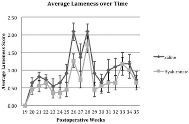
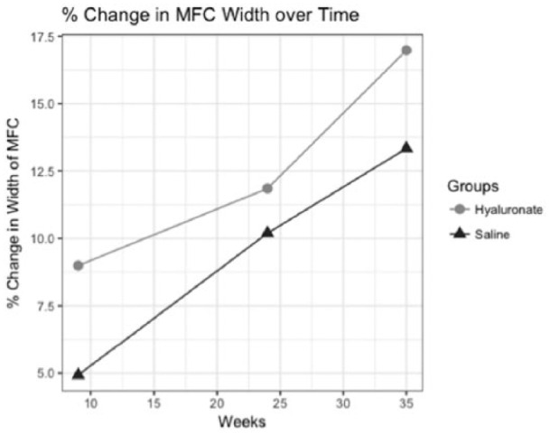
Methods: Twenty-two sheep underwent arthroscopic meniscal destabilization to induce OA. Serial computed tomography (CT) imaging and arthroscopy were used to monitor osteoarthritis progression at 3-month intervals over 9 months. Eleven sheep received 1 intra-articular injection of hyaluronate 3 months after OA induction and another group of 11 received saline. A linear mixed model was used to define the trajectory of shape change in the medial joint compartment. Ordinal logistic regression was used to investigate the association between morphological changes and sclerosis.
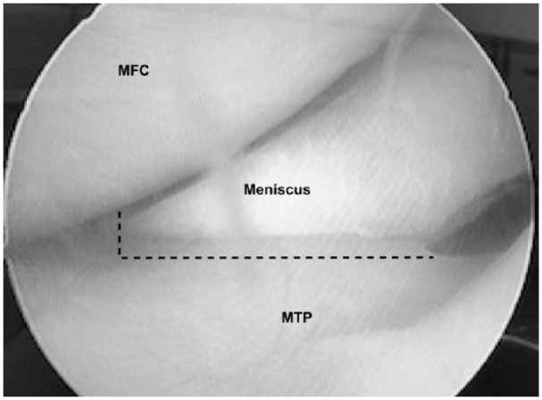
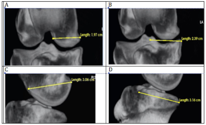
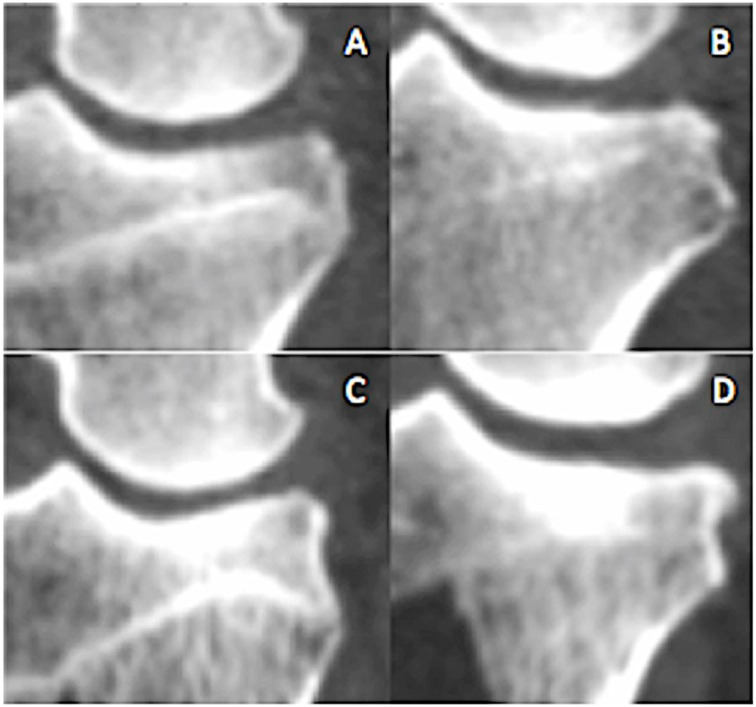
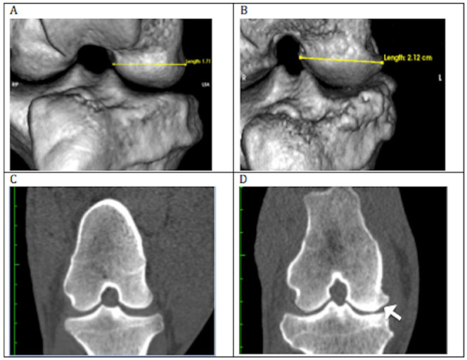
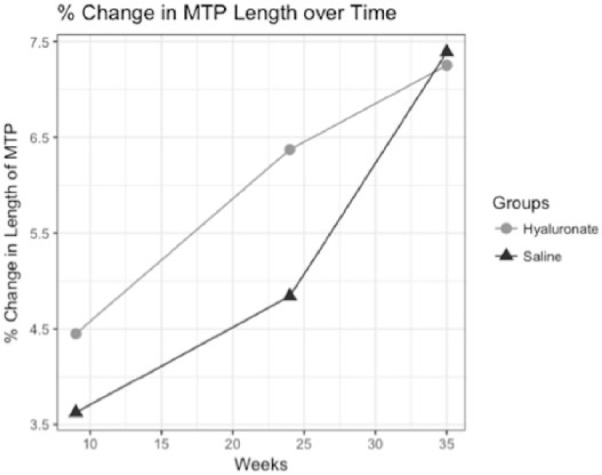
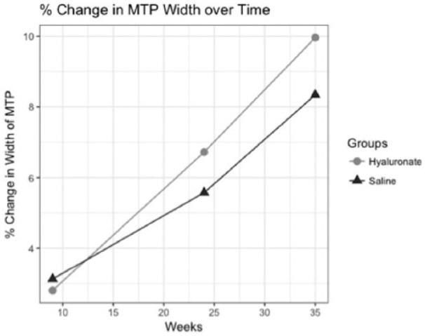
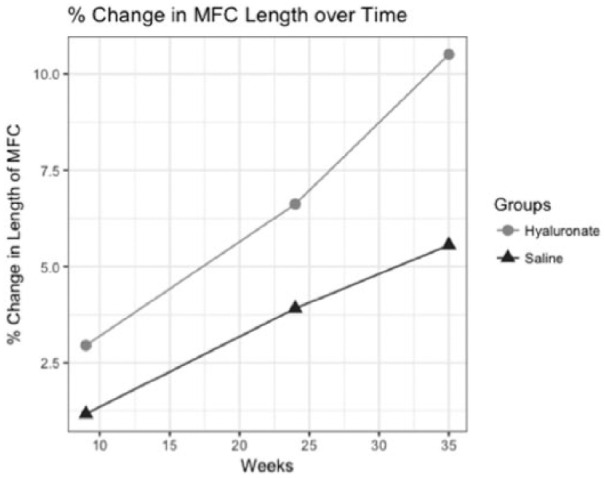
Results: Three months after meniscal destabilization there were early bipolar chondral lesions in the medial compartment of the knee, as well as osteophytes and bone remodeling. Superficial fissures and cartilage cracks progressed to discrete areas of cartilage thinning and fibrillation on the medial tibial plateau by 6 months that became cartilage erosions by nine months. A linear mixed effect model demonstrated significant change in medial compartment length and width with over time (P < 0.05) for both groups. A significant association between severity of sclerosis and medial compartment morphology was also observed.
Conclusions: The induction of osteoarthritic lesions with meniscal release model can be followed using noninvasive and minimally invasive procedures allowing for real-time decisions about redosing therapies, or other changes such as extending trial timelines without sacrificing animals to conduct assessments.
Keywords: animal model; intervention; osteoarthritis; trajectory.
Conflict of interest statement
Figures
Similar articles
-
Risk factors associated with the loss of cartilage volume on weight-bearing areas in knee osteoarthritis patients assessed by quantitative magnetic resonance imaging: a longitudinal study.Arthritis Res Ther. 2007;9(4):R74. doi: 10.1186/ar2272. Arthritis Res Ther. 2007. PMID: 17672891 Free PMC article.
-
Correlations of Medial Joint Space Width on Fixed-Flexed Standing Computed Tomography and Radiographs With Cartilage and Meniscal Morphology on Magnetic Resonance Imaging.Arthritis Care Res (Hoboken). 2016 Oct;68(10):1410-6. doi: 10.1002/acr.22888. Arthritis Care Res (Hoboken). 2016. PMID: 26991547 Free PMC article.
-
Tibiofemoral joint structural change from 2.5 to 4.5 years following ACL reconstruction with and without combined meniscal pathology.BMC Musculoskelet Disord. 2019 Jul 4;20(1):312. doi: 10.1186/s12891-019-2687-9. BMC Musculoskelet Disord. 2019. PMID: 31272448 Free PMC article.
-
Early osteoarthritis of the knee.Knee Surg Sports Traumatol Arthrosc. 2016 Jun;24(6):1753-62. doi: 10.1007/s00167-016-4068-3. Epub 2016 Mar 21. Knee Surg Sports Traumatol Arthrosc. 2016. PMID: 27000393 Review.
-
New imaging tools for mouse models of osteoarthritis.Geroscience. 2022 Apr;44(2):639-650. doi: 10.1007/s11357-022-00525-3. Epub 2022 Feb 7. Geroscience. 2022. PMID: 35129777 Free PMC article. Review.
Cited by
-
Inhibition of a signaling modality within the gp130 receptor enhances tissue regeneration and mitigates osteoarthritis.Sci Transl Med. 2023 Mar 22;15(688):eabq2395. doi: 10.1126/scitranslmed.abq2395. Epub 2023 Mar 22. Sci Transl Med. 2023. PMID: 36947594 Free PMC article.
-
Deep Learning-Based CT Imaging to Evaluate the Therapeutic Effects of Acupuncture and Moxibustion Therapy on Knee Osteoarthritis.Comput Math Methods Med. 2022 May 21;2022:1135196. doi: 10.1155/2022/1135196. eCollection 2022. Comput Math Methods Med. 2022. PMID: 35637844 Free PMC article.
-
Anionic Contrast-Enhanced MicroCT Imaging Correlates with Biochemical and Histological Evaluations of Osteoarthritic Articular Cartilage.Cartilage. 2021 Dec;13(2_suppl):1388S-1397S. doi: 10.1177/1947603520924748. Epub 2020 May 26. Cartilage. 2021. PMID: 32456450 Free PMC article.
References
MeSH terms
LinkOut - more resources
Full Text Sources
Other Literature Sources
Medical
