. 2018 Sep 26;92(20):e00680-18.
doi: 10.1128/JVI.00680-18. Print 2018 Oct 15.

Porcine Adenovirus Type 3 E3 Encodes a Structural Protein Essential for Capsid Stability and Production of Infectious Progeny Virions

Affiliations

Porcine Adenovirus Type 3 E3 Encodes a Structural Protein Essential for Capsid Stability and Production of Infectious Progeny Virions

Abdelrahman Said et al. J Virol. .

Abstract

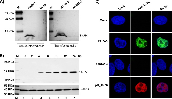
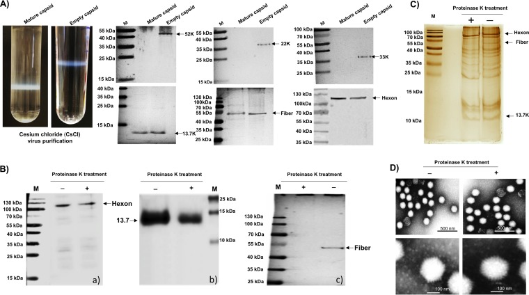
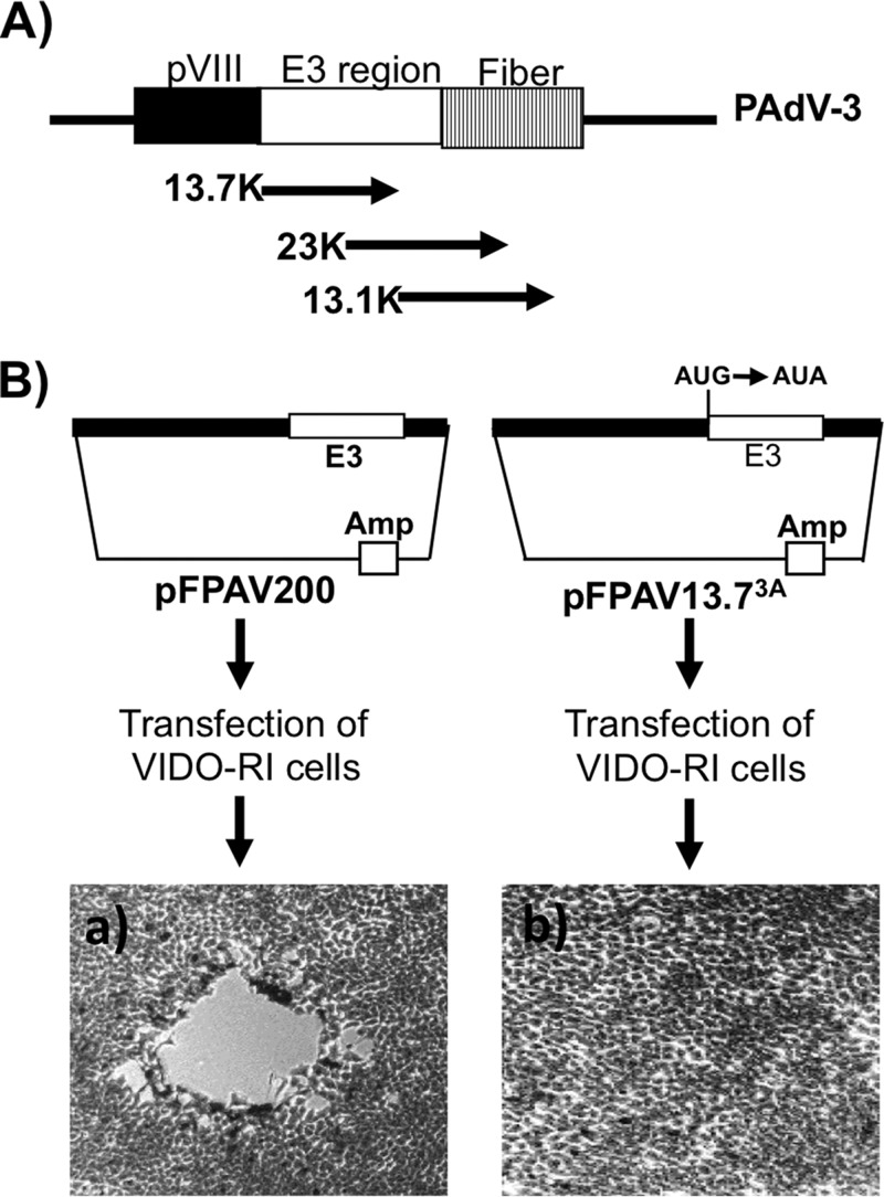
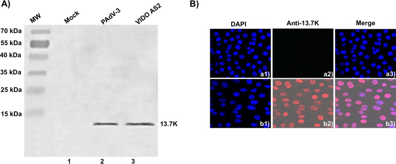
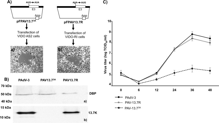
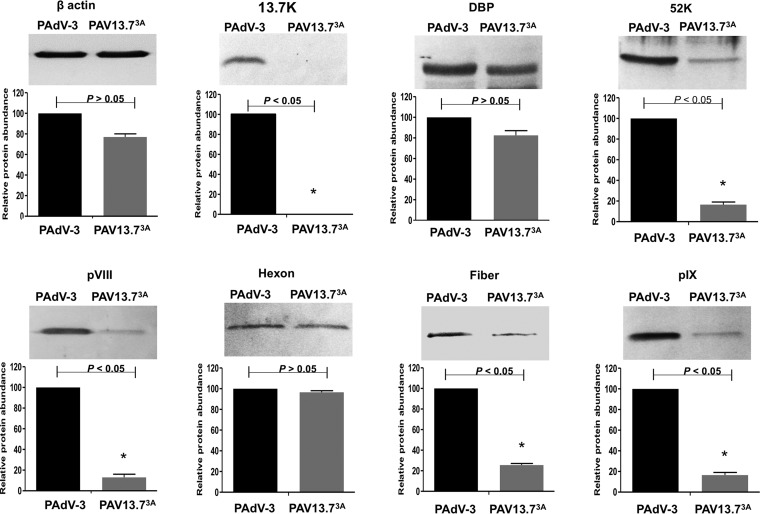
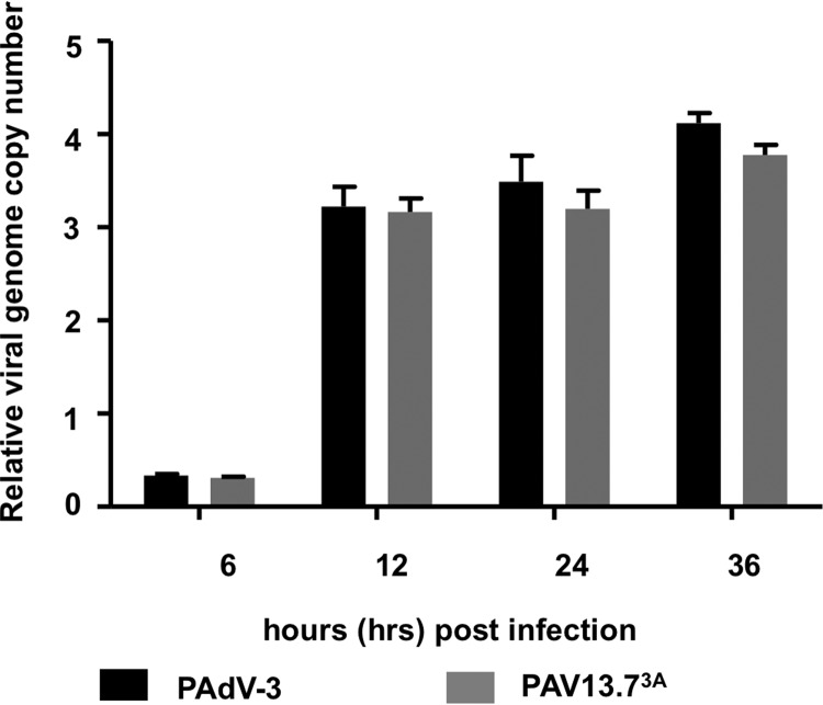
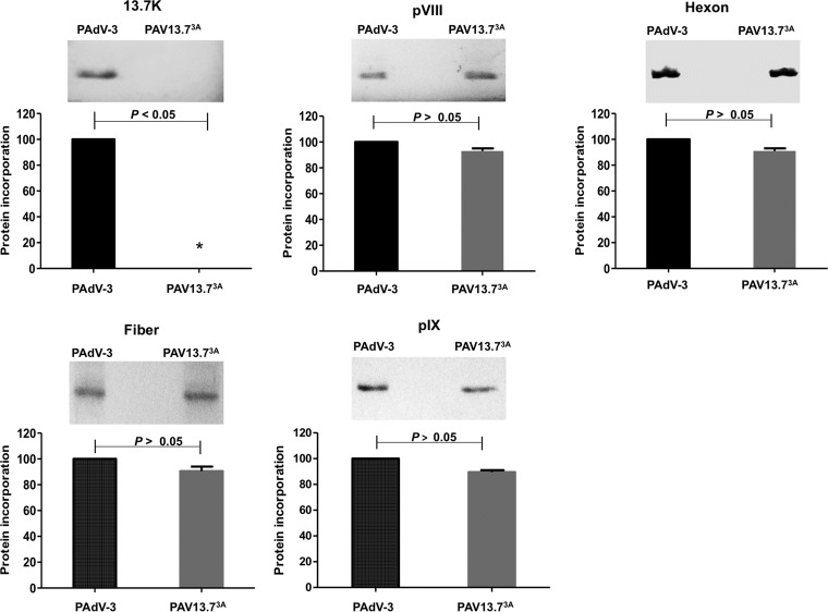
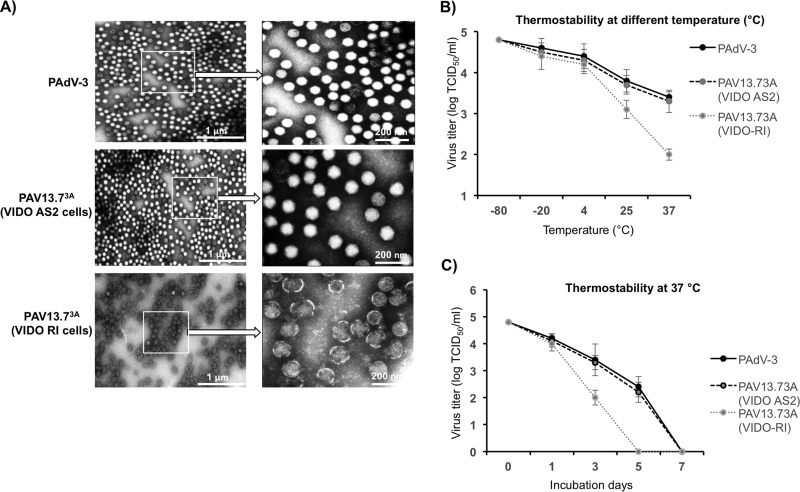
The adenovirus E3 region encodes proteins that are not essential for viral replication in vitro The porcine adenovirus type 3 (PAdV-3) E3 region encodes three proteins, including 13.7K. Here, we report that 13.7K is expressed as an early protein, which localizes to the nucleus of infected cells. The 13.7K protein is a structural protein, as it is incorporated in CsCl-purified virions. The 13.7K protein appears to be essential for PAdV-3 replication, as mutant PAV13.73A expressing a mutated 13.7K could be isolated only in VIDO AS2 cells expressing the 13.7K protein. Analysis of PAV13.73A suggested that even in the presence of reduced levels of some late viral proteins, there appeared to be no effect on virus assembly and production of mature virions. Further analysis of CsCl-purified PAV13.73A by transmission electron microscopy revealed the presence of disrupted/broken capsids, suggesting that inactivation of 13.7K protein expression may produce fragile capsids. Our results suggest that the PAdV-3 E3 region-encoded 13.7K protein is a capsid protein, which appears to be essential for the formation of stable capsids and production of infectious progeny virions.IMPORTANCE Although E3 region-encoded proteins are involved in the modulation of leukocyte functions (N. Arnberg, Proc Natl Acad Sci U S A 110:19976-19977, 2013) and inducing a lytic infection of lymphocytes (V. K. Murali, D. A. Ornelles, L. R. Gooding, H. T. Wilms, W. Huang, A. E. Tollefson, W. S. Wold, and C. Garnett-Benson, J Virol 88:903-912, 2014), none of the E3 proteins appear to be a component of virion capsid or required for replication of adenovirus. Here, we demonstrate that the 13.7K protein encoded by the E3 region of porcine adenovirus type 3 is a component of progeny virion capsids and appears to be essential for maintaining the integrity of virion capsid and production of infectious progeny virions. To our knowledge, this is the first report to suggest that an adenovirus E3-encoded protein is an essential structural protein.

Keywords: 13.7K; E3; PAdV-3; essential protein; porcine adenovirus; structural protein.


Figures

FIG 1
FIG 1
Expression of 13.7K. (A, B) Western blots. Proteins from the lysates of mock-infected cells (lanes 1), PAdV-3-infected cells (panel A, lane 2; panel B, lanes 2 to 7), plasmid pcDNA-3 DNA-transfected cells (panel A, lane 3), or plasmid pC_13.7 DNA-transfected VIDO R1 cells (6) (panel A, lane 4) were separated by 12% SDS-PAGE, transferred to nitrocellulose, and analyzed by Western blotting using anti-13.7K serum. Expression of β-actin using an anti-β-actin MAb was used as a loading control. The positions of molecular weight markers in kilodaltons are shown on the left. The molecular weights (in kilodaltons) of observed proteins are shown on the right. (C) Subcellular localization of PAdV-3 13.7K. Monolayers of VIDO R1 (6) cells mock infected (top row), infected with PAdV-3 (second row), transfected with plasmid pcDNA-3 DNA (third row), or transfected with plasmid pC_13.7K DNA (bottom row) were analyzed by indirect immunofluorescence using anti 13.7K serum and Alexa Fluor 488-conjugated (top and second rows) or TRITC-conjugated (third and bottom rows) goat anti-rabbit serum. The cells were mounted in a Vectashield mounting medium with DAPI (Vector Laboratories) to stain nuclei and monitored by confocal microscope.
FIG 2
FIG 2
Analysis of CsCl-purified PAdV-3 virions. (A) CsCl density gradient purification of PAdV-3. The upper band contains empty capsids (EC), and the lower band contains mature capsid (MC) (upper panels). Two to five micrograms of PAdV-3 empty capsids or mature capsids was used with anti-13.7K, anti-52K (23), antifiber (23), antihexon (23) anti-22K (23), or anti-33K (23) serum. (B) Proteinase K treatment. Proteins from purified PAdV-3 untreated (lane −) or treated (lane +) with 20 μg of proteinase K were separated by 10% to 12% SDS-PAGE, transferred to nitrocellulose, and probed with antihexon serum (a), anti-13.7K serum (b), and antifiber serum (c). The molecular weight markers (M) in kilodaltons are shown. (C) Silver staining. Proteins from purified PAdV-3 untreated (lane −) or treated (lane +) with 20 μg of proteinase K were separated by 12% SDS-PAGE, and the gel was stained with silver staining. The positions of the molecular weights (M) in kilodaltons are shown on the left. (D) Transmission electron microscopy. Purified PAdV-3 virions untreated (lane −) or treated (lane +) with 20 μg proteinase K were negatively stained with 2% aqueous phosphotungstic acid and analyzed by transmission electron microscopy.
FIG 3
FIG 3
Isolation of PAV13.73A in VIDO R1 cells (6). (A) Schematic illustration of the genomic organization of pVIII, early (E) region 3, and fiber in the PAdV-3 genome (8, 12). The E3 region encoding three overlapping proteins (13.7K, 23K, and 13.1K) is shown. (B) Schematic representation of plasmids pFPAV200 (7) and pFPAV13.73A. PAdV-3 genomic DNA (black filled box). E3 region (unfilled box). Plasmid DNA is represented by a thin line). Amp, ampicillin resistance gene. Mutation of 13.7K start codon AUG to AUA in plasmid pFPAV13.73A DNA is depicted. Micrographs represent cells showing cytopathic effect (a) and no cytopathic effect (b).
FIG 4
FIG 4
Expression of 13.7K in VIDO AS2 cell line. (A) Western blot. Proteins from the lysates (10 μl) of VIDO R1 cells (6) mock infected (lane 1) or infected with PAdV-3 at an MOI of 2 (lane 2) and of VIDO AS2 cells (lane 3) were analyzed by Western blotting using anti-13.7K serum. (B) Indirect immunofluorescence. Monolayers of VIDO-R1 cells (a1 to a3) or VIDO AS2 cells (expressing protein 13.7K) (b1 to b3) were fixed with 3.7% paraformaldehyde and visualized by indirect immunostaining using anti-13.7K serum followed by TRITC-conjugated goat anti-rabbit IgG using a confocal microscope. The nuclei were stained by DAPI.
FIG 5
FIG 5
Isolation of PAV13.73A in VIDO R1 cells. (A) Schematic representation of plasmid pFPAV13.73A and plasmid pFPAV13.7R DNAs as described for Fig. 3B. (B) Proteins from the lysates of cells infected with indicated viruses were analyzed by Western blotting using anti-DBP serum (24) as described for Fig. 1A and B. (C) Virus growth. Confluent monolayers of VIDO R1 were infected with PAdV-3 (wild-type), PAV13.73A (13.7K point mutation), or PAV13.7R (PAV13.73A revertant). At indicated times postinfection, the cell pellets were collected, freeze-thawed, and virus titrated on VIDO R1 (6) cells using TCID50 assay (6, 7). Values represent averages from two independent experiments, each with triplicate samples, and error bars represent standard deviations.
FIG 6
FIG 6
Analysis of viral protein expression in virus-infected cells. Proteins from the lysates of PAdV-3- or PAV13.73A-infected cells were analyzed by Western blotting using anti-pIX (23), anti-DBP (30), anti-13.7K (this study), anti-52K (23), antihexon (23), anti-pVIII (50), and anti-fiber (22) antibodies followed by alkaline phosphatase-conjugated secondary antibodies. The β-actin was detected using anti-β-actin MAb followed by alkaline phosphatase-conjugated secondary antibodies. The BCIP/NBT solution was used as a substrate to visualize proteins. The results were quantified using ImageJ software (http://rsb.info.nih.gov/ij/). Values represent the means from two independent experiments, and error bars indicate SD. Significant differences (P < 0.05) are indicated with an asterisk (*).
FIG 7
FIG 7
Analysis of mutant PAV13.73A. Analysis of genome replication in virus-infected cells. VIDO R1 cells were infected with PAdV-3 or PAV13.73A at an MOI of 2 and harvested at indicated times postinfection. The cell pellets were collected, and genomic DNA was extracted. The viral genome copy number was determined by quantitative PCR using primers (Table 3) as described previously (46).
FIG 8
FIG 8
Analysis of viral protein incorporation in purified virions. Proteins from CsCl-purified PAdV-3 or PAV13.73A were separated on 10 to 12% SDS-PAGE, transferred to nitrocellulose, and probed by Western blotting using anti-13.7K (this study), anti-pVIII (50), anti-hexon (23), anti-fiber (23), and anti-pIX (23) sera. All data were analyzed using GraphPad Prism, version 6 (GraphPad Software, Inc., La Jolla, CA, USA). The values represent averages from three independent experiments, and errors bars represent standard deviations.
FIG 9
FIG 9
Purified virus analysis and interaction of protein 13.7K with other PADV-3 protein(s). (A) Electron microscopic analysis. Purified PAdV-3 and PAV13.73A grown in VIDO AS2 cells and PAV13.73A grown in VIDO R-1 cells (6) are shown as indicated at a magnification of ×30,000. The arrows indicate enlargements of selected boxed regions of each virus (magnification, ×1,000,000). (B, C) Thermostability assay. Purified virions (105 TCID50) of PAdV-3 and PAV13.73A grown in either VIDO R1 or VIDO AS2 (expressing protein 13.7K) cells were incubated at different temperatures for 3 days, and the residual viral infectivity was determined by TCID50 (B). Purified virions (105 TCID50) of PAdV-3 and PAV13.73A grown in either VIDO R1 or VIDO AS2 cells were incubated at 37°C for the indicated time points (0, 1, 3, or 7 days postinfection), and the residual viral infectivity was determined by TCID50 (C).

Similar articles

Cited by

References

    1. Haig DA, Clarke MC, Pereira MS. 1964. Isolation of an adenovirus from a pig. J Compar Pathol 74:81–84. doi:10.1016/S0368-1742(64)80009-6. - DOI - PubMed
    1. Kadoi K, Inoue Y, Ikeda T, Kamata H, Yukawa M, Iwabuchi M, Inaba Y. 1995. Isolation of porcine adenovirus as a candidate of 5th serotype. J Basic Microbiol 35:195–204. doi:10.1002/jobm.3620350314. - DOI - PubMed
    1. Kadoi K, Iwabuchi M, Satoh T, Katase T, Kawaji T, Morichi T. 1997. Adenovirus isolation from spleen lymphocytes of apparently healthy pigs. New Microbiol 20:215–220. - PubMed
    1. Hirahara T, Yasuhara H, Matsui O, Yamanaka M, Tanaka M, Fukuyama S, Izumida A, Yoshiki K, Kodama K, Nakai M, et al. . 1990. Isolation of porcine adenovirus from the respiratory tract of pigs in Japan. Nihon juigaku zasshi. Jpn J Vet Sci 52:407–409. doi:10.1292/jvms1939.52.407. - DOI - PubMed
    1. Derbyshire JB, Clarke MC, Collins AP. 1975. Serological and pathogenicity studies with some unclassified porcine adenoviruses. J Compar Pathol 85:437–443. doi:10.1016/0021-9975(75)90031-6. - DOI - PubMed

Publication types

LinkOut - more resources