. 2018 Oct 1;217(10):3698-3714.
doi: 10.1083/jcb.201802018. Epub 2018 Jul 30.

GSK3-β promotes calpain-1-mediated desmin filament depolymerization and myofibril loss in atrophy

Affiliations

GSK3-β promotes calpain-1-mediated desmin filament depolymerization and myofibril loss in atrophy

Dina Aweida et al. J Cell Biol. .

Abstract

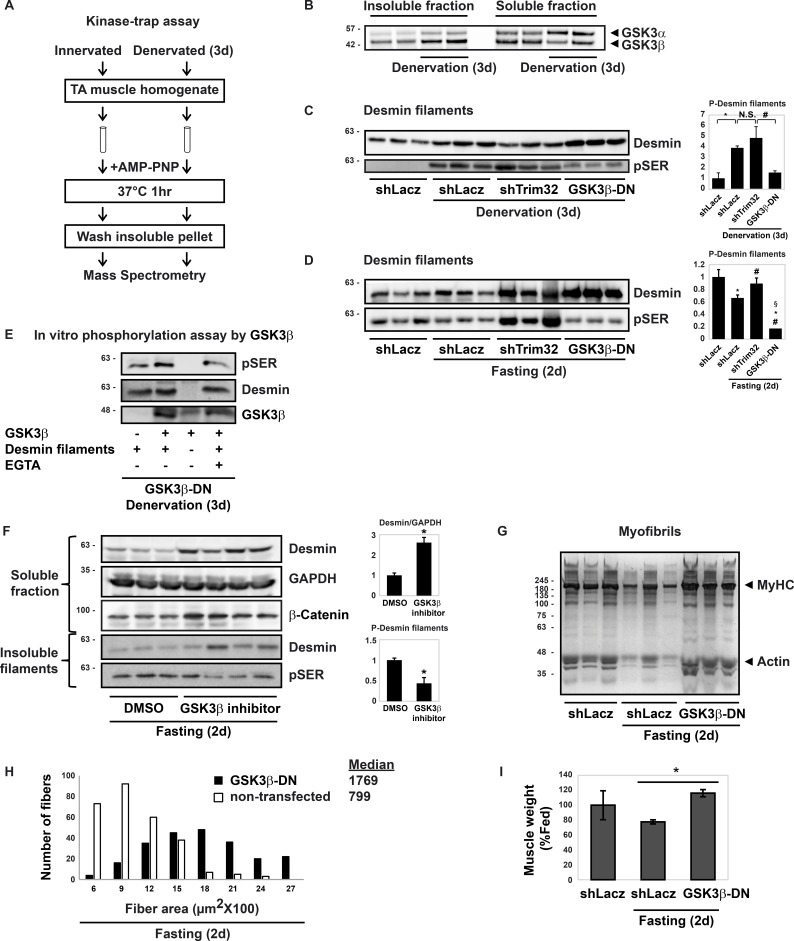
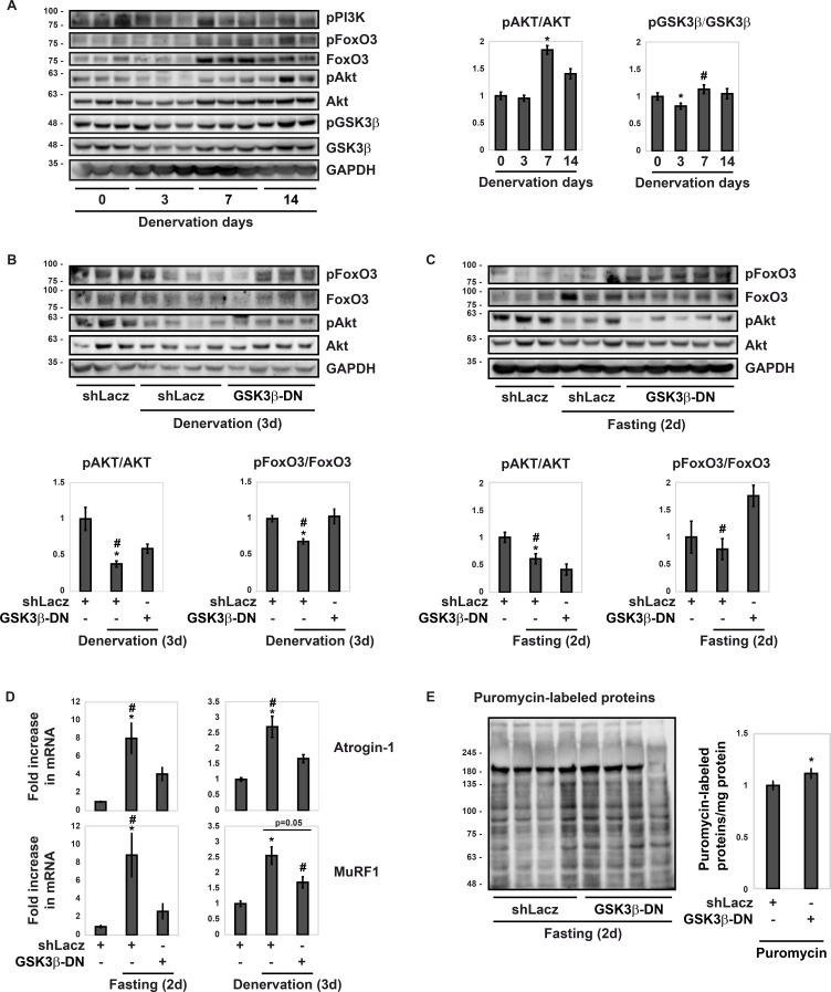
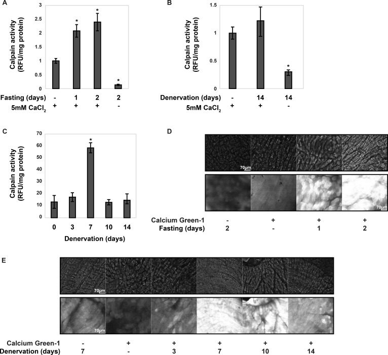
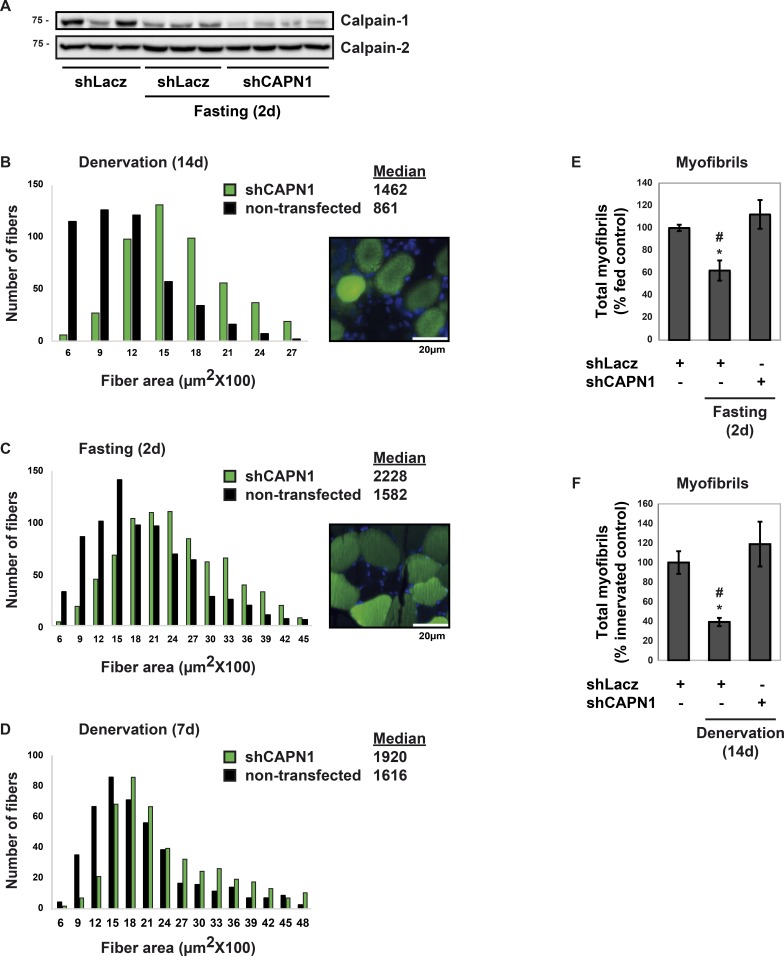
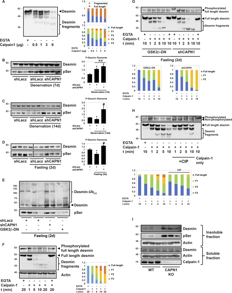
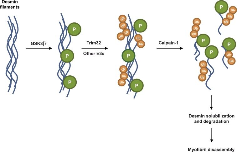
Myofibril breakdown is a fundamental cause of muscle wasting and inevitable sequel of aging and disease. We demonstrated that myofibril loss requires depolymerization of the desmin cytoskeleton, which is activated by phosphorylation. Here, we developed a mass spectrometry-based kinase-trap assay and identified glycogen synthase kinase 3-β (GSK3-β) as responsible for desmin phosphorylation. GSK3-β inhibition in mice prevented desmin phosphorylation and depolymerization and blocked atrophy upon fasting or denervation. Desmin was phosphorylated by GSK3-β 3 d after denervation, but depolymerized only 4 d later when cytosolic Ca2+ levels rose. Mass spectrometry analysis identified GSK3-β and the Ca2+-specific protease, calpain-1, bound to desmin and catalyzing its disassembly. Consistently, calpain-1 down-regulation prevented loss of phosphorylated desmin and blocked atrophy. Thus, phosphorylation of desmin filaments by GSK3-β is a key molecular event required for calpain-1-mediated depolymerization, and the subsequent myofibril destruction. Consequently, GSK3-β represents a novel drug target to prevent myofibril breakdown and atrophy.


Figures

Figure 1.
Figure 1.
GSK3-β promotes atrophy by catalyzing desmin filament phosphorylation and loss. (A) A scheme for kinase-trap assay. (B) GSK3-β binds the myofibrillar pellet 3 d after denervation. Soluble and insoluble fractions from control and 3-d-denervated TA muscles were incubated with AMP-PNP and analyzed by SDS-PAGE and immunoblotting. (C and D) During atrophy, inhibition of GSK3-β markedly reduces desmin phosphorylation. Desmin filaments isolated from atrophying muscles expressing GSK3-β-DN, shTrim32, or shLacz were analyzed by immunoblotting. Right: Densitometric measurement of presented blots. n = 3. *, P < 0.05 vs. control; #, P < 0.05 vs. shLacz in atrophy; §, P < 0.05 vs. shTrim32 in atrophy. (E) GSK3-β phosphorylates desmin filaments in vitro. Pellets from denervated (3 d) muscles expressing GSK3-β-DN were subjected to in vitro phosphorylation reaction by recombinant GSK3-β. (F) Inhibition of GSK3-β with specific inhibitor prevents desmin phosphorylation and loss during fasting. TA muscles from fasted mice injected with GSK3-β inhibitor (L803mts) or vehicle were analyzed by immunoblotting. Right: Densitometric measurement of presented blots. n = 3 for DMSO. n = 4 for GSK3-β inhibitor. *, P < 0.05. (G) During fasting, GSK3-β inhibition with a dominant negative prevents myofibril destruction. Equal fractions (0.1%) of myofibrils from GSK3-β-DN– or shLacz-expressing muscles from fed or fasted mice were analyzed by SDS-PAGE and Coomassie blue staining. (H) GSK3-β inhibition markedly reduces fiber atrophy during fasting. Measurements of cross-sectional areas of 500 fibers expressing GSK3-β (black bars) versus 500 nontransfected fibers (open bars) in the same muscle. n = 5 mice. (I) Inhibition of GSK3-β blocks the loss of muscle mass during fasting. Mean weights of electroporated muscles are plotted as percentage of fed control. n = 3. *, P < 0.05 vs. shLacz in atrophy.
Figure 2.
Figure 2.
GSK3-β is activated at an early phase during atrophy when PI3K–Akt–FoxO signaling is low. (A) Time course of PI3K-Akt activation after muscle denervation. Right: Densitometric measurement of presented blots. n = 3. *, P < 0.05 vs. innervated; #, P < 0.05 vs. 3-d-denervated muscles. (B and C) Inhibition of GSK3-β with GSK3-β-DN activates FoxO3 on denervation (B) or fasting (C). Transfected muscles were analyzed by immunoblotting. Bottom: Densitometric measurement of presented blots. n = 3 for shLacz. n = 4 for GSK3-β-DN. *, P < 0.05 vs. control; #, P < 0.05 vs. GSK3-β-DN. (D) GSK3-β inhibition reduces MuRF1 and Atrogin1 expression during atrophy. RT-PCR of mRNA preparations from atrophying and control muscles expressing shLacz or GSK3-β-DN using primers for MuRF1 and Atrogin1. Data are plotted as the mean fold change relative to control ± SEM. n = 5. *, P < 0.05 vs. control. #, P < 0.05 vs. GSK3-β-DN. (E) During fasting, inhibition of GSK3-β increases rates of protein synthesis. Mice were injected with puromycin, and soluble fractions of electroporated muscles were analyzed by immunoblotting. Right: Densitometric measurement of the presented blot. n = 4. *, P < 0.05 vs. shLacz.
Figure 3.
Figure 3.
During atrophy, calpain activity increases when cellular Ca2+ levels rise. (A–C) Calpain activity was measured in the insoluble fraction of normal and atrophying muscles during fasting (A) or after denervation (B and C) using the fluorogenic substrate SLY-AMC. n = 3. *, P < 0.05 vs. control. (D and E) Cellular Ca2+ levels were measured in strips of control and atrophying muscles using calcium green-1-AM. n = 3. Top, transmitted light; bottom, fluorescence (480 nm). Scale bar: 70 µm.
Figure 4.
Figure 4.
Calpain-1 down-regulation blocks myofibril loss and atrophy. TA muscles were electroporated with shCAPN1 or shLacz, and atrophy was induced by fasting (2 d) or denervation (7 or 14 d). (A) shRNA-mediated knockdown of calpain-1 in TA muscles from fasted mice. Soluble extracts were analyzed by immunoblotting. (B–D) Calpain-1 down-regulation reduces atrophy upon denervation (B and D) or fasting (C). Measurements of cross-sectional areas of 500 fibers transfected with shCAPN1 (and expressing GFP, green bars) versus 500 nontransfected fibers (black bars) in the same muscle. n = 3 mice. Scale bar: 20 µm. (E and F) Calpain-1 down-regulation blocks myofibril loss upon fasting (E) or denervation (F). The mean content of myofibrils per electroporated muscle is presented as percentage of control. n = 4. *, P < 0.05 vs. control; #, P < 0.05 vs. shCAPN1.
Figure 5.
Figure 5.
Desmin filament phosphorylation is essential for depolymerization by calpain-1. (A) Desmin is a direct substrate of calpain-1. In vitro cleavage of desmin filaments by recombinant calpain-1. Right: Densitometric measurement of presented blots. Full-length desmin and desmin fragments (F) are depicted as a fraction of the total amount of desmin. (B) Down-regulation of calpain-1 7 d after denervation does not affect desmin integrity. Desmin filaments were analyzed by immunoblotting. Right: Densitometric measurement of presented blots. n = 3. *, P < 0.05 vs. innervated control. (C and D) Down-regulation of calpain-1 prevents loss of phosphorylated desmin upon denervation (C) or fasting (D). Desmin filaments were analyzed by immunoblotting. Right: Densitometric measurement of presented blots. n = 3. *, P < 0.05 vs. control; #, P < 0.05 vs. shLacz in atrophy. Black line indicates the removal of an intervening lane for presentation purposes. (E) GSK3-β-mediated desmin filament phosphorylation precedes depolymerization by calpain-1. Insoluble fractions of muscles expressing shCAPN1, GSK3-β-DN, or shLacz were analyzed by immunoblotting. (F–H) Insoluble fractions from transfected muscles were incubated with purified calpain-1 for the indicated times. Protein cleavage was detected by immunoblotting. In densitometric measurement of presented blots, full-length desmin and desmin fragments (F) are depicted as a fraction of the total amount of desmin. (F) Desmin filaments from muscles expressing shCAPN1 from fasted mice are selectively cleaved by calpain-1. (G) Phosphorylation of desmin filaments facilitates cleavage by calpain-1. Cleavage of desmin filaments from atrophying muscles expressing GSK3-β-DN or shCAPN1 was analyzed in parallel. (H) Desmin filaments from muscles expressing shCAPN1 during fasting were treated with CIP (lanes 6–10) or left untreated (lanes 1–5) and then subjected to cleavage by recombinant calpain-1. (I) In muscles from calpain-1 null mice, desmin filaments accumulate. Insoluble and soluble fractions of muscles from WT and calpain-1 knockout mice were analyzed by immunoblotting.
Figure 6.
Figure 6.
Proposed mechanism for desmin filament loss during atrophy. Phosphorylation of desmin filaments by GSK3-β precedes and promotes ubiquitination by Trim32 and depolymerization by calpain-1, ultimately leading to myofibril loss and atrophy. Thus, phosphorylation by GSK3-β is a critical early step for desmin filament destruction and overall protein degradation during atrophy. Consequently, GSK3-β and calpain-1 may represent new therapeutic targets to reduce myofibril breakdown and muscle wasting during aging or disease.

Similar articles

Cited by

References

    1. Adams J.A. 2001. Kinetic and catalytic mechanisms of protein kinases. Chem. Rev. 101:2271–2290. 10.1021/cr000230w - DOI - PubMed
    1. Agnetti G., Halperin V.L., Kirk J.A., Chakir K., Guo Y., Lund L., Nicolini F., Gherli T., Guarnieri C., Caldarera C.M., et al. . 2014. Desmin modifications associate with amyloid-like oligomers deposition in heart failure. Cardiovasc. Res. 102:24–34. 10.1093/cvr/cvu003 - DOI - PMC - PubMed
    1. Albrecht L.V., Zhang L., Shabanowitz J., Purevjav E., Towbin J.A., Hunt D.F., and Green K.J.. 2015. GSK3- and PRMT-1-dependent modifications of desmoplakin control desmoplakin-cytoskeleton dynamics. J. Cell Biol. 208:597–612. 10.1083/jcb.201406020 - DOI - PMC - PubMed
    1. Blunt B.C., Creek A.T., Henderson D.C., and Hofmann P.A.. 2007. H2O2 activation of HSP25/27 protects desmin from calpain proteolysis in rat ventricular myocytes. Am. J. Physiol. Heart Circ. Physiol. 293:H1518–H1525. 10.1152/ajpheart.00269.2006 - DOI - PubMed
    1. Bodine S.C., Latres E., Baumhueter S., Lai V.K., Nunez L., Clarke B.A., Poueymirou W.T., Panaro F.J., Na E., Dharmarajan K., et al. . 2001. Identification of ubiquitin ligases required for skeletal muscle atrophy. Science. 294:1704–1708. 10.1126/science.1065874 - DOI - PubMed

Publication types