. 2018 Jul 30;14(7):e1007533.
doi: 10.1371/journal.pgen.1007533. eCollection 2018 Jul.

Metagenomic sequencing suggests a diversity of RNA interference-like responses to viruses across multicellular eukaryotes

Affiliations

Metagenomic sequencing suggests a diversity of RNA interference-like responses to viruses across multicellular eukaryotes

Fergal M Waldron et al. PLoS Genet. .

Erratum in

Abstract

RNA interference (RNAi)-related pathways target viruses and transposable element (TE) transcripts in plants, fungi, and ecdysozoans (nematodes and arthropods), giving protection against infection and transmission. In each case, this produces abundant TE and virus-derived 20-30nt small RNAs, which provide a characteristic signature of RNAi-mediated defence. The broad phylogenetic distribution of the Argonaute and Dicer-family genes that mediate these pathways suggests that defensive RNAi is ancient, and probably shared by most animal (metazoan) phyla. Indeed, while vertebrates had been thought an exception, it has recently been argued that mammals also possess an antiviral RNAi pathway, although its immunological relevance is currently uncertain and the viral small RNAs (viRNAs) are not easily detectable. Here we use a metagenomic approach to test for the presence of viRNAs in five species from divergent animal phyla (Porifera, Cnidaria, Echinodermata, Mollusca, and Annelida), and in a brown alga-which represents an independent origin of multicellularity from plants, fungi, and animals. We use metagenomic RNA sequencing to identify around 80 virus-like contigs in these lineages, and small RNA sequencing to identify viRNAs derived from those viruses. We identified 21U small RNAs derived from an RNA virus in the brown alga, reminiscent of plant and fungal viRNAs, despite the deep divergence between these lineages. However, contrary to our expectations, we were unable to identify canonical (i.e. Drosophila- or nematode-like) viRNAs in any of the animals, despite the widespread presence of abundant micro-RNAs, and somatic transposon-derived piwi-interacting RNAs. We did identify a distinctive group of small RNAs derived from RNA viruses in the mollusc. However, unlike ecdysozoan viRNAs, these had a piRNA-like length distribution but lacked key signatures of piRNA biogenesis. We also identified primary piRNAs derived from putatively endogenous copies of DNA viruses in the cnidarian and the echinoderm, and an endogenous RNA virus in the mollusc. The absence of canonical virus-derived small RNAs from our samples may suggest that the majority of animal phyla lack an antiviral RNAi response. Alternatively, these phyla could possess an antiviral RNAi response resembling that reported for vertebrates, with cryptic viRNAs not detectable through simple metagenomic sequencing of wild-type individuals. In either case, our findings show that the antiviral RNAi responses of arthropods and nematodes, which are highly divergent from each other and from that of plants and fungi, are also highly diverged from the most likely ancestral metazoan state.


Conflict of interest statement

The authors have declared that no competing interests exist.

Figures
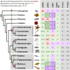
Fig 1
Fig 1. Distribution of small RNA pathways across the Metazoa.
Phylogeny of selected metazoan (animal) phyla (topology follows [180]) with a table recording the reported range of modal lengths for miRNAs, piRNAs, and viRNAs detectable by bulk sequencing from wild-type organisms (miRNA modes taken from miRbase). Entries marked ‘No’ have been reported to be absent, and those marked ‘?’ are untested. Focal taxa in this study are marked in colour, and the target table entries are outlined. Vertebrate viRNAs are marked ‘(×)’ as mammalian virus-derived small RNAs are only detectable in tissues and experimental systems lacking viral suppressors of RNAi and/or an interferon response [–35]. Note that piRNAs are absent from some, but not all, nematodes [57]. The column ‘dsRNA KD’ records whether dsRNA knockdown of gene expression using long dsRNA (i.e. a Dicer substrate) has been reported, as this may suggest the presence of an RNAi pathway capable of producing viRNAs from replicating viruses. The ‘Dcrs’ and ‘Agos’ columns record the inferred number of Dicers and (non-Piwi) Argonautes ancestrally present in each phylum, although the number of Dicers in Platyhelminthes is contentious as the putative second Dicer lacks the majority of expected Dicer domains. Broadly speaking, there are two competing hypotheses for the histories of Dicers and (non-Piwi) Argonautes in animals [47,50,181]. The first (labelled H1), posits that an early duplication in Dicer and/or Argonaute (marked D+ and A+ in dark green on the phylogeny) gave rise to at least two very divergent homologues of each gene in the lineage leading to the Metazoa, followed by subsequent losses (D- and A- in dark red). The second (H2), suggests that divergent homologues are the result of more recent duplications (D+ and A+ in pale green), and where homologs have high divergence it is as a result of rapid evolution. Note that these hypotheses are independent for Argonautes and Dicers, and one may be ancient but the other recent. For Dicers, at least, the ‘ancient’ duplication is arguably better supported [47], although it remains extremely difficult to determine orthology between the duplicates. In addition, Dicers and Argonautes have unambiguously diversified within some phyla (important examples marked A+ and D+ in grey)—as seen for the large nematode-specific WAGO clade of Argonautes (reviewed in [141]), and the multiple Argonautes in vertebrates.
Fig 2
Fig 2. Phylogenetic relationships of virus-like contigs from the dog whelk.
Mid-point rooted maximum likelihood phylogenetic trees for each of the virus-like contigs associated with viRNAs in the dog whelk (Nucella lapillus). New virus-like contigs described here are marked in red, sequences marked ‘TSA’ are derived from public transcriptome assemblies of the species named, and the scale is given in amino acid substitutions per site. Panels are: (A) rhabdoviruses related to lyssaviruses, inferred using the protein sequence of the nucleoprotein (the only open reading frame available from this contig, which is likely an EVE); (B) orthomyxoviruses related to influenza and thogoto viruses, inferred using the protein sequence of PB1; (C) rhabdoviruses and chuviruses, inferred from the RNA polymerase. Support values and accession identifiers are presented in S2 Fig and S3 Data, and alignments in S2 Data. Given the high level of divergence, alignments and inferred trees should be treated as tentative.
Fig 3
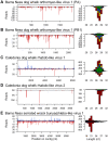
Fig 3. Small RNAs from RNA virus-like contigs.
Panels to the left show the distribution of 20-30nt small RNAs along the length of the virus-like contig, and panels to the right show the size distribution small RNA reads coloured by the 5' base (U red, G yellow, C blue, A green). Read counts above the x-axis represent reads mapping to the positive sense (coding) sequence and counts below the x-axis represent reads mapping to the complementary sequence. For the dog whelk (A-D), only reads from the oxidised library are shown. Other dog whelk libraries display similar distributions and the small-RNA ‘hotspot’ pattern along the contig is highly repeatable (S6 Fig). Small RNAs from the two segments of the orthomyxovirus (A and B) show strong strand bias to the negative strand and no 5' base composition bias. Those from the first rhabdo-like virus (C) display little strand bias and no base composition bias, and those from the second rhabdo virus-like contig, which is a probable EVE (D), derive only from the negative strand and display a very strong 5' U bias. There were insufficient reads from the positive strand of this virus to detect a ping-pong signature. Small RNAs from the four dog whelk contigs all display 28nt peaks. Small RNAs from the bunya/phlebo-like virus identified in the brown alga (E) derive from both strands, and show a strong 5' U bias with a peak size of 21nt. The data required to plot the size distributions are provided in S5 Table.
Fig 4
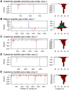
Fig 4. Small RNAs from DNA parvo/densovirus-like contigs.
Panels to the left show the distribution of 20-30nt small RNAs along the length of the parvo/densovirus-like contigs from sea anemone (A) and starfish (B-E), and panels to the right show the size distribution small RNA reads coloured by the 5' base (U red, G yellow, C blue, A green). Read counts above the x-axis represent reads mapping to the positive sense (coding) sequence, and counts below the x-axis represent reads mapping to the complementary sequence. Only reads from the oxidised library are shown, but other libraries display similar distributions, and the small-RNA ‘hotspot’ pattern is highly repeatable (S6 Fig). For all but one of the parvo/denso-like virus contigs, the small RNAs derived exclusively from the negative sense strand and showed a strong 5'U bias, consistent with piRNAs derived from endogenous copies (see main text). For one contig (B: Millport starfish parvo-like virus 1) reads derived predominantly from the positive strand and did not display a 5' U bias. Although the number of unique small RNA sequences from this virus was small, the positive-sense small RNAs showed a slight bias to A at position 10, consistent with ping-pong (S6 Fig). The data required to plot these size distributions is provided in S5 Table.
Fig 5
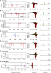
Fig 5. Small RNAs from TE-like contigs.
The threecolumns show (left to right): the distribution of 20-30nt small RNAs along the length of a TE-like contig; the size distribution of small RNA reads (U red, G yellow, C blue, A green); and the sequence ‘logo’ of unique sequences for the dominant sequence length. Read counts above the x-axis represent reads mapping to the positive sense (coding) sequence, and counts below the x-axis represent reads mapping to the complementary sequence. For the sequence logos, the upper and lower plots show positive and negative sense reads respectively, and the y-axis of each measures relative information content in bits. Where available, reads from the oxidised library are shown (A-F), but other libraries display similar distributions (S8 Fig). These examples from sponge (A), sea anemone (B), starfish (C), earthworm (D), dog whelk (E-F) and brown alga (G) were chosen to best illustrate the presence of the ‘ping pong’ signature, but other examples are shown in S8 Fig. Note that the size distribution of TE-derived small RNAs varies substantially among species, and that the dog whelk (E and F) displays at least two distinct patterns, one (F) reminiscent of that seen for some RNA virus contigs (Fig 3C). The data required to plot these figures is provided in S5 Table.

Similar articles

Cited by

References

    1. Obbard DJ, Gordon KHJ, Buck AH, Jiggins FM (2009) The evolution of RNAi as a defence against viruses and transposable elements. Philos Trans R Soc Lond B Biol Sci 364: 99–115. 10.1098/rstb.2008.0168 - DOI - PMC - PubMed
    1. Cerutti H, Casas-Mollano JA (2006) On the origin and functions of RNA-mediated silencing: from protists to man. Curr Genet 50: 81–99. 10.1007/s00294-006-0078-x - DOI - PMC - PubMed
    1. Ding SW, Li H, Lu R, Li F, Li WX (2004) RNA silencing: A conserved antiviral immunity of plants and animals. Virus Res 102: 109–115. 10.1016/j.virusres.2004.01.021 - DOI - PubMed
    1. Buchon N, Vaury C (2006) RNAi: a defensive RNA-silencing against viruses and transposable elements. Heredity (Edinb) 96: 195–202. 10.1038/sj.hdy.6800789 - DOI - PubMed
    1. Segers GC, Zhang X, Deng F, Sun Q, Nuss DL (2007) Evidence that RNA silencing functions as an antiviral defense mechanism in fungi. Proc Natl Acad Sci 104: 12902–12906. 10.1073/pnas.0702500104 - DOI - PMC - PubMed

Publication types

MeSH terms

LinkOut - more resources