

Anti-Inflammatory, Antioxidative, and Hepatoprotective Effects of Trans Δ9-Tetrahydrocannabinol/Sesame Oil on Adjuvant-Induced Arthritis in Rats
- PMID: 30046349
- PMCID: PMC6036806
- DOI: 10.1155/2018/9365464
Anti-Inflammatory, Antioxidative, and Hepatoprotective Effects of Trans Δ9-Tetrahydrocannabinol/Sesame Oil on Adjuvant-Induced Arthritis in Rats
Abstract
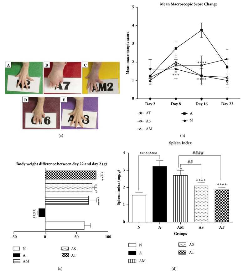
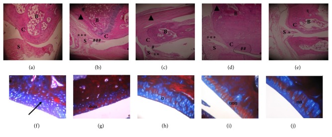
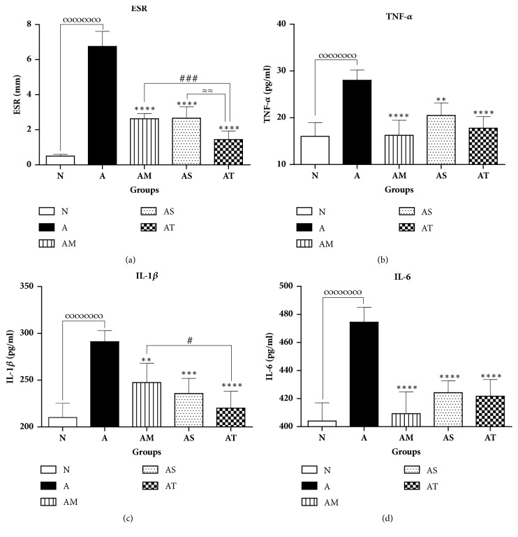
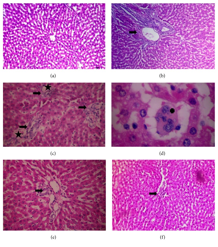
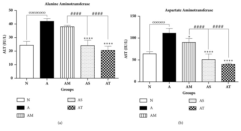
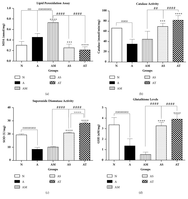
Rheumatoid arthritis (RA) is a painful chronic autoimmune disease affecting the joints. Its first-line therapy, Methotrexate (MTX), although effective in ameliorating the progress of the disease, induces hepatotoxicity over long-term usage. Thus, seeking natural compounds with fewer side effects could be an alternative therapeutic approach. This study aimed to investigate the anti-inflammatory, antiarthritic, and antioxidative effects of synthetic trans-Δ9-tetrahydrocannabinol (Δ9-THC) dissolved in sesame oil (Dronabinol) against MTX in adjuvant-induced arthritis (AIA) rat model. Daily oral administration of Δ9-THC/sesame oil, over a period of 21 days, was well tolerated in arthritic rats with no particular psychoactive side effects. It markedly attenuated the severity of clinical manifestations, recovered the histopathological changes in tibiotarsal joints, and repressed the splenomegaly in arthritic rats. Δ9-THC/sesame oil therapy showed similar effects to MTX in neutralizing the inflammatory process of AIA, through attenuating erythrocyte sedimentation rate (ESR) scores and proinflammatory cytokines, including tumor necrosis factor-alpha (TNF-α), interleukin 1-beta (IL-1β), and interleukin-6 (IL-6) levels, to normal values. As opposed to MTX, this natural combination markedly protected the liver of arthritic rats and downregulated the induced oxidative stress by increasing the antioxidant defense system such as activities of catalase and superoxide dismutase (SOD) and levels of glutathione (GSH). These results suggest promising effects for the clinical use of Δ9-THC/sesame oil therapy in alleviating arthritic clinical signs as well as arthritis-induced liver injury.
Figures
Similar articles
-
Silibinin's Effects against Methotrexate-Induced Hepatotoxicity in Adjuvant-Induced Arthritis Rat Model.Pharmaceuticals (Basel). 2024 Mar 28;17(4):431. doi: 10.3390/ph17040431. Pharmaceuticals (Basel). 2024. PMID: 38675395 Free PMC article.
-
Therapeutic benefits of Indole-3-Carbinol in adjuvant-induced arthritis and its protective effect against methotrexate induced-hepatic toxicity.BMC Complement Altern Med. 2018 Dec 19;18(1):337. doi: 10.1186/s12906-018-2408-1. BMC Complement Altern Med. 2018. PMID: 30567538 Free PMC article.
-
Evaluation of the effect of andrographolide and methotrexate combined therapy in complete Freund's adjuvant induced arthritis with reduced hepatotoxicity.Biomed Pharmacother. 2018 Oct;106:637-645. doi: 10.1016/j.biopha.2018.07.001. Epub 2018 Jul 11. Biomed Pharmacother. 2018. PMID: 29990853
-
Combination of coenzyme Q10 with methotrexate suppresses Freund's complete adjuvant-induced synovial inflammation with reduced hepatotoxicity in rats: Effect on oxidative stress and inflammation.Int Immunopharmacol. 2015 Jan;24(1):80-7. doi: 10.1016/j.intimp.2014.11.018. Epub 2014 Dec 3. Int Immunopharmacol. 2015. PMID: 25488045
-
Anti-arthritic effect of β-caryophyllene and its ameliorative role on methotrexate and/or leflunomide-induced side effects in arthritic rats.Life Sci. 2019 Sep 15;233:116750. doi: 10.1016/j.lfs.2019.116750. Epub 2019 Aug 10. Life Sci. 2019. PMID: 31408659
Cited by
-
Cannabis sativa L. (var. indica) Exhibits Hepatoprotective Effects by Modulating Hepatic Lipid Profile and Mitigating Gluconeogenesis and Cholinergic Dysfunction in Oxidative Hepatic Injury.Front Pharmacol. 2021 Dec 21;12:705402. doi: 10.3389/fphar.2021.705402. eCollection 2021. Front Pharmacol. 2021. PMID: 34992528 Free PMC article.
-
Protective Effect of α-Linolenic Acid on Non-Alcoholic Hepatic Steatosis and Interleukin-6 and -10 in Wistar Rats.Nutrients. 2019 Dec 18;12(1):9. doi: 10.3390/nu12010009. Nutrients. 2019. PMID: 31861497 Free PMC article.
-
Cannabinoids as Key Regulators of Inflammasome Signaling: A Current Perspective.Front Immunol. 2021 Jan 28;11:613613. doi: 10.3389/fimmu.2020.613613. eCollection 2020. Front Immunol. 2021. PMID: 33584697 Free PMC article. Review.
-
Cannabis sativa demonstrates anti-hepatocellular carcinoma potentials in animal model: in silico and in vivo studies of the involvement of Akt.J Cannabis Res. 2023 Jul 12;5(1):27. doi: 10.1186/s42238-023-00190-z. J Cannabis Res. 2023. PMID: 37434213 Free PMC article.
-
Analysis of Anti-Cancer and Anti-Inflammatory Properties of 25 High-THC Cannabis Extracts.Molecules. 2022 Sep 16;27(18):6057. doi: 10.3390/molecules27186057. Molecules. 2022. PMID: 36144796 Free PMC article.
References
LinkOut - more resources
Full Text Sources
Other Literature Sources
Research Materials
Miscellaneous
