. 2018 Jul 9:9:1573.
doi: 10.3389/fimmu.2018.01573. eCollection 2018.

Candida albicans-Induced NETosis Is Independent of Peptidylarginine Deiminase 4

Affiliations

Candida albicans-Induced NETosis Is Independent of Peptidylarginine Deiminase 4

Eva Guiducci et al. Front Immunol. .

Abstract

Neutrophils are the most abundant innate immune cells and the first line of defense against many pathogenic microbes, including the human fungal pathogen Candida albicans. Among the neutrophils' arsenal of effector functions, neutrophil extracellular traps (NETs) are thought to be of particular importance for trapping and killing the large fungal filaments by means of their web-like structures that consist of chromatin fibers decorated with proteolytic enzymes and host defense proteins. Peptidylarginine deiminase 4 (PAD4)-mediated citrullination of histones in activated neutrophils correlates with chromatin decondensation and extrusion and is widely accepted to act as an integral process of NET induction (NETosis). However, the requirement of PAD4-mediated histone citrullination for NET release during C. albicans infection remains unclear. In this study, we show that although PAD4-dependent neutrophil histone citrullination is readily induced by C. albicans, PAD4 is dispensable for NETosis in response to the fungus and other common NET-inducing stimuli. Moreover, PAD4 is not required for antifungal immunity during mucosal and systemic C. albicans infection. Our results demonstrate that PAD4 is dispensable for C. albicans-induced NETosis, and they highlight the limitations of using histone citrullination as a marker for NETs and PAD4-/- mice as a model of NET-deficiency.

Keywords: Candida albicans; host–fungus interaction; neutrophil extracellular traps; peptidylarginine deiminase 4.


Figures
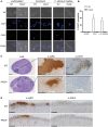
Figure 1
Figure 1
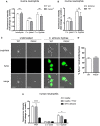
Candida albicans induces peptidylarginine deiminase 4 (PAD4)-mediated citrullination of histone H3. (A,B) Bone marrow neutrophils from WT and PAD4−/− mice were stimulated for 2.5 h with C. albicans hyphae or ionomycin or left unstimulated and then stained for DNA (DAPI; blue) and citrullinated histone H3 (magenta); brightfield. Images were acquired by confocal microscopy (A) and the fraction of CitH3+ neutrophils was determined (B). In (B), the bars are the mean with standard error of the mean (SEM) of 10–30 cells analyzed per condition. (C,D) WT and PAD4−/− mice were infected with C. albicans via the tail vein (C) or sublingually (D). Consecutive kidney (C) or tongue (D) sections were stained with periodic-acid schiff reagent (C), α-Ly6G or with α-CitH3 on day 3 postinfection (C) or on day 1 postinfection (D), respectively. Representative images are shown. Scale bar = 50 µm [in (A)], 1 mm [in (C), left panel], 250 µm [in (C) middle and right panel and in (D)]. See also Figure S1 in Supplementary Material.
Figure 2
Figure 2
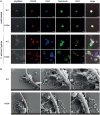
NETosis in response to Candida albicans hyphae is independent of Peptidylarginine deiminase 4 (PAD4). (A) The release of extracellular DNA from murine bone marrow neutrophils was detected by Sytox green after stimulation for 2.5 h with the yeast-locked strain Δhgc1 (C.a. yeast), preformed hyphae of the control strain Δhgc1 + HGC1 (C.a. hyphae), or ionomycin as indicated. The increase in fluorescence intensity from stimulated relative to unstimulated neutrophils is shown. Each bar represents the mean with standard error of the mean (SEM) of each group (n = 3) with data pooled from three independent experiments. (B) Bone marrow neutrophils were stimulated with C. albicans yeast cells or preformed hyphae of the highly virulent lab strain SC5413 and the low-virulent strain 101 as indicated. The release of extracellular DNA was detected by Sytox green as in (A). Data are the mean with SEM of each group (n = 3) with data pooled from three independent experiments. (C,D) The release of DNA from WT and PAD4−/− neutrophils that were stimulated and stained with Sytox as described in (A) was visualized by immunofluorescence microscopy. Representative images for each condition are shown in (C). Scale bar = 20 µm. The frequency of Sytox+ neutrophils among all C. albicans hyphae-associated neutrophils is shown in (D). The bars are the mean with SEM of 15 images analyzed per condition. (E) Human peripheral blood neutrophils from healthy donors that were treated with the PAD inhibitor Cl-amidine or left untreated and from patients with acquired myeloperoxidase-deficiency were stimulated for 2.5 h with the yeast-locked strain Δhgc1 (C.a. yeast), preformed hyphae of the control strain Δhgc1 + HGC1 (C.a. hyphae), or phorbol 12-myristate 13-acetate. Sytox was detected as described in (A). Each bar represents the mean with SEM of three technical replicates of each group. Data are representative of one out of two independent experiments.
Figure 3
Figure 3
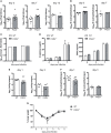
Characterization of neutrophil extracellular traps released from WT and PAD4−/− neutrophils. (A) Bone marrow neutrophils isolated from WT and PAD4−/− mice were stimulated for 2.5 h with Candida albicans hyphae or left unstimulated and then stained for membrane lipids (PKH26; red), DNA (DAPI; blue), calprotectin (anti-S100A8 and anti-S100A9, green), and myeloperoxidase (yellow). Images were acquired by confocal microscopy. Representative images of each group are shown. Scale bar = 20 µm. (B) Bone marrow neutrophils isolated from WT and PAD4−/− mice were stimulated for 2.5 h with C. albicans hyphae and phorbol 12-myristate 13-acetate/ionomycin. Cells were fixed and imaged by scanning electron microscopy. Representative images of each group are shown. Scale bar = 1 µm. Red arrowheads show C. albicans hyphae.
Figure 4
Figure 4
Peptidylarginine deiminase 4 (PAD4)-deficiency results in increased clustering of neutrophils to Candida albicans hyphae but does not affect their antifungal activity. (A–C) Neutrophils were isolated from the bone marrow of naïve WT and PAD4−/− mice (A,B) or from peripheral blood of a healthy donor and treated or not with the PAD inhibitor Cl-amidine (C). Neutrophils were incubated with C. albicans hyphae and then stained with DAPI (blue). The number of neutrophils interacting with each C. albicans hypha was quantified after 30 min (A) or 2.5 h (B,C) of stimulation. Representative images are shown on the left (Scale bar = 20 µm), summary plots are shown on the right. Bars are the mean with standard error of the mean (SEM) of each group with n = 2–3. (D) The cell wall integrity of C. albicans hyphae was assessed by transmission electron microscopy after 2.5 h incubation with WT or PAD4−/− bone marrow neutrophils. Representative images of each group are shown in the upper row. Lower row: some hyphae exposed to neutrophils showed membrane retraction and extensive cytoplasm disintegration (Di) or vacuolation, nuclear alterations, and swelling of the cell wall (Dii). Scale bar = 1 µm. (E) The frequency of C. albicans hyphae that were affected by WT and PAD4−/− neutrophils as described in (Di,Dii) was quantified. Bars are the mean with SEM of fungal elements from 18 EM images analyzed per condition. (F) WT and PAD4−/− bone marrow neutrophils were incubated with GFP-expressing C. albicans yeast cells and the degree of Candida uptake was determined by flow cytometry. Bars are the mean with standard error of the mean (SEM) of each group with n = 2. (G) Reactive oxygen species (ROS) production by WT and PAD4−/− bone marrow neutrophils in response to C. albicans yeast or hyphae was detected by chemiluminescence using the cell-impermeable substrate lucigenin to detect extracellular ROS (left) or the cell-permeable substrate luminol to detect total ROS (right). (H) WT and PAD4−/− bone marrow neutrophils were incubated for 3 h with opsonized or unopsonized C. albicans hyphae. The percentage of C. albicans killing was assessed by alamar blue assay. Bars are the mean with SEM of each group with n = 4. Data are pooled from (A,B,H) or representative of (G) two independent experiments.
Figure 5
Figure 5
The role of peptidylarginine deiminase 4 (PAD4) during systemic and oropharyngeal candidiasis. (A–D) WT and PAD4−/− mice were infected with Candida albicans via the tail vein. (A) The kidney fungal burden was determined on day 3, day 7, and day 13 postinfection. (B) CD45+ Ly6Cint Ly6G+ neutrophils were quantified in the kidney by flow cytometry on day 3 and day 7 postinfection. (C) Kidney injury molecule-1 (KIM-1) transcripts were quantified in kidney homogenates by qRT-PCR on day 3 and day 7 postinfection. (D) Creatinine and blood urea nitrogen levels were determined in the serum on day 3 and day 7 postinfection. (E–G) WT and PAD4−/− mice were infected with C. albicans sublingually. (E) The tongue fungal burden was determined on day 1 and day 3 postinfection. (F) CD45+ Ly6Cint Ly6G+ neutrophils were quantified in the tongue by flow cytometry on day 1 and day 3 postinfection. (G) The body weight was monitored during the course of infection. In (A,B,E,F), each symbol represents an individual mouse. The geometric mean (A–C,E,F) of each group is indicated. In (D), the bars are the mean with standard error of the mean (SEM) of each group (n = 8–10). Data are pooled from two to three independent experiments. The dotted lines in (A,E) represent the detection limit. See also Figures S2 and S3 in Supplementary Material.

Similar articles

Cited by

References

    1. Brown GD, Denning DW, Gow NA, Levitz SM, Netea MG, White TC. Hidden killers: human fungal infections. Sci Transl Med (2012) 4:165rv13.10.1126/scitranslmed.3004404 - DOI - PubMed
    1. Noble SM, Gianetti BA, Witchley JN. Candida albicans cell-type switching and functional plasticity in the mammalian host. Nat Rev Microbiol (2017) 15:96–108.10.1038/nrmicro.2016.157 - DOI - PMC - PubMed
    1. Berman J, Sudbery PE. Candida albicans: a molecular revolution built on lessons from budding yeast. Nat Rev Genet (2002) 3:918–30.10.1038/nrg948 - DOI - PubMed
    1. Jacobsen ID, Wilson D, Wachtler B, Brunke S, Naglik JR, Hube B. Candida albicans dimorphism as a therapeutic target. Expert Rev Anti Infect Ther (2012) 10:85–93.10.1586/eri.11.152 - DOI - PubMed
    1. Zhu W, Filler SG. Interactions of Candida albicans with epithelial cells. Cell Microbiol (2010) 12:273–82.10.1111/j.1462-5822.2009.01412.x - DOI - PMC - PubMed

LinkOut - more resources