. 2018 Aug;3(8):920-931.
doi: 10.1038/s41564-018-0191-x. Epub 2018 Jul 23.

The type VI secretion system deploys antifungal effectors against microbial competitors

Affiliations

The type VI secretion system deploys antifungal effectors against microbial competitors

Katharina Trunk et al. Nat Microbiol. 2018 Aug.

Abstract

Interactions between bacterial and fungal cells shape many polymicrobial communities. Bacteria elaborate diverse strategies to interact and compete with other organisms, including the deployment of protein secretion systems. The type VI secretion system (T6SS) delivers toxic effector proteins into host eukaryotic cells and competitor bacterial cells, but, surprisingly, T6SS-delivered effectors targeting fungal cells have not been reported. Here we show that the 'antibacterial' T6SS of Serratia marcescens can act against fungal cells, including pathogenic Candida species, and identify the previously undescribed effector proteins responsible. These antifungal effectors, Tfe1 and Tfe2, have distinct impacts on the target cell, but both can ultimately cause fungal cell death. 'In competition' proteomics analysis revealed that T6SS-mediated delivery of Tfe2 disrupts nutrient uptake and amino acid metabolism in fungal cells, and leads to the induction of autophagy. Intoxication by Tfe1, in contrast, causes a loss of plasma membrane potential. Our findings extend the repertoire of the T6SS and suggest that antifungal T6SSs represent widespread and important determinants of the outcome of bacterial-fungal interactions.


Conflict of interest statement

Competing Financial Interests

The authors declare no competing financial interests.

Figures

Figure 1
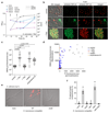
Figure 1. Cross-kingdom targeting by the Type VI secretion system of S. marcescens depends on the anti-fungal effectors Tfe1 and Tfe2.
(a) Number of recovered viable cells of S. cerevisiae K699 following co-culture with wild type (WT) or a T6SS-inactive mutant (ΔtssE) strain of S. marcescens Db10 as attacker, or with sterile media alone (media), in the absence or presence of a separating membrane. (b) Recovery of C. albicans SC5314 and C. glabrata ATCC2001 following co-culture with wild type or a T6SS-inactive mutant of S. marcescens. (c) Recovery of C. albicans, left, S. cerevisiae, middle, and C. glabrata, right, following co-culture with wild type or mutant (ΔtssE, Δtfe1, Δtfe2, Δtfe1Δtfe2 and ΔSMDB11_3457) S. marcescens. (d) Recovery of C. albicans, S. cerevisiae and C. glabrata following co-culture with wild type or mutant strains of S. marcescens Db10 carrying either the vector control plasmid (+VC, pSUPROM), or a plasmid directing the expression of Tfe1 with the immunity protein Sip3 (+Tfe1), or Tfe2 (+Tfe2). (e) Immunoblot detection of Hcp1 and Ssp2 in cellular and secreted fractions of wild type or mutant S. marcescens as indicated. The data are representative of two independent experiments and the full, uncropped blots can be found in Supplementary Fig. 18. In parts a-d, individual data points are overlaid with the mean +/- SEM (n=4 biological replicates).
Figure 2
Figure 2. Quantitative cellular proteomics identifies known and previously-unidentified T6SS-secreted proteins.
(a) Volcano plot summarising the proteomic comparison of total intracellular proteins between the wild type (WT) and a T6SS-inactive mutant (ΔtssE) strain of S. marcescens Db10 using label-free quantitation. The log2 of the ratios of protein intensities between the wild type and the ΔtssE mutant are plotted against the –log10 of t-test p-values (unpaired t-test, two-sided; n=6 biological replicates). Proteins significantly increased in abundance in the ΔtssE mutant compared with the wild type (ΔtssE/WT > 1.4-fold, p < 0.05) are depicted as circles, with magenta circles representing those secreted effectors (SE) identified as anti-fungal effectors in this study, and blue circles representing other hits, including secreted core components (SCC) or previously identified secreted effectors. Grey squares represent proteins significantly decreased in abundance in the ΔtssE mutant compared with the wild type (ΔtssE/WT < -1.4-fold, p < 0.05) and black diamonds correspond to proteins without significant changes. Numbers in brackets represent genomic identifiers for the proteins (SMDB11_xxxx). (b) Schematic depiction of the genetic loci containing the tfe1, tfe2 and SMDB11_3457 genes in S. marcescens Db10. The sip3 gene encodes the previously identified immunity protein for Tfe1/Ssp3.
Figure 3
Figure 3. Tfe1 and Tfe2 are anti-fungal toxins that impact fungal cell integrity.
(a) DIC microscopy of target strains C. albicans SC5314, S. cerevisiae K699 and C. glabrata ATCC2001, following co-culture with wild type or mutant (ΔtssE, Δtfe1, Δtfe2, Δtfe1Δtfe2) strains of S. marcescens Db10. ‘None’ indicates co-culture of the target with media alone. Representative of three independent experiments. (b) Percentage of live fungal cells of C. albicans, C. glabrata and S. cerevisiae, following co-culture with strains of S. marcescens, as indicated by propidium iodide (PI) staining. Bars show mean +/- SEM (n=3 independent experiments, with individual data points shown). (c) DIC microscopy of C. albicans SC5314 (control) and derivatives expressing GFP, Tfe1 or Tfe2, upper panel, and S. cerevisiae K699 carrying the control plasmid pRB1438 or GFP-, Tfe1- or Tfe2-expression plasmids, lower panel. Representative of two independent experiments. (d) Growth of S. cerevisiae K699 chromosomal integration strains harbouring the empty promoter construct (control) or constructs for expression of GFP, Tfe1 or Tfe2, on non-inducing and inducing media (gal, galactose). Representative of two independent experiments. In parts a and c, scale bars represent 10 µm and scale is the same across each row of images.
Figure 4
Figure 4. T6SS-mediated effector delivery disrupts fungal metabolic activity and membrane potential, and can act against hyphal cells.
(a) Growth of S. cerevisiae expressing Tfe2 as determined by cell density (OD600) and viable cell counts (cfu/ml). Time-course presented includes an induction period in non-inducing (none, 2% raffinose) or inducing media (induce, 2% raffinose 2% galactose), followed by either relief (none, 2% raffinose), repression (repress, 2% raffinose 2% glucose) or ongoing (induce, 2% raffinose 2% galactose) expression of Tfe2. Points show mean +/- SEM (n=3). (b) Fluorescence microscopy of FUN1-stained S. cerevisiae cells expressing GFP, Tfe1 or Tfe2. The single and merged green and red fluorescence channels, and their overlays on the corresponding DIC image are shown. Representative of two independent experiments. (c) Quantification of fluorescence from S. cerevisiae cells expressing Tfe1 or Tfe2, following staining with the potential-sensitive dye DiBAC4(3). Control cells were additionally treated with 10 µM mellitin (membrane depolarization through pore formation) or 3 µg/ml amphotericin B (membrane depolarization without pore formation). Points represent individual cells and bars show mean (p values are indicated; unpaired, two-sided t-test; n=100-116). (d) Quantification of cellular DiBAC4(3) and propidium iodide (PI) fluorescence for cells expressing Tfe1 or Tfe2, or treated with mellitin or amphotericin B. Scatter plots depict fluorescence intensity values of individual cells for both dyes (au, arbitary units; n=95-128). Example microscopy images and separate scatter plots are in Supplementary Fig. 8. (a)-(d) S. cerevisiae strains were K699 chromosomal integration strains harbouring the empty promoter construct (control) or constructs for galactose-inducible expression of GFP, Tfe1 or Tfe2. (e) Representative images and (f) quantification of PI staining of hyphae of the constitutively filamentous tup1Δ mutant of C. albicans, following co-culture with the strains of S. marcescens Db10 indicated (none, media only; WT, wild type). Bars show mean +/- SEM (n=3 independent experiments, individual data points shown), images are representative of four independent experiments. In parts b and e, scale bars represent 10 µm.
Figure 5
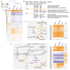
Figure 5. ‘In competition’ quantitative proteomics reveals interference of Tfe2 with fungal nutrient uptake and metabolism.
(a) Heat map showing hierarchical clustering of the differential proteome of C. albicans SC5314 following co-culture with wild type S. marcescens Db10 compared with mutant (ΔtssE, Δtfe1, Δtfe2 and Δtfe1Δtfe2) strains of Db10. All 1667 proteins with significant ANOVA scores (p<0.05, n=6 biological replicates) are included and the full cluster analysis with gene identifiers is available as Supplementary Data 4. (b) GO terms significantly enriched (p<0.05) in the set of 30 C. albicans SC5314 proteins identified as significantly responsive to Tfe2 (Supplementary Table 1). Where different, the protein name in S. cerevisiae is given in brackets. (c,d) Schematic illustration (left panels) and corresponding heat-maps of the pathway-associated proteins (right panels) of the sulfate assimilation and arginine biosynthesis pathways in fungal cells. Proteins increased in abundance following co-culture with wild type S. marcescens as compared with the mutant strains indicated are shown in purple, and proteins decreased are shown in orange.
Figure 6
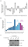
Figure 6. Tfe2 alters the cellular amino acid pool and induces autophagy in fungal cells.
(a) Relative levels of total intracellular amino acids in S. cerevisiae K699 expressing plasmid-borne Tfe2 (+Tfe2) compared with the vector control (Cit, citrulline; Orn, ornithine). Bars show log2 of the mean +/- SEM (n=3 biological replicates), with individual data points shown. (b) Differential expression analysis of genes involved in nutrient sensing and starvation in S. cerevisiae expressing Tfe2 relative to the vector control. qRT-PCR analysis was performed on transcripts of genes for amino acid permease and dipeptide transporters that are under the control of the amino acid sensor complex SPS (AGP1, GNP1, BAP2, BAP3, DIP5, CAN1, PTR2), the SPS sensor component (SSY1), the SPS-activated transcriptional regulators (STP1, STP2), the sulfate transporter (SUL1, SUL2) and the general amino acid control response regulator (GCN4). Bars show log2 of the mean relative transcript abundance +/- SEM (n=3 biological replicates). (c) Immunoblot detection of GFP-Atg8 processing in S. cerevisiae K699 following co-culture with wild type (WT) or mutant (ΔtssE and Δtfe2) strains of S. marcescens Db10 using anti-GFP antibody. Yeast cells carry the reporter plasmid GFP-ATG8(416) directing expression of GFP-Atg8 from the endogenous Atg8 promoter. GFP-Atg8 has a MW of 41 kDa and cleaved GFP is 27 kDa. Pgk1 was used as control cellular protein and dashed line indicates where the blot membrane was cut to allow detection with the two different antibodies; the full, uncropped blots can be found in Supplementary Fig. 18. These data are representative of three independent experiments.

Comment in

  • The needle and the damage done.
    Brunke S, Hube B. Brunke S, et al. Nat Microbiol. 2018 Aug;3(8):860-861. doi: 10.1038/s41564-018-0194-7. Nat Microbiol. 2018. PMID: 30046168 No abstract available.

Similar articles

Cited by

References

    1. Boer W, Folman LB, Summerbell RC, Boddy L. Living in a fungal world: impact of fungi on soil bacterial niche development. FEMS Microbiol Rev. 2005;29:795–811. - PubMed
    1. Peleg AY, Hogan DA, Mylonakis E. Medically important bacterial-fungal interactions. Nat Rev Microbiol. 2010;8:340–349. - PubMed
    1. Costa TR, et al. Secretion systems in Gram-negative bacteria: structural and mechanistic insights. Nat Rev Microbiol. 2015;13:343–359. - PubMed
    1. Basler M. Type VI secretion system: secretion by a contractile nanomachine. Philos Trans R Soc Lond B Biol Sci. 2015;370 - PMC - PubMed
    1. Cianfanelli FR, Monlezun L, Coulthurst SJ. Aim, Load, Fire: The Type VI Secretion System, a Bacterial Nanoweapon. Trends Microbiol. 2016;24:51–62. - PubMed

Publication types

MeSH terms

Substances