

Distinct mechanisms for PDGF and FGF signaling in primitive endoderm development
- PMID: 30026121
- PMCID: PMC6163042
- DOI: 10.1016/j.ydbio.2018.07.010
Distinct mechanisms for PDGF and FGF signaling in primitive endoderm development
Abstract
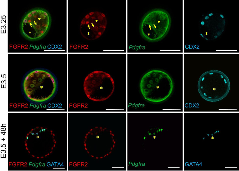
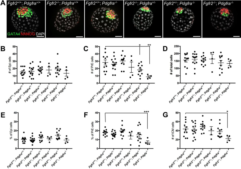
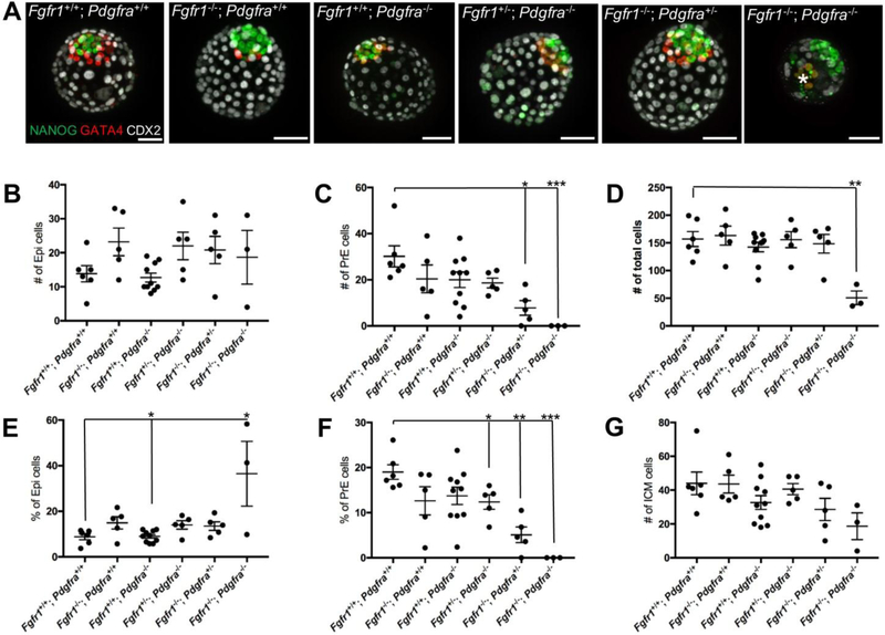
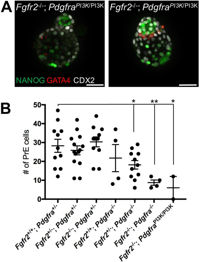
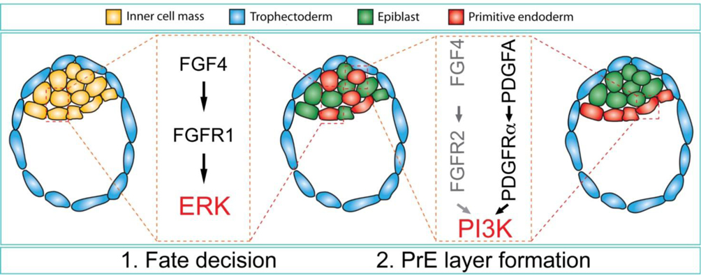
FGF signaling is known to play a critical role in the specification of primitive endoderm (PrE) and epiblast (Epi) from the inner cell mass (ICM) during mouse preimplantation development, but how FGFs synergize with other growth factor signaling pathways is unknown. Because PDGFRα signaling has also been implicated in the PrE, we investigated the coordinate functions of PDGFRα together with FGFR1 or FGFR2 in PrE development. PrE development was abrogated in Pdgfra; Fgfr1 compound mutants, or significantly reduced in Pdgfra; Fgfr2 or PdgfraPI3K; Fgfr2 compound mutants. We provide evidence that both Fgfr2 and Pdgfra play roles in PrE cell survival while Fgfr1 controls PrE cell specification. Our results suggest a model where FGFR1-engaged ERK1/2 signaling governs PrE specification while PDGFRα- and by analogy possibly FGFR2- engaged PI3K signaling regulates PrE survival and positioning in the embryo. Together, these studies indicate how multiple growth factors and signaling pathways can cooperate in preimplantation development.
Keywords: Cell specification; ERK1/2; PI3K; Preimplantation; Survival.
Copyright © 2018 Elsevier Inc. All rights reserved.
Figures
Similar articles
-
PDGF Signaling in Primitive Endoderm Cell Survival Is Mediated by PI3K-mTOR Through p53-Independent Mechanism.Stem Cells. 2019 Jul;37(7):888-898. doi: 10.1002/stem.3008. Epub 2019 Apr 10. Stem Cells. 2019. PMID: 30913328
-
Distinct Requirements for FGFR1 and FGFR2 in Primitive Endoderm Development and Exit from Pluripotency.Dev Cell. 2017 Jun 5;41(5):511-526.e4. doi: 10.1016/j.devcel.2017.05.004. Epub 2017 May 25. Dev Cell. 2017. PMID: 28552557 Free PMC article.
-
Lineage Establishment and Progression within the Inner Cell Mass of the Mouse Blastocyst Requires FGFR1 and FGFR2.Dev Cell. 2017 Jun 5;41(5):496-510.e5. doi: 10.1016/j.devcel.2017.05.003. Epub 2017 May 25. Dev Cell. 2017. PMID: 28552559 Free PMC article.
-
[Epiblast and primitive endoderm cell specification during mouse preimplantation development: a combination between biology and mathematical modeling].Med Sci (Paris). 2016 Feb;32(2):192-7. doi: 10.1051/medsci/20163202013. Epub 2016 Mar 2. Med Sci (Paris). 2016. PMID: 26936177 Review. French.
-
Primitive endoderm differentiation: from specification to epithelium formation.Philos Trans R Soc Lond B Biol Sci. 2014 Dec 5;369(1657):20130537. doi: 10.1098/rstb.2013.0537. Philos Trans R Soc Lond B Biol Sci. 2014. PMID: 25349446 Free PMC article. Review.
Cited by
-
A PDGFRβ-PI3K signaling axis mediates periosteal cell activation during fracture healing.PLoS One. 2019 Oct 30;14(10):e0223846. doi: 10.1371/journal.pone.0223846. eCollection 2019. PLoS One. 2019. PMID: 31665177 Free PMC article.
-
FGF20-FGFR1 signaling through MAPK and PI3K controls sensory progenitor differentiation in the organ of Corti.Dev Dyn. 2021 Feb;250(2):134-144. doi: 10.1002/dvdy.231. Epub 2020 Sep 9. Dev Dyn. 2021. PMID: 32735383 Free PMC article.
-
MED20 is essential for early embryogenesis and regulates NANOG expression.Reproduction. 2019 Mar;157(3):215-222. doi: 10.1530/REP-18-0508. Reproduction. 2019. PMID: 30571656 Free PMC article.
-
A pendulum of induction between the epiblast and extra-embryonic endoderm supports post-implantation progression.Development. 2022 Oct 15;149(20):dev192310. doi: 10.1242/dev.192310. Epub 2022 Aug 22. Development. 2022. PMID: 35993866 Free PMC article.
-
Platelet-Derived Growth Factor Receptor Type α Activation Drives Pulmonary Vascular Remodeling Via Progenitor Cell Proliferation and Induces Pulmonary Hypertension.J Am Heart Assoc. 2022 Apr 5;11(7):e023021. doi: 10.1161/JAHA.121.023021. Epub 2022 Mar 29. J Am Heart Assoc. 2022. PMID: 35348002 Free PMC article.
References
-
- Chazaud C, Yamanaka Y, Pawson T, Rossant J, 2006. Early lineage segregation between epiblast and primitive endoderm in mouse blastocysts through the Grb2-MAPK pathway. Developmental cell 10, 615–624. - PubMed
Publication types
MeSH terms
Substances
Grants and funding
LinkOut - more resources
Full Text Sources
Other Literature Sources
Molecular Biology Databases
Miscellaneous
