The S2 Subunit of Infectious Bronchitis Virus Beaudette Is a Determinant of Cellular Tropism
- PMID: 30021894
- PMCID: PMC6146808
- DOI: 10.1128/JVI.01044-18
The S2 Subunit of Infectious Bronchitis Virus Beaudette Is a Determinant of Cellular Tropism
Abstract
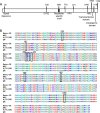
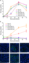
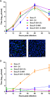
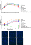
The spike (S) glycoprotein of the avian gammacoronavirus infectious bronchitis virus (IBV) is comprised of two subunits (S1 and S2), has a role in virulence in vivo, and is responsible for cellular tropism in vitro We have previously demonstrated that replacement of the S glycoprotein ectodomain from the avirulent Beaudette strain of IBV with the corresponding region from the virulent M41-CK strain resulted in a recombinant virus, BeauR-M41(S), with the in vitro cell tropism of M41-CK. The IBV Beaudette strain is able to replicate in both primary chick kidney cells and Vero cells, whereas the IBV M41-CK strain replicates in primary cells only. In order to investigate the region of the IBV S responsible for growth in Vero cells, we generated a series of recombinant IBVs expressing chimeric S glycoproteins, consisting of regions from the Beaudette and M41-CK S gene sequences, within the genomic background of Beaudette. The S2, but not the S1, subunit of the Beaudette S was found to confer the ability to grow in Vero cells. Various combinations of Beaudette-specific amino acids were introduced into the S2 subunit of M41 to determine the minimum requirement to confer tropism for growth in Vero cells. The ability of IBV to grow and produce infectious progeny virus in Vero cells was subsequently narrowed down to just 3 amino acids surrounding the S2' cleavage site. Conversely, swapping of the 3 Beaudette-associated amino acids with the corresponding ones from M41 was sufficient to abolish Beaudette growth in Vero cells.IMPORTANCE Infectious bronchitis remains a major problem in the global poultry industry, despite the existence of many different vaccines. IBV vaccines, both live attenuated and inactivated, are currently grown on embryonated hen's eggs, a cumbersome and expensive process due to the fact that most IBV strains do not grow in cultured cells. The reverse genetics system for IBV creates the opportunity for generating rationally designed and more effective vaccines. The observation that IBV Beaudette has the additional tropism for growth on Vero cells also invokes the possibility of generating IBV vaccines produced from cultured cells rather than by the use of embryonated eggs. The regions of the IBV Beaudette S glycoprotein involved in the determination of extended cellular tropism were identified in this study. This information will enable the rational design of a future generation of IBV vaccines that may be grown on Vero cells.
Keywords: S2′; cellular tropism; coronavirus; infectious bronchitis virus; reverse genetic analysis.
Copyright © 2018 Bickerton et al.
Figures


Similar articles
-
The spike protein of the apathogenic Beaudette strain of avian coronavirus can elicit a protective immune response against a virulent M41 challenge.PLoS One. 2024 Jan 24;19(1):e0297516. doi: 10.1371/journal.pone.0297516. eCollection 2024. PLoS One. 2024. PMID: 38265985 Free PMC article.
-
Contributions of the S2 spike ectodomain to attachment and host range of infectious bronchitis virus.Virus Res. 2013 Nov 6;177(2):127-37. doi: 10.1016/j.virusres.2013.09.006. Epub 2013 Sep 13. Virus Res. 2013. PMID: 24041648 Free PMC article.
-
Recombinant Infectious Bronchitis Viruses Expressing Chimeric Spike Glycoproteins Induce Partial Protective Immunity against Homologous Challenge despite Limited Replication In Vivo.J Virol. 2018 Nov 12;92(23):e01473-18. doi: 10.1128/JVI.01473-18. Print 2018 Dec 1. J Virol. 2018. PMID: 30209177 Free PMC article.
-
Severe acute respiratory syndrome vaccine development: experiences of vaccination against avian infectious bronchitis coronavirus.Avian Pathol. 2003 Dec;32(6):567-82. doi: 10.1080/03079450310001621198. Avian Pathol. 2003. PMID: 14676007 Free PMC article. Review.
-
The avian coronavirus spike protein.Virus Res. 2014 Dec 19;194:37-48. doi: 10.1016/j.virusres.2014.10.009. Epub 2014 Oct 17. Virus Res. 2014. PMID: 25451062 Free PMC article. Review.
Cited by
-
Comparative Analysis of Gene Expression in Virulent and Attenuated Strains of Infectious Bronchitis Virus at Subcodon Resolution.J Virol. 2019 Aug 28;93(18):e00714-19. doi: 10.1128/JVI.00714-19. Print 2019 Sep 15. J Virol. 2019. PMID: 31243124 Free PMC article.
-
Revealing Novel-Strain-Specific and Shared Epitopes of Infectious Bronchitis Virus Spike Glycoprotein Using Chemical Linkage of Peptides onto Scaffolds Precision Epitope Mapping.Viruses. 2023 Nov 20;15(11):2279. doi: 10.3390/v15112279. Viruses. 2023. PMID: 38005955 Free PMC article.
-
Lithium chloride inhibits infectious bronchitis virus-induced apoptosis and inflammation.Microb Pathog. 2022 Jan;162:105352. doi: 10.1016/j.micpath.2021.105352. Epub 2021 Dec 7. Microb Pathog. 2022. PMID: 34883226 Free PMC article.
-
The V617I Substitution in Avian Coronavirus IBV Spike Protein Plays a Crucial Role in Adaptation to Primary Chicken Kidney Cells.Front Microbiol. 2020 Dec 18;11:604335. doi: 10.3389/fmicb.2020.604335. eCollection 2020. Front Microbiol. 2020. PMID: 33391226 Free PMC article.
-
The spike protein of the apathogenic Beaudette strain of avian coronavirus can elicit a protective immune response against a virulent M41 challenge.PLoS One. 2024 Jan 24;19(1):e0297516. doi: 10.1371/journal.pone.0297516. eCollection 2024. PLoS One. 2024. PMID: 38265985 Free PMC article.
References
-
- Cavanagh D, Gelb J Jr. 2008. Infectious bronchitis, p 117–135. In Saif YM. (ed), Diseases of poultry, 12th ed Blackwell Publishing, Ames, IA.
Publication types
MeSH terms
Substances
Grants and funding
- BBS/E/I/00007034/BB_/Biotechnology and Biological Sciences Research Council/United Kingdom
- BBS/E/I/00007038/BB_/Biotechnology and Biological Sciences Research Council/United Kingdom
- BBS/E/I/00007039/BB_/Biotechnology and Biological Sciences Research Council/United Kingdom
- BBS/E/I/00001524/BB_/Biotechnology and Biological Sciences Research Council/United Kingdom
LinkOut - more resources
Full Text Sources
Other Literature Sources
Research Materials

