. 2018 Sep 14;430(18 Pt B):3170-3189.
doi: 10.1016/j.jmb.2018.07.008. Epub 2018 Jul 12.

Structural and Functional Characterization of the BcsG Subunit of the Cellulose Synthase in Salmonella typhimurium

Affiliations

Structural and Functional Characterization of the BcsG Subunit of the Cellulose Synthase in Salmonella typhimurium

Lei Sun et al. J Mol Biol. .

Abstract

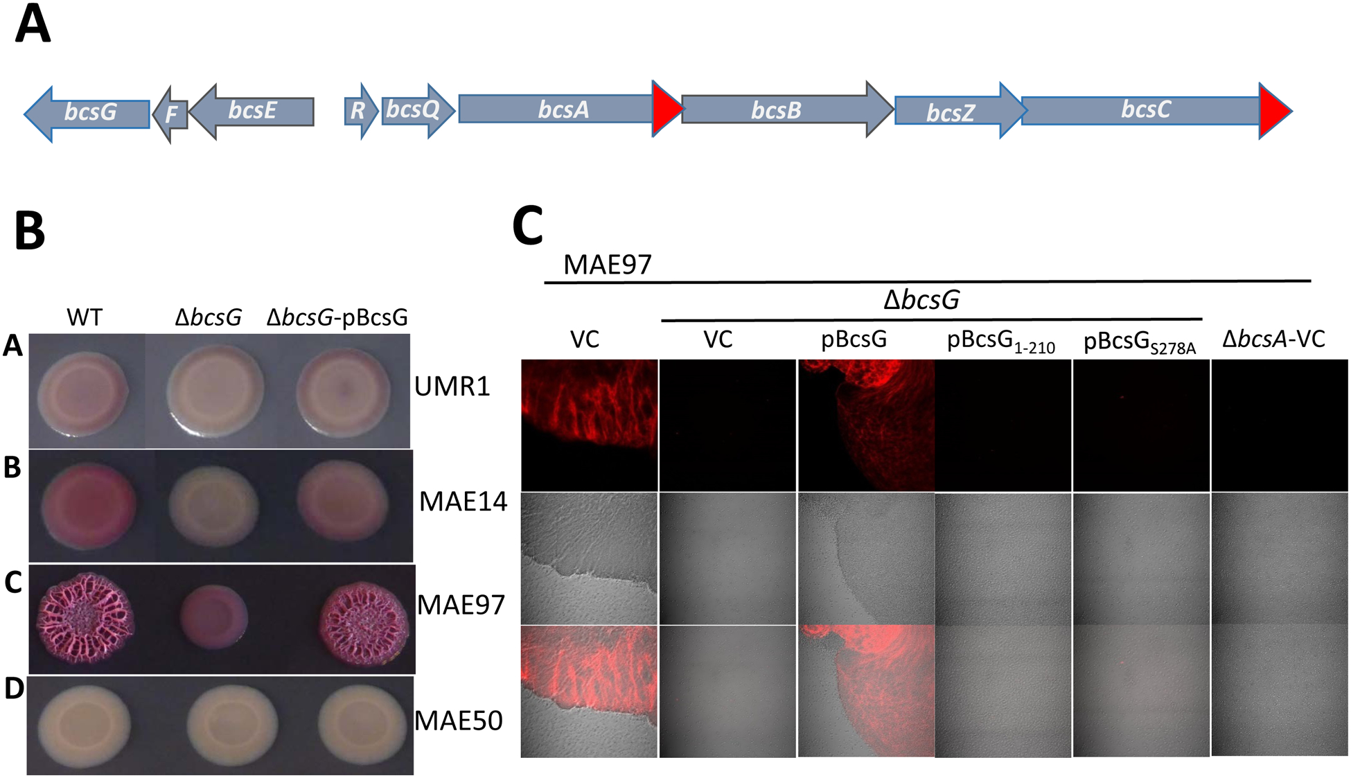
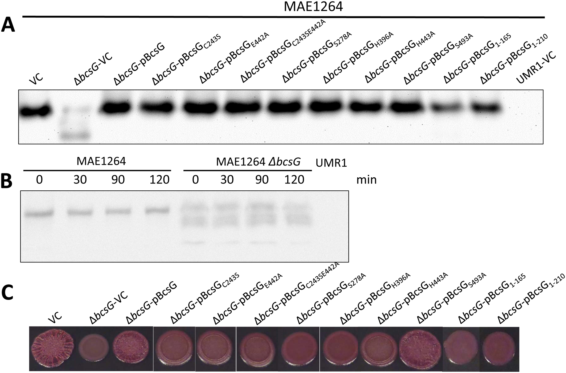
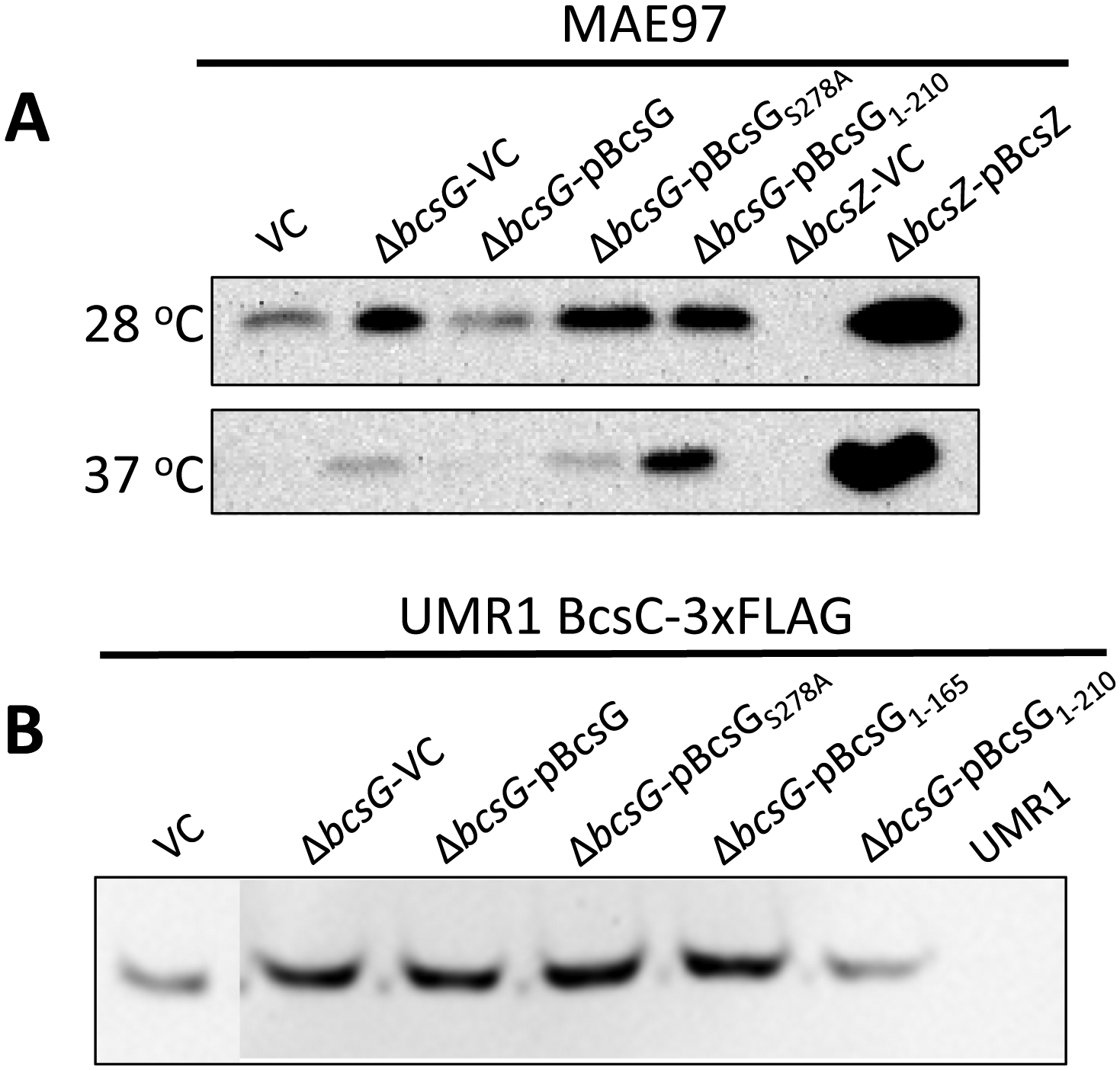
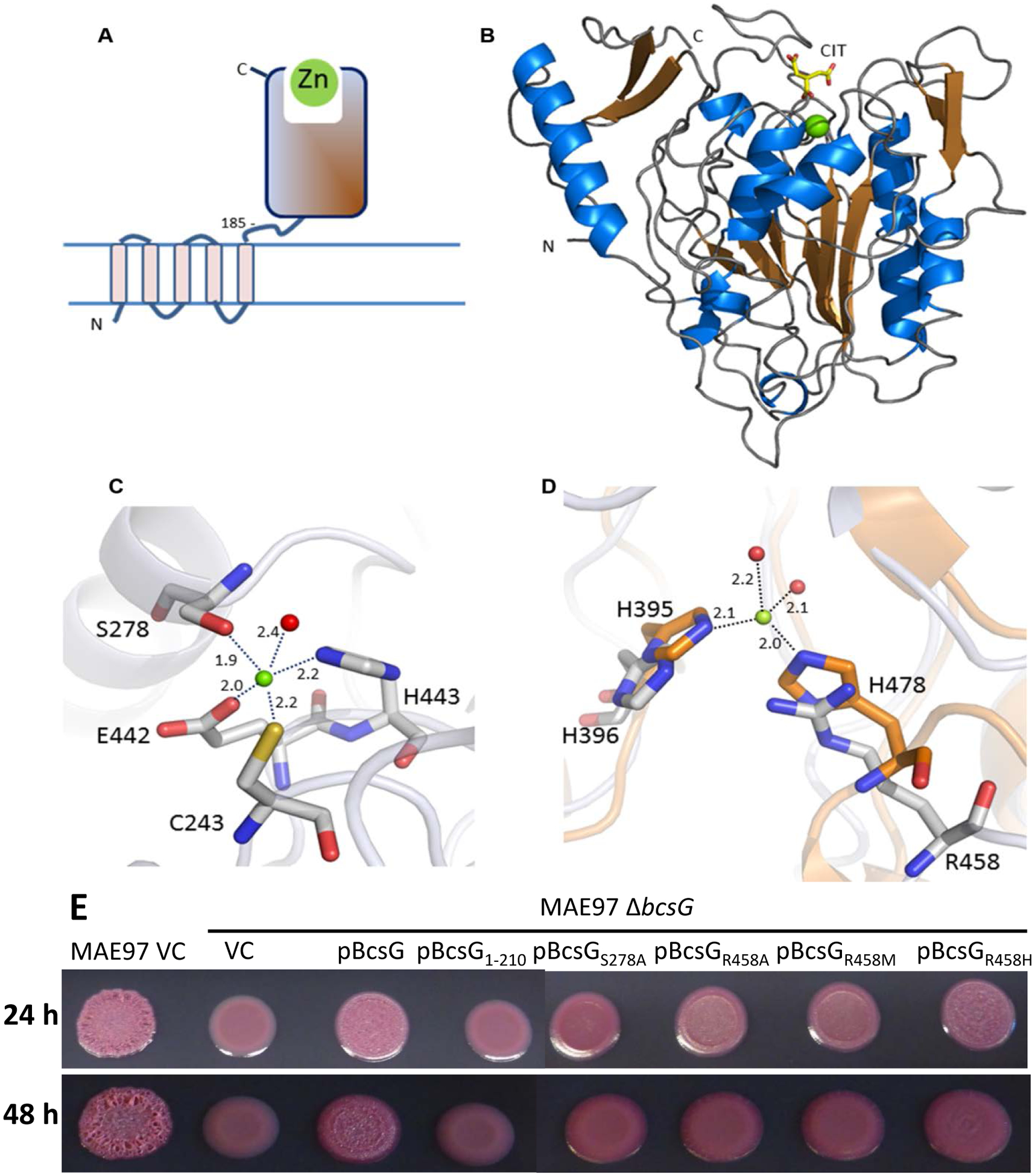
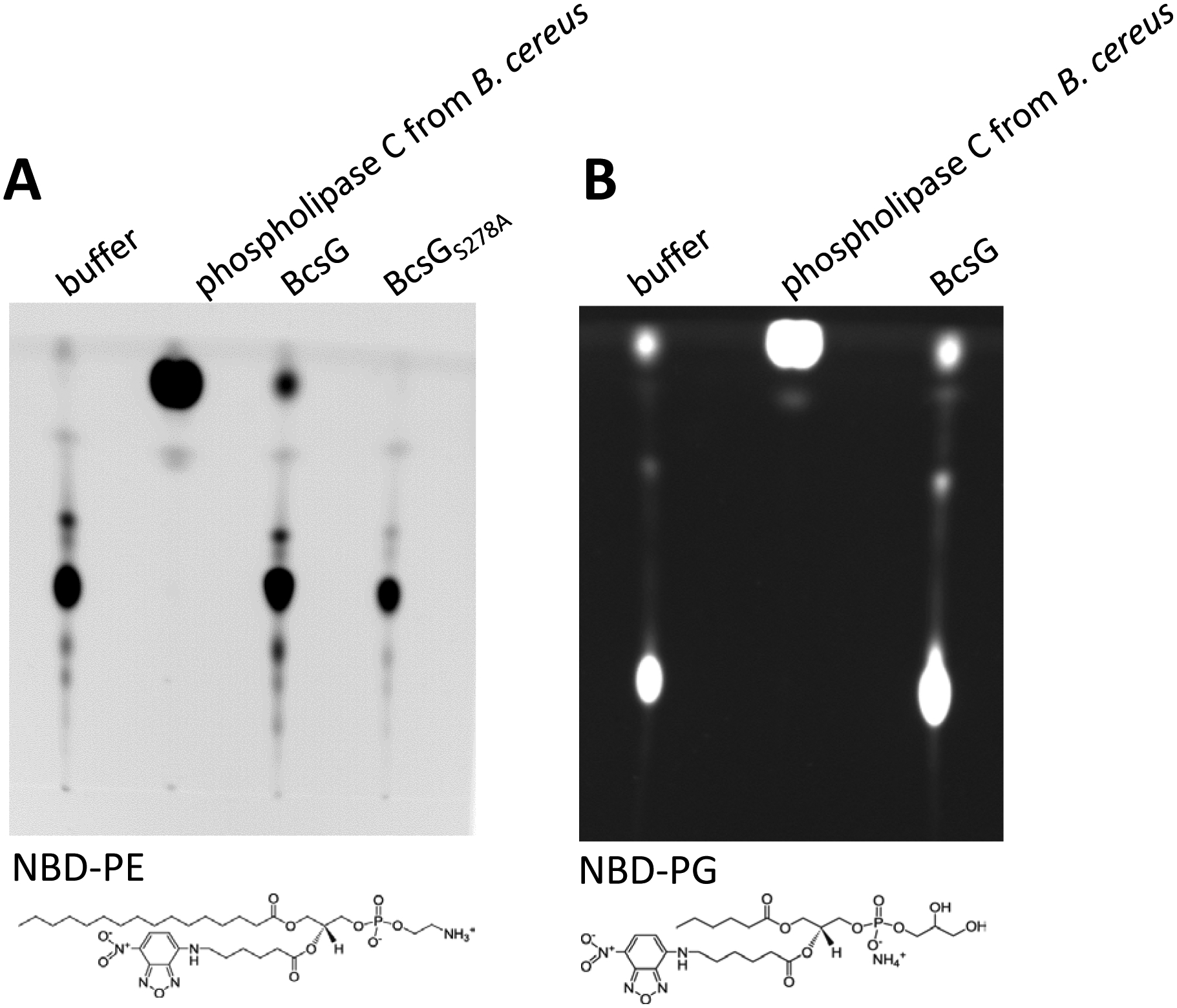
Many bacteria secrete cellulose, which forms the structural basis for bacterial multicellular aggregates, termed biofilms. The cellulose synthase complex of Salmonella typhimurium consists of the catalytic subunits BcsA and BcsB and several auxiliary subunits that are encoded by two divergently transcribed operons, bcsRQABZC and bcsEFG. Expression of the bcsEFG operon is required for full-scale cellulose production, but the functions of its products are not fully understood. This work aimed to characterize the BcsG subunit of the cellulose synthase, which consists of an N-terminal transmembrane fragment and a C-terminal domain in the periplasm. Deletion of the bcsG gene substantially decreased the total amount of BcsA and cellulose production. BcsA levels were partially restored by the expression of the transmembrane segment, whereas restoration of cellulose production required the presence of the C-terminal periplasmic domain and its characteristic metal-binding residues. The high-resolution crystal structure of the periplasmic domain characterized BcsG as a member of the alkaline phosphatase/sulfatase superfamily of metalloenzymes, containing a conserved Zn2+-binding site. Sequence and structural comparisons showed that BcsG belongs to a specific family within alkaline phosphatase-like enzymes, which includes bacterial Zn2+-dependent lipopolysaccharide phosphoethanolamine transferases such as MCR-1 (colistin resistance protein), EptA, and EptC and the Mn2+-dependent lipoteichoic acid synthase (phosphoglycerol transferase) LtaS. These enzymes use the phospholipids phosphatidylethanolamine and phosphatidylglycerol, respectively, as substrates. These data are consistent with the recently discovered phosphoethanolamine modification of cellulose by BcsG and show that its membrane-bound and periplasmic parts play distinct roles in the assembly of the functional cellulose synthase and cellulose production.

Keywords: alkaline phosphatase superfamily; biofilm formation; cellulose biosynthesis; extracellular matrix; virulence.


Figures

Fig. 1.
Fig. 1.. Cellulose biosynthesis operon structure and colony morphotypes of the bcsG deletion mutants in different backgrounds of Salmonella typhimurium ATCC14028–1s.
A. Organization of the bcsRQABZC and bcsEFG operons in both E. coli and S. typhimurium. Red arrows indicate addition of 3xFLAG-tags to the open reading frames of S. typhimurium used in this work. B. Congo Red-stained colony morphotypes of S. typhimurium strains and their ΔbcsG derivatives. Strains used were UMR1 (wild type, A); MAE14 (cellulose positive/curli negative, B); MAE97 (semi-constitutive cellulose positive/curli negative, C), and MAE50 (UMR1 ΔcsgD, D), see Table S2 for the complete genotypes. WT indicates the wild type with respect to the bcsG gene, pBcsG indicates complementation with pBAD30 carrying the bcsG gene under the control of arabimose-dependent promoter. Cells were grown on salt-free LB agar plates for 24 h at 28 °C. C. Cellulose production by agar-grown colonies of S. typhimurium strain MAE97 (bcsG+) and its ΔbcsG derivative as assessed by Calcofluor white staining and observed by confocal laser scanning microscopy. Cells grown on agar as in panel B were gently submerged in 0.001% Calcofluor white to preserve colony structure and aggregation patterns. The wild type MAE97 and the ΔbcsG mutant complemented with wild-type bcsG gene show major cell aggregation and cellulose production (displayed as false color image). The ΔbcsG mutant with pBAD30 alone (vector control, VC) or pBAD30 carrying either truncated (pBcsG1–210) or mutated (pBcsGS278A) bcsG gene dispersed into single cells with residual or no cellulose production. Top panel, fluorescence microscopy; middle panel, phase contrast; bottom panel, overlay of the two. 63x (oil) magnification. pBcsG, pBcsG1–210, and pBcsGS278A are wild-type bcsG and its variants cloned in pBAD30 with a C-terminal 8xHis tag. ΔbcsA strain was used as negative control.
Fig. 2.
Fig. 2.. Effects of bcsG variants on the expression of the cellulose synthase subunit BcsA and cellulose production.
A. Expression of BcsA-3xFLAG in S. typhimurium strain MAE1264 (bcsG+ [5]) and its ΔbcsG mutant upon overexpression of bcsG and its variants. VC, pBcsG, pBcsG1–210, and pBcsGS278A are as in Fig. 1. pBcsGC243S, pBcsGE442A, pBcsGC243SE442A, pBcsGS278A, pBcsGH396A, pBcsGH443A, and pBcsGS493A are bcsG variants with mutations in predicted active site residues of the AlkP superfamily cloned in pBAD30 with a C-terminal 8x-His tag. Strain UMR1 with plasmid pBAD30 (UMR1-VC) without the 3xFLAG tag was used as a negative control. All samples contained equal amounts of cell extracts as judged by Coomassie staining. B. Stability of chromosomally encoded BcsA in the presence (MAE1264) and the absence of bcsG. After translation was inhibited with chloramphenicol, the amount of the 3xFLAG-tagged BcsA subunit was quantified using anti-FLAG antibodies at indicated time points. Strain UMR1 without the 3xFLAG tag was the negative control. Owing to the lower expression of BcsA in the ΔbcsG mutant, 10-fold higher amounts of its cell extract than those for MAE1264 were applied on the gel. C. Pdar colony morphotype (cellulose biosynthesis) of strains listed in panel A. Despite similar levels of the BcsA subunit (panel A), the wild-type (VC) level of cellulose production could only be observed upon overexpression of the wild-type BcsG or the BcsGS493A mutant. Cells were grown on CR salt-free LB agar plates for 24 h at 28 °C.
Fig. 3.
Fig. 3.. Effects of bcsG variants on the expression of the cellulose synthase auxiliary subunits BcsZ and BcsC.
A. Production of the cellulase BcsZ in the ΔbcsG mutants of the cellulose synthase-positive strain MAE97. MAE97 bcsZ deletion strains with pBAD30 vector control (ΔbcsZ-VC) and with overexpressed BcsZ (ΔbcsZ-pBcsZ) were used as negative and positive controls, respectively. Detection of BcsZ production was by Western blot using a rabbit anti-BcsZ antibody. B. Production of the putative outer membrane pore BcsC in S. typhimurium wild type and bcsG derivatives. Wild-type S. typhimurium UMR1 with BcsC-3xFLAG and its ΔbcsG mutant were complemented by the same plasmids as in panel A with the addition of the second truncated variant, pBcsG1–165. Detection of the 132.7 kDa BcsC-3xFLAG was by Western blot using a mouse anti-FLAG-tag antibody. UMR1 BcsC-3xFLAG with pBAD30 (vector control, left lane) and UMR1 without the FLAG tag (right lane) were used as positive and negative controls, respectively. Cells were grown on salt-free LB agar plates for 24 h at either 28 °C (A, top lane) or 37 °C (A, bottom lane), or for 16 h at 28 °C (panel B).
Fig. 4.
Fig. 4.. Structure of BcsG.
A. Domain structure of BcsG. The N-terminal transmembrane domain (residues 1–165) is linked to the C-terminal alkaline phosphatase-like domain (residues 185–559) via flexible inter-domain linker (residues 166–210). The fragment from aa 185 to 559 was used in the construct designed for crystallization. B. Schematic cartoon of the C-terminal alkaline phosphatase-type domain of BcsG. The zinc ion is shown as a green sphere and the citrate molecule bound close to the metal binding site as a stick-model. C. Metal binding site in BcsG. Distances between the Zn2+ and the coordinating atoms are indicated in Å. The water molecule is shown as a red sphere. D. View of the second Zn2+-binding site in the alkaline phosphatase family illustrated after superimposition of the phosphoethanolamine transferase MCR-2 (PDB code 5MX9) (orange carbon atoms) with BcsG (grey carbon atoms). While NmEptA contains a fully functional second Zn2+ site [35], one of the metal ligands, His478 in NmEptA, is replaced by Arg458 in BcsG, making binding of Zn2+ to this site less likely. E. Cellulose production in S. typhimurium strain MAE97 and its ΔbcsG mutants complemented with wild-type BcsG, the mutant lacking the alkaline phosphatase-type domain, and the mutants affecting the residues Ser278 and Arg458, shown in panels C and D. Other labels are as in Fig. 2. Cells were grown on salt-free LB agar plates for 24 h at 37 °C.
Fig. 5.
Fig. 5.. Enzymatic activity of BcsG.
Hydrolysis of NBD-phosphatidylethanolamine (PE) by BcsG, but not the BcsGS278A variant. B. NBD-phosphatidylglycerol (PG) is not hydrolyzed by BcsG. The reaction was carried out in 300 μl of 10 mM sodium succinate pH 6.5/50 mM NaCl, 20 mM MnCl2 with 4.2 ng of purified NBD-PE or NBD-PG lipid and 400 μg MBP-BcsG fusion protein (or 2.5 units of phospholipase C from Bacillus cereus used as positive control) at 37 °C for 3 h. Substrates NBD-PE and NBD-PG are shown below the TLC plates. Each experiment was performed twice.

Similar articles

Cited by

References

    1. Römling U, Galperin MY, Bacterial cellulose biosynthesis: diversity of operons, subunits, products, and functions. Trends Microbiol. 23 (2015) 545–557. - PMC - PubMed
    1. Matthysse AG, Role of bacterial cellulose fibrils in Agrobacterium tumefaciens infection. J. Bacteriol 154 (1983) 906–915. - PMC - PubMed
    1. Robledo M, Rivera L, Jimenez-Zurdo JI, Rivas R, Dazzo F, Velazquez E, Martinez-Molina E, Hirsch AM, Mateos PF, Role of Rhizobium endoglucanase CelC2 in cellulose biosynthesis and biofilm formation on plant roots and abiotic surfaces. Microb. Cell Fact 11 (2012) 125. - PMC - PubMed
    1. Pontes MH, Lee EJ, Choi J, Groisman EA, Salmonella promotes virulence by repressing cellulose production. Proc. Natl. Acad. Sci. USA 112 (2015) 5183–5188. - PMC - PubMed
    1. Ahmad I, Rouf SF, Sun L, Cimdins A, Shafeeq S, Le Guyon S, Schottkowski M, Rhen M, Römling U, BcsZ inhibits biofilm phenotypes and promotes virulence by blocking cellulose production in Salmonella enterica serovar Typhimurium. Microb. Cell Fact 15 (2016) 177. - PMC - PubMed

Publication types

MeSH terms

LinkOut - more resources