. 2018 Nov 15;27(22):3827-3839.
doi: 10.1093/hmg/ddy252.

PIN1 is a new therapeutic target of craniosynostosis

Affiliations

PIN1 is a new therapeutic target of craniosynostosis

H R Shin et al. Hum Mol Genet. .

Abstract

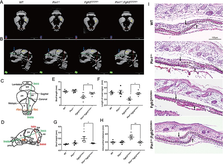
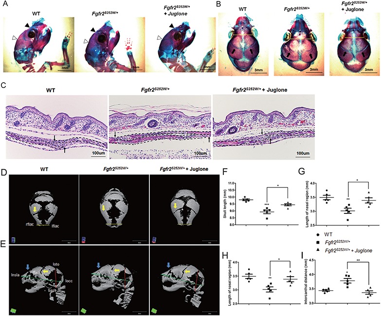
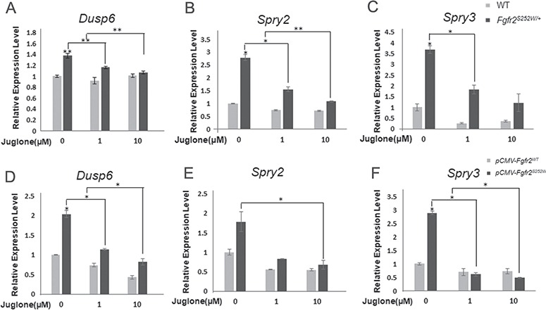
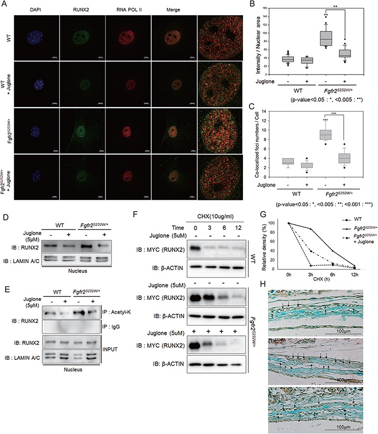
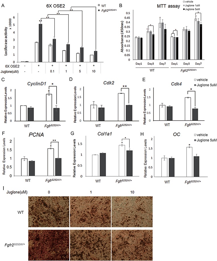
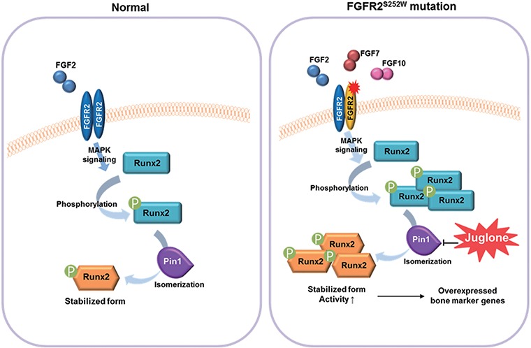
Gain-of-function mutations in fibroblast growth factor receptors (FGFRs) cause congenital skeletal anomalies, including craniosynostosis (CS), which is characterized by the premature closure of craniofacial sutures. Apert syndrome (AS) is one of the severest forms of CS, and the only treatment is surgical expansion of prematurely fused sutures in infants. Previously, we demonstrated that the prolyl isomerase peptidyl-prolyl cis-trans isomerase interacting 1 (PIN1) plays a critical role in mediating FGFR signaling and that Pin1+/- mice exhibit delayed closure of cranial sutures. In this study, using both genetic and pharmacological approaches, we tested whether PIN1 modulation could be used as a therapeutic regimen against AS. In the genetic approach, we crossbred Fgfr2S252W/+, a mouse model of AS, and Pin1+/- mice. Downregulation of Pin1 gene dosage attenuated premature cranial suture closure and other phenotypes of AS in Fgfr2S252W/+ mutant mice. In the pharmacological approach, we intraperitoneally administered juglone, a PIN1 enzyme inhibitor, to pregnant Fgfr2S252W/+ mutant mice and found that this treatment successfully interrupted fetal development of AS phenotypes. Primary cultured osteoblasts from Fgfr2S252W/+ mutant mice expressed high levels of FGFR2 downstream target genes, but this phenotype was attenuated by PIN1 inhibition. Post-translational stabilization and activation of Runt-related transcription factor 2 (RUNX2) in Fgfr2S252W/+ osteoblasts were also attenuated by PIN1 inhibition. Based on these observations, we conclude that PIN1 enzyme activity is important for FGFR2-induced RUNX2 activation and craniofacial suture morphogenesis. Moreover, these findings highlight that juglone or other PIN1 inhibitors represent viable alternatives to surgical intervention for treatment of CS and other hyperostotic diseases.


Figures

Figure 1
Figure 1
Rescue of premature fusion of coronal suture in Fgfr2S252W/+ mice by removal of one allele of Pin1. (A, B) Representative micro-CTs of skulls from WT, Pin1+/−, Fgfr2S252W/+ and Pin1+/−;Fgfr2S252W/+ mice (n = 5). Superior views (A) and lateral views (B) of skull calvaria. Removal of one allele of Pin1 rescued premature fusion of coronal suture (yellow arrows), a CS calvarial phenotype, and the frontal–nasal suture (blue arrows). (C, D) Schematic diagram and landmarks of mouse skull vault. The mouse skull consists of paired frontal bones (fr), paired parietal bones (pa) and the interparietal bone (ipa), intervened by the coronal, sagittal, metopic and lambdoid sutures. The 3D coordinates of specific craniofacial landmarks, shown in (C) and (D), were used for morphometric analyses of the skulls. Green lines represent linear distances corresponding to the length of the nasal region (lnsla–lflac) and the skull length (rnsla–rocc). Red line indicates skull height (lpto–bas). Orange lines indicating intercanthal distance (lflac–rflac) show linear distances corresponding to facial width. (EH) Representative micro-CT results of skulls, shown as bar graphs (n = 5 in each group; *P < 0.05; **P < 0.005; ***P < 0.001). (I) Histological sections of coronal sutures in newborn mice. Arrows indicate growing fronts of frontal and parietal bones. The calvarial bone is highlighted with dotted lines.
Figure 2
Figure 2
Craniosynostosis phenotypes in AS model mice are rescued by treatment with the PIN1 inhibitor juglone. Pregnant mice were intraperitoneally injected with 1 mg/kg juglone (Fgfr2S252W/+ + juglone) or vehicle (Fgfr2S252W/+) once a day from E14.5 to E18.5. WT and Fgfr2S252W/+ mice were sacrificed at birth. (A, B) Mouse calvaria were examined after Alizarin Red S and Alcian Blue staining. Black arrowheads, frontal–nasal suture; white arrowheads, coronal suture (A). Both sutures were already closed in Fgfr2S252W/+ mice, whereas those of WT and Fgfr2S252W/+ + juglone mice remained open. Coronal views of the heads of the same animals are shown in (B). Intercanthal distance (yellow lines) was significantly wider in Fgfr2S252W/+ mice than in control mice. Arrows indicate a coronal suture that is almost overlapping in Fgfr2S252W/+, whereas sutures in the other group remain open. (C) Histological sections of coronal sutures in newborn mice. Arrows indicate growing fronts of frontal and parietal bones. Note that suture overlap is much more pronounced in Fgfr2S252W/+ mice than in the other groups. The calvarial bone is highlighted with dotted lines. (D, E) Representative micro-CTs of skulls from WT, Fgfr2S252W/+ and juglone-treated Fgfr2S252W/+ mice (n = 5). Superior views (D) and lateral views (F) of skull calvaria. Yellow arrows indicate rescue of early fusion of coronal suture (5/5, 100%), and blue arrows indicate rescue of the distorted frontal–nasal suture (2/5, 40%) of mutant mice by juglone. Green lines represent linear distances corresponding to the lengths of the nasal region and the skull. The red line indicates skull height, whereas the yellow lines, such as the neurocranial width at intercanthal distance (lflac–rflac), indicate linear distances corresponding to facial width. (FI) Representative micro-CT results of skulls are shown in bar graphs (n = 5 in each group; *P < 0.05; **P < 0.005; ***P < 0.001). Statistically significant differences indicate rescue of the CS calvarial phenotype.
Figure 3
Figure 3
Expression of target genes changes upon treatment with juglone. (A, B) Relative expression levels of FGFR2 signaling modulators and downstream genes, based on qPCR analysis of WT and Fgfr2S252W/+ calvaria cells (n = 3) following treatment with different dosages of juglone for 1 day. (CE) 70% confluent MC3T3-E1 cells were transfected with Fgfr2WT or Fgfr2S252W mutant expression plasmids. After 24 h, cells were treated with 0, 1 and 10 μm of juglone for 1 day. mRNA levels of FGFR signaling modulators and downstream genes, such as Dusp6 (C) and the Sprouty family genes (Spry2, 3) (D, E), were analyzed quantitatively (n = 3 in each group; *P < 0.01; **P < 0.005; ***P < 0.001). The level of each mRNA was normalized to that of Gapdh in the same sample.
Figure 4
Figure 4
RUNX2 is overactivated in Fgfr2S252W/+ mice, and juglone destabilizes RUNX2 by decreasing acetylation. (A) Endogenous RUNX2 was detected by immunofluorescence and immunocytochemistry. Primary mouse calvarial cells from WT and Fgfr2S252W/+ mice were treated with 5 μm juglone for 24 h. Cells were fixed, and endogenous proteins were immunostained with anti-RUNX2 (green) and anti-RNA POL II (red) antibodies, as well as 4′,6-diamidino-2-phenylindole (blue). In the merged image, RUNX2 and RNA POL II colocalization (yellow) was observed as foci, particularly in Fgfr2S252W mutant-expressing cells. (B) Endogenous intranuclear RUNX2 was quantified, and results are shown as a sigma plot (n = 20 in each group; *P < 0.01; **P < 0.005; ***P < 0.001). (C) The numbers of RUNX2/RNA POL II-colocalized foci were quantified (n = 10 in each group; *P < 0.01; **P < 0.005; ***P < 0.001). (D) Abundance of endogenous RUNX2 in dissociated nuclear extracts, as determined by immunoblot assay. (E) Degree of acetylation of RUNX2, as determined by IP with anti-acetyl-lysine antibody from dissociated nuclear extracts. (F, G) RUNX2 stability analysis. Myc-tagged Runx2 plasmid was transfected into primary mouse calvaria cells. After 24 h, cells were pre-treated with or without 5 μm juglone for 1 h and then treated with 10 μg/ml CHX for the indicated times. Cells were harvested and followed by immunoblotting with the indicated antibodies. β-Actin was used as a loading control. (G) Band intensities of RUNX2 in F were quantitated and plotted against time. (H) RUNX2 in calvaria tissue from coronal sutures of newborn mice was detected by IHC using Alcian Blue counterstaining.
Figure 5
Figure 5
Effects of juglone treatment on osteoblast proliferation and differentiation. (A) Relative RUNX2 transacting activity was assessed using a 6XOSE2 luciferase reporter construct. Following transfection with the 6XOSE2 reporter gene, cells were treated with juglone at various concentrations for 24 h. Luciferase assays were performed on both WT and Fgfr2S252W/+ mouse calvarial cells. (B) Proliferation of WT and Fgfr2S252W/+ mouse calvarial cells following treatment with various doses of juglone for 1, 3 and 7 days. Cell proliferation was assessed by water-soluble tetrazolium assay. (CF) Relative expression of proliferation-related genes in WT and Fgfr2S252W/+ mouse calvarial cells, as determined by qPCR. (GH) Relative expression of osteoblast differentiation markers in both genotypes of primary mouse calvarial cells, as determined by qPCR. Cells were treated with vehicle or 5 μm juglone for 24 h and were more cultured for 2 days in osteogenic media. Expression of each marker gene was normalized against that of Gapdh in the same sample. (I) Late-stage primary osteoblast differentiation was confirmed by Alizarin Red S staining. Both genotypes of primary calvarial cells were cultured for 3 weeks in osteogenic media after 48 h culture with the indicated concentrations of juglone (n = 3 in each group; *P < 0.01; **P < 0.005; ***P < 0.001).
Figure 6
Figure 6
Mechanisms underlying restoration of the normal phenotype by juglone in calvaria with gain-of-function mutations in FGFR2. Juglone treatment lowers abnormally elevated RUNX2 levels in Fgfr2S252W/+ mice by inhibiting RUNX2 isomerization, thereby restoring proper downstream transcriptional activity (9,83). The S252W mutation of FGFR2 increases ligand affinity and alters ligand specificity, resulting in hyperactive FGF signaling. In this genetic background, RUNX2, one of the targets of FGF signaling, undergoes excessive PTM, such as isomerization and acetylation. Repression of RUNX2 isomerization by juglone, a PIN1 inhibitor, decreases the RUNX2 level and normalizes the expression of RUNX2 target genes, thereby rescuing the abnormal calvaria phenotypes of Fgfr2S252W/+ mice.

Similar articles

Cited by

References

    1. Johnson D. and Wilkie A.O. (2011) Craniosynostosis. Eur. J. Hum. Genet., 19, 369–376. - PMC - PubMed
    1. Clendenning D.E. and Mortlock D.P. (2012) The BMP ligand Gdf6 prevents differentiation of coronal suture mesenchyme in early cranial development. PloS One, 7, e36789. - PMC - PubMed
    1. Jin S.W., Sim K.B. and Kim S.D. (2016) Development and growth of the normal cranial vault: an embryologic review. J. Korean Neurosurg. Soc., 59, 192–196. - PMC - PubMed
    1. Derderian C. and Seaward J. (2012) Syndromic craniosynostosis. Semin. Plast. Surg., 26, 64–75. - PMC - PubMed
    1. Bagheri-Fam S., Ono M., Li L., Zhao L., Ryan J., Lai R., Katsura Y., Rossello F.J., Koopman P., Scherer G. et al. (2015) FGFR2 mutation in 46,XY sex reversal with craniosynostosis. Hum. Mol. Genet., 24, 6699–6710. - PMC - PubMed

Publication types

MeSH terms