

Organotypic Slice Cultures to Study Oligodendrocyte Proliferation, Fate, and Myelination
- PMID: 30006707
- PMCID: PMC6372239
- DOI: 10.1007/978-1-4939-7862-5_11
Organotypic Slice Cultures to Study Oligodendrocyte Proliferation, Fate, and Myelination
Abstract
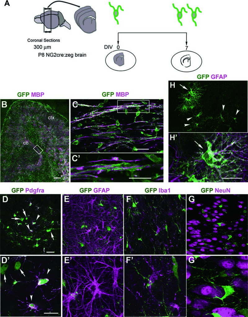
Oligodendrocyte development and myelination are processes in the central nervous system that are regulated by cell intrinsic and extrinsic mechanisms. Organotypic slice cultures provide a simple method for studying factors that affect oligodendrocyte proliferation, differentiation, and myelination in the context of the local cellular environment. Here we show that major glial cell types and neurons are preserved in slice cultures from postnatal mouse forebrain, and their morphological characteristics are retained. We further demonstrate that cellular processes requiring interactions with neighboring cells such as myelination can proceed in slice culture.
Keywords: Myelin; NG2; Oligodendrocyte; Oligodendrocyte precursor; Organotypic slice culture; PDGF; Pdgfrα; Proliferation.
Figures
Similar articles
-
Organotypic slice cultures to study oligodendrocyte dynamics and myelination.J Vis Exp. 2014 Aug 25;(90):e51835. doi: 10.3791/51835. J Vis Exp. 2014. PMID: 25177825 Free PMC article.
-
Oligodendrocyte precursor cell transplantation into organotypic cerebellar shiverer slices: a model to study myelination and myelin maintenance.PLoS One. 2012;7(7):e41237. doi: 10.1371/journal.pone.0041237. Epub 2012 Jul 20. PLoS One. 2012. PMID: 22911763 Free PMC article.
-
Neuronal activity and AMPA-type glutamate receptor activation regulates the morphological development of oligodendrocyte precursor cells.Glia. 2015 Jun;63(6):1021-35. doi: 10.1002/glia.22799. Epub 2015 Mar 4. Glia. 2015. PMID: 25739948
-
Recent Advances in Live Imaging of Cells of the Oligodendrocyte Lineage.Methods Mol Biol. 2019;1936:275-294. doi: 10.1007/978-1-4939-9072-6_16. Methods Mol Biol. 2019. PMID: 30820905 Review.
-
Temporal oligodendrocyte lineage progression: in vitro models of proliferation, differentiation and myelination.Biochim Biophys Acta. 2014 Sep;1843(9):1917-29. doi: 10.1016/j.bbamcr.2014.04.018. Epub 2014 Apr 21. Biochim Biophys Acta. 2014. PMID: 24768715 Review.
Cited by
-
Transcription factor Tcf4 is the preferred heterodimerization partner for Olig2 in oligodendrocytes and required for differentiation.Nucleic Acids Res. 2020 May 21;48(9):4839-4857. doi: 10.1093/nar/gkaa218. Nucleic Acids Res. 2020. PMID: 32266943 Free PMC article.
-
Microglial neuropilin-1 promotes oligodendrocyte expansion during development and remyelination by trans-activating platelet-derived growth factor receptor.Nat Commun. 2021 Apr 15;12(1):2265. doi: 10.1038/s41467-021-22532-2. Nat Commun. 2021. PMID: 33859199 Free PMC article.
-
Organotypic slice culture based on in ovo electroporation for chicken embryonic central nervous system.J Cell Mol Med. 2019 Mar;23(3):1813-1826. doi: 10.1111/jcmm.14080. Epub 2018 Dec 18. J Cell Mol Med. 2019. PMID: 30565384 Free PMC article.
-
Novel guanidine compounds inhibit platelet-derived growth factor receptor alpha transcription and oligodendrocyte precursor cell proliferation.Glia. 2021 Mar;69(3):792-811. doi: 10.1002/glia.23930. Epub 2020 Oct 24. Glia. 2021. PMID: 33098183 Free PMC article.
References
-
- Dawson MR, Polito A, Levine JM, Reynolds R (2003) NG2-expressing glial progenitor cells: an abundant and widespread population of cycling cells in the adult rat CNS. Mol Cell Neurosci 24(2):476–488 - PubMed
-
- Nishiyama A, Komitova M, Suzuki R, Zhu X (2009) Polydendrocytes (NG2 cells): multifunctional cells with lineage plasticity. Nat Rev Neurosci 10(1):9–22 - PubMed
Publication types
MeSH terms
Grants and funding
LinkOut - more resources
Full Text Sources
Other Literature Sources
