. 2018 Jul;559(7714):419-422.
doi: 10.1038/s41586-018-0323-8. Epub 2018 Jul 11.

Prespliceosome structure provides insights into spliceosome assembly and regulation

Affiliations

Prespliceosome structure provides insights into spliceosome assembly and regulation

Clemens Plaschka et al. Nature. 2018 Jul.

Abstract

The spliceosome catalyses the excision of introns from pre-mRNA in two steps, branching and exon ligation, and is assembled from five small nuclear ribonucleoprotein particles (snRNPs; U1, U2, U4, U5, U6) and numerous non-snRNP factors1. For branching, the intron 5' splice site and the branch point sequence are selected and brought by the U1 and U2 snRNPs into the prespliceosome1, which is a focal point for regulation by alternative splicing factors2. The U4/U6.U5 tri-snRNP subsequently joins the prespliceosome to form the complete pre-catalytic spliceosome. Recent studies have revealed the structural basis of the branching and exon-ligation reactions3, however, the structural basis of the early events in spliceosome assembly remains poorly understood4. Here we report the cryo-electron microscopy structure of the yeast Saccharomyces cerevisiae prespliceosome at near-atomic resolution. The structure reveals an induced stabilization of the 5' splice site in the U1 snRNP, and provides structural insights into the functions of the human alternative splicing factors LUC7-like (yeast Luc7) and TIA-1 (yeast Nam8), both of which have been linked to human disease5,6. In the prespliceosome, the U1 snRNP associates with the U2 snRNP through a stable contact with the U2 3' domain and a transient yeast-specific contact with the U2 SF3b-containing 5' region, leaving its tri-snRNP-binding interface fully exposed. The results suggest mechanisms for 5' splice site transfer to the U6 ACAGAGA region within the assembled spliceosome and for its subsequent conversion to the activation-competent B-complex spliceosome7,8. Taken together, the data provide a working model to investigate the early steps of spliceosome assembly.


Conflict of interest statement

The authors declare no competing financial interests.

Figures

Extended Data Figure 1
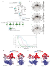
Extended Data Figure 1. Biochemical characterization and cryo-EM of the prespliceosome A complex.
a. Mutation of the UBC4 pre-mRNA branch point sequence (UACUAAC to UACAAAC, where A is the BP adenosine) stalls splicing before the first step, as described. Splicing reactions were carried out for 30 min at 23 ºC in yeast extract using wild-type (lane 1) or mutant (U/A, lane 2) pre-mRNA (see Methods for details). This experiment was performed three times. The asterisk indicates a degradation product. For gel source data see Supplementary Fig. 1a. b. Protein analysis of purified A complex (SDS-PAGE stained with Coomassie blue). The U2-associated Prp5 protein is sub-stoichiometric and not observed in the A complex structure. The purification and analysis of protein compositions were performed at least five times with similar results. For gel source data see Supplementary Fig. 1b. c. Cryo-EM micrograph of the A complex. Scale bar, 100 nm. d. 2D class averages of the A complex were determined in RELION 2.1 (refs 39,40), and reveal a bipartite architecture, comprising the U1 snRNP and the U2 snRNP 3' and 5' regions, respectively. e. Composite cryo-EM density of the A complex shown in two orthogonal views (compare Fig. 1). The respective densities used for modeling the U1 snRNP (A2, gray), the U2 3' region (A1, cyan), and the U2 5' region (A3, green) are coloured and superimposed on a transparent outline of the full A3 map (Methods). The overall resolution of each map as well as the percentage from the cleaned dataset of 153,556 particles are shown in parentheses. Non-modelled regions are indicated and putatively assigned. f. Composite cryo-EM density with the final A complex model superimposed in a cartoon representation. The path of 40 nucleotides of the disordered UBC4 pre-mRNA intron are indicated. A complex components are coloured as in Fig. 1. Views as in panel e.
Extended Data Figure 2
Extended Data Figure 2. Cryo-EM image classification and refinement.
a. Image processing workflow for analysis of the A complex cryo-EM data set (Methods). To visualize differences between the reconstructions the U1 snRNP (gray), U2 3' (cyan) and U2 5' regions (green) are coloured. For each round of three-dimensional classification, the percentage of the data and the type of soft-edged mask are indicated. The type of mask and overall resolution are indicated for each 3D refinement (blue box). b. Orientation distribution plots for all particles that contribute to the respective A1, A2, and A3 cryo-EM reconstructions. c. Gold-standard Fourier shell correlation (FSC = 0.143) of the respective A1, A2, and A3 cryo-EM reconstructions. d. Two views of the composite A complex cryo-EM density (maps A1, A2, and A3) coloured by local resolution as determined by ResMap. e. As panel d, but for a central slice through the composite A complex cryo-EM map.
Extended Data Figure 3
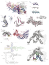
Extended Data Figure 3. Details of the U1 snRNP.
a. U1 snRNP structure with subunits coloured as in Fig. 1, except for Nam8 (orange), Snu56 (light blue), Snu71 (blue), Luc7 (dark purple), Mud1 (red) and U1 snRNA (various). The pre-mRNA nucleotides are labelled relative to the first nucleotide (+1) of the intron. The Nam8 RRM1 and RRM2 domains are flexible and project downstream of the 5'SS. The protein attributed to Luc7 in the free U1 snRNP structure was re-assigned to Snu71. Stem loop, SL; RNA recognition motif, RRM; Zn-finger, ZnF; N-terminus, N-term; C-terminus, C-term. In the structure we do not observe any evidence that the C-terminal tails of SmB, SmD1, and SmD3 interact with the 5’SS, consistent with their absence in the human 5’SS–minimal U1 snRNP crystal structure. b. Representative regions of the sharpened U1 snRNP density determined at 4 Å resolution (map A2) are superimposed on the refined coordinate model. The density reveals side-chain details, and here segments from the Prp42 N-terminus (TPR repeat 1), the Sm ring subunit SmB, and the Snu56 α-helical domain are shown. c. The A2 cryo-EM density is shown superimposed on the coordinate models of a selection of U1 snRNP proteins: Luc7, Snu71, Yhc1, and Prp39. In the structure most of Snu71 is disordered, except for a small N-terminal domain (residues 2-43) that binds between the Prp42 N-terminus and the Snu56 KH-like fold, consistent with protein crosslinking. Functional regions and disordered domains are indicated. d. The U1 snRNA–pre-mRNA 5' splice site (U1–5'SS) model is superimposed on its cryo-EM density (map A2). A secondary structure diagram of the U1–5'SS interaction is shown underneath the model. The register of the 5'SS is shifted by one nucleotide compared to the minimal human 5'SS–U1 snRNP crystal structure, due to an additional nucleotide in yeast U1 snRNA (U11). Lines indicate Watson–Crick base pairs and dots pseudouridine (ψ)-containing base pairs. e. The Prp39-Prp42 heterodimer is coloured to indicate each of their respective TPR repeats. f. Cryo-EM density of U1 snRNA from maps A2 (dark gray) and A3 (light gray) without (top) and with the superimposed coordinate model of yeast U1 snRNA (bottom). The model is labelled and coloured according to functional regions of U1 snRNA (5' end, pink; H helix, cyan; SL1, dark blue; SL2-1, green; SL3-1, light blue; SL2-2 and SL3-2 to -7, gray; 3’end and Sm site, yellow). Stem loop, SL. g. Secondary structure diagram of U1 snRNA. Bold letters indicate residues included in the model, lines indicate Watson–Crick base pairs, and dots G–U wobble and pseudouridine (ψ)-containing base pairs. Compare panel e. The conserved U1 snRNA ‘core’ is outlined with a gray box. The region of the putative phosphate backbone model of part of the U1 SL3-7 region is indicated with a gray box.
Extended Data Figure 4
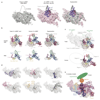
Extended Data Figure 4. Comparisons of yeast and human U1 snRNPs and implications for alternative splicing.
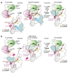
a. Formation of the U1–5'SS helix induces stable binding of Luc7. In the absence of a pre-mRNA 5'SS in the free U1 snRNP density (left, EMD-8622), Luc7 and the U1 5' end are disordered. Upon 5'SS recognition at the U1 5' end (center, map A2), Luc7 becomes ordered and stabilizes the U1–5'SS interaction, suggesting a mechanism for the selection of weak 5'SS sequences. The free U1 snRNP and the 5'SS-bound (map A2) cryo-EM densities are superimposed on the right. Although the long α-helical density next to Luc7 cannot be assigned with confidence, protein-protein crosslinking data and protein secondary structure prediction are consistent with either to Prp40 or Snu71. Based on additional biochemical data on the interaction between the α-helical Prp40 FF1 domain and Luc7 ZnF2 (ref. 52), we would speculate that the Prp40 FF1 domain is the most likely candidate for this density. b. Comparison of the yeast U1 snRNP ‘core’ with the human U1 snRNP crystal structure (PDB ID 3CW1). Protein and RNA (top) and RNA only (bottom) are shown side-by-side (left and center) and superimposed by a global alignment in PyMOL (right). Coloured as in Extended Data Fig. 3a. c. The yeast U1 snRNP model suggests regulatory mechanisms for human alternative splicing factors. The human homologues of the peripheral yeast U1 proteins may function through stabilization of the U1–5'SS interaction (region 1), of the U1-U2 3' region interface (region 2), or the U1-U2 5' interface (region 3). The yeast U1 snRNP ‘core’ is shown superimposed on a surface representation of the U1 snRNP model (top), compared with the similarly coloured human U1 snRNP (below). Interaction sites with the U2 snRNP are labelled (top). d. The location of yeast U1 snRNP components with homology to human splicing factors are indicated in the U1 snRNP structure. The Prp39-Prp42 heterodimer (human PRPF39 homodimer), Nam8 (ref. 18) (human TIA-1 and TIA-R), Luc7 (ref. 53) (human LUC7-like 1-3), and the Yhc1 C-terminus (human U1C) have clear counterparts in the human system. The yeast-specific U1 snRNA insertions may be replaced in the human system by alternative splicing factors that modulate interactions with the U2 5' region. e. Schematic model of the yeast E complex based on the U1 snRNP structure and biochemical data. Luc7, Snu71, and Prp40 form a heterotrimer in vitro, and their interacting regions may be located near unassigned density (compare Extended Data Fig. 1e) at the tip of an unassigned 40-residue α-helix next to Luc7 ZnF2. This helix is likely to belong to the U1 subunit Snu71 or Prp40, consistent with protein crosslinking and protein secondary structure prediction. Prp40 could then bind the yeast branch point binding protein (BBP, human SF1), which in turn interacts with Mud2 (human U2AF65) to tether the pre-mRNA branch point sequence in the E complex.
Extended Data Figure 5
Extended Data Figure 5. Conformational flexibility of the U2 snRNP.
a. Two defined positions of the U1 snRNP-U2 3' region could be identified relative to the U2 5' region. A complex models were fitted into class 2 and 4 from Round 2 of three-dimensional image classification (compare Extended Data Fig. 2a). The classes are aligned via their U2 5' region, illustrating their relative flexibility. b. Cartoon schematic of observed positions of the U2 3' region relative to the U2 5' region in A complex (left), B complex (center), and Bact complex (right, modelled from ref. 54). While in B complex the U2 3' region is free, in A and Bact complexes its position is influenced by interactions with Prp39 as well as Syf1 and Clf1, respectively. c. The U2 snRNP subunit Lea1 (human U2A’) aids to position the U2 snRNP 3' domain in different spliceosome states. In our A complex structure, the Prp39 TPR repeat T1 contacts the helical C-terminus of Lea1. In the yeast C complex structure, non-modeled density for the Syf1 N-terminus binds a neighboring but non-overlapping surface of Lea1 (PDB ID 5LJ5). In C*/P complex (PDB ID 6EXN), the Syf1 N-terminus binds yet another Lea1 surface and the U2 3' domain is repositioned relative to its C complex location. Together, this suggests that the Lea1 provides multiple interfaces that can be used to position the U2 3' domain in different spliceosomal complexes. d. Fit of the U2 3’ region coordinate model to the A1 cryo-EM density. The dashed black separates the U2 3’domain (Sm ring, Msl1 and Lea1 subunits, and U2 snRNA) and the SF3a subcomplex (Prp9, Prp11, and Prp21). Two orthogonal views are shown. See Supplementary Video 2. e. Fit of the U2 5’ region coordinate model to the A3 cryo-EM density. Density consistent with U2 snRNA stem IIa/b and the branch helix is observed. Two density thresholds are shown side-by-side (left, 0.0163; right, 0.0121), and orthogonal views are shown underneath. See Supplementary Video 2.
Extended Data Figure 6
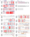
Extended Data Figure 6. Luc7 and Nam8 sequence alignments.
a. The Luc7 (human LUC7-like) amino acid sequence alignment comparing Saccharmoyces cerevisiae, Kluyveromyces lactis, Schizosaccharomyces pombe, Danio rerio, Xenopus tropicalis, Mus musculus, Bos taurus, and Homo sapiens was generated with Clustal Omega and visualized with ESPript 3 (refs 56,57). For the human sequence, LUC7-like 1 was used. Secondary structure elements are indicated above the sequence and derive from the A complex structure (purple) or PSIPRED secondary structure prediction (gray). Modelled regions (dashed line) and the Zn-coordinating residues of Zn-finger 1 and 2 (ZnF, asterisk) are indicated. Invariant or conserved residues are highlighted with a red box or red letter font, respectively. b. As panel a but for Nam8 (human TIA-1) comparing Saccharmoyces cerevisiae, Kluyveromyces lactis, Schizosaccharomyces pombe, Drosophila melanogaster, Danio rerio, Xenopus tropicalis, Mus musculus, Bos taurus, and Homo sapiens amino acid sequences. RNA recognition motif, RRM; ribonucleoprotein domain, RNP.
Extended Data Figure 7
Extended Data Figure 7. Details of the pre-B complex model.
a. Multiple views of the pre-B complex model, generated by combining functional and structural data from yeast and human systems,. The mobility of the U1 snRNP relative to the U2 snRNP in A complex (this study) as well as of the U2 snRNP relative to tri-snRNP in the B complex structure are indicated (left). The pre-B model contained only minor clashes, and a clash between the highly flexible Prp28 C-terminal RecA-2 lobe (from human tri-snRNP25) and the highly flexible U6 snRNA 5' Stem loop (from yeast B complex8) may be resolved by small movements of either domain. See Methods for details on the pre-B model. b. Structural comparisons of the yeast pre-B model (this study) and the yeast B complex structure (PDB ID 5NRL, ref. 8) suggest the existence of a molecular checkpoint to couple 5'SS transfer to U1 snRNP release and formation of the activation-competent B complex. In the pre-B model (left) Sad1 tethers Brr2 through its interaction with the conserved Brr2 PWI domain, and the U1 snRNP and its U1–5'SS helix are positioned near the U6 ACAGAGA region and the helicase Prp28. Subsequent to Prp28-mediated 5'SS transfer, Brr2 is repositioned onto its U4 snRNA substrate, guided by the B complex-specific proteins (right). In this conformation the Brr2 helicase and its associated factors would clash with the U1 snRNP, consistent with U1 snRNP destabilization and release yeast and human B complexes,. Brr2 is now ready to initiate spliceosome activation and formation of the active site in the Bact complex. Regions that are changed between pre-B and B complex models (black outline) and the clash between the Brr2-containing ‘helicase’ domain and the U1 snRNP in B complex (red ‘X’) are indicated. The lower right panel would conform to the alternative ‘U1-B complex’ model.
Extended Data Figure 8
Extended Data Figure 8. Model for early splicing events.
a. Cartoon schematic of proposed early splicing events, detailing (I) assembly of pre-B complex spliceosome from A complex and the U4/U6.U5 tri-snRNP and (II) the subsequent conversion to the pre-catalytic B complex spliceosome. In the pre-B model the mobile U1 snRNP is next to Prp28, which is bound at the Prp8N domain. To initiate 5'SS transfer, Prp28 could clamp the pre-mRNA at or next to the U1–5'SS helix to destabilize it and to hand over the 5'SS to the U6 ACAGAGA region of tri-snRNP, consistent with protein-RNA crosslinks. Formation of the U6–5'SS interaction may induce the binding of the B complex proteins to replace Prp28 at the Prp8N domain and induce the large movement of Brr2 to its B complex location on U4 snRNA (Fig. 5). The U1 snRNP, now loosely tethered to U2, may dissociate from B complex due to the steric clash with the Brr2-containing ‘helicase’ domain (Extended Data Fig. 7b). In agreement with this, the human pre-B complex converts to a B complex-like state in presence of a 5'SS oligonucleotide, which coincides with U1 snRNP release. This model can explain how Brr2 is kept inactive to prevent premature U4/U6 duplex unwinding. The model thereby implies the existence of a molecular checkpoint, coupling 5'SS transfer from U1 to U6 snRNA with Brr2 helicase repositioning and U1 snRNP release to generate the activation-competent B complex spliceosome. b. Cartoon schematic of an alternative model for spliceosome assembly and 5'SS transfer that relies only on the yeast A complex (this work), tri-snRNP,, and B complex structures. In this model the tri-snRNP that associates with A complex already contains the Brr2 helicase bound to the U4 snRNA substrate and the yeast B complex proteins at the Prp8 N-terminal (Prp8N) domain. The tri-snRNP then binds the A complex (transition I, ‘Assembly’), requiring a significant readjustment to avoid a steric clash of the Brr2-containing ‘helicase’ domain and the U1 snRNP (‘U1-B complex’). The Prp28 helicase is then recruited to U1 snRNP directly as the Prp28-binding site on the Prp8 N-terminal domain in human tri-snRNP is occupied by B complex proteins. Prp28 then disrupts the U1-5'SS helix, leading to 5'SS transfer (transition II, ‘Transfer’). Similar to the ‘pre-B complex’ assembly model in panel a, the U1 snRNP, now freed from the 5'SS, may then be released due to a steric clash with the Brr2-containing ‘helicase’ domain. This model does not require Sad1. Compare to panel a.
Extended Data Figure 9
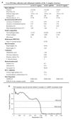
Extended Data Figure 9. Data collection, refinement statistics, and validation.
a. Cryo-EM data collection and refinement statistics of the A complex structure. Maps A1 and A3 were used to position the U2 snRNP 3' and 5' regions, respectively (see Methods). b. FSC between the A2 cryo-EM density and the refined A complex U1 snRNP coordinate model.
Figure 1
Figure 1. Prespliceosome A complex structure.
Two orthogonal views of the yeast A complex structure. Subunits are coloured according to snRNP identity (U1, shades of purple, U2, shades of green), and the pre-mRNA intron (black) and its 5' exon (orange) are highlighted. Orthologous human proteins are shown after the backslash. The location of the cap-binding complex (CBC) is indicated with a brown oval (Extended Data Figure 1d,e).
Figure 2
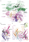
Figure 2. 5'SS recognition and implications for alternative splicing.
a. The A complex U1-U2 snRNP interfaces (A and B) and RNA network are shown as cartoons, and are superimposed on transparent surfaces of prespliceosome proteins. The U2 subunit Hsh155 surface (gray oval), which interacts with the tri-snRNP in the B complex, is freely accessible in the A complex. The U1 snRNP proteins Nam8 (orange, human TIA-1), Luc7 (purple, human LUC7-like, LUC7L) and Yhc1 (magenta, human U1C) are shown as ribbons. The remaining densities are likely accounted for by Nam8 (see c) and cap binding complex (CBC). Branch point, BP; β-propeller B and C, BPB and BPC; RNA-recognition motif, RRM. b. The pre-mRNA 5'SS is recognized by the U1 snRNA 5' end, and is stabilized by Luc7 and Yhc1. Notably, the Yhc1 ZnF and Luc7 ZnF2 domains are arranged with pseudo-C2 symmetry around the U1-5'SS helix. c. Nam8 binds the U1 snRNP through its linker (yellow), RNA recognition motif 3 (RRM3, light orange) and C-terminal regions (orange), while its RRM1 and RRM2 domains are mobile and project towards the intron, to bind uridine-rich sequences downstream of the pre-mRNA 5’SS (dashed line), like its human counterpart TIA-1 (ref. 18). Nam8 contacts the Yhc1 (human U1C) C-terminus, and human TIA-1 biochemically also interacts with human U1C. Snu56 (blue), Prp39 (magenta), Prp42 (violet), and Hsh49 (light green) are shown as transparent ribbon models and other protein and U1 snRNA elements were removed for clarity.
Figure 3
Figure 3. Spliceosome assembly and 5'SS transfer.
a. One of the two alternative pre-B-complex models, suggesting that the U2 snRNP orients the U1 snRNP to deliver the pre-mRNA 5'SS to the U6 ACAGAGA stem. The model was obtained by superposing the yeast A (this study) and B complex structures (PDB 5NRL) and by modifying the locations of Brr2, U4 Sm ring, Sad1, and Prp28 to resemble a human-like pre-B complex conformation based on biochemical data and the human U4/U6.U5 tri-snRNP structure (PDB 3JCR) (see Methods). Coloured as in Fig. 1 and ref. . b. The pre-B complex RNA network and the Prp28 helicase are shown as cartoons and are superimposed on transparent surfaces of spliceosome proteins. Prp28 is positioned at the Prp8 N-terminal domain as in human tri-snRNP and may clamp onto the pre-mRNA near the U1–5'SS helix to destabilize it and transfer the 5'SS from U1 snRNA to the U6 snRNA ACAGAGA stem (red arrow), which are separated by ~20 Å in the pre-B model.

Similar articles

Cited by

References

    1. Will CL, Lührmann R. Spliceosome structure and function. Cold Spring Harb Perspect Biol. 2011;3 a003707. - PMC - PubMed
    1. Papasaikas P, Valcarcel J. The spliceosome: The ultimate RNA chaperone and sculptor. Trends Biochem Sci. 2016;41:33–45. - PubMed
    1. Wilkinson ME, Lin PC, Plaschka C, Nagai K. Cryo-EM Studies of Pre-mRNA Splicing: From Sample Preparation to Model Visualization. Annu Rev Biophys. 2018 doi: 10.1146/annurev-biophys-070317-033410. - DOI - PubMed
    1. Behzadnia N, et al. Composition and three-dimensional EM structure of double affinity-purified, human prespliceosomal A complexes. EMBO J. 2007;26:1737–1748. - PMC - PubMed
    1. Gao G, Dudley SC., Jr RBM25/LUC7L3 function in cardiac sodium channel splicing regulation of human heart failure. Trends Cardiovasc Med. 2013;23:5–8. - PMC - PubMed

Publication types

MeSH terms

Substances