. 2018 Sep 14;38(19):e00084-18.
doi: 10.1128/MCB.00084-18. Print 2018 Oct 1.

Histone Citrullination Represses MicroRNA Expression, Resulting in Increased Oncogene mRNAs in Somatolactotrope Cells

Affiliations

Histone Citrullination Represses MicroRNA Expression, Resulting in Increased Oncogene mRNAs in Somatolactotrope Cells

Stanley B DeVore et al. Mol Cell Biol. .

Abstract

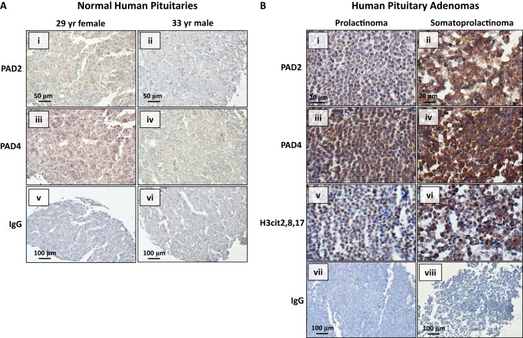
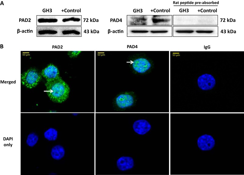
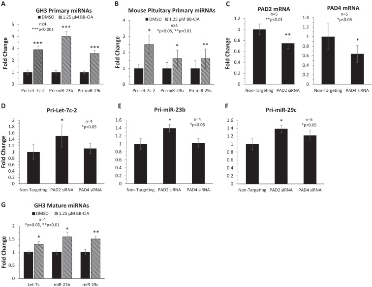
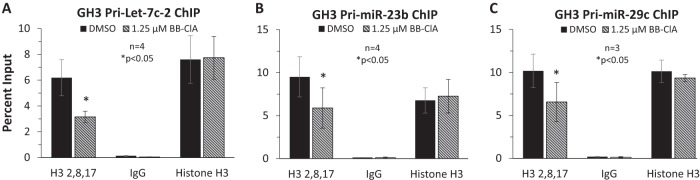
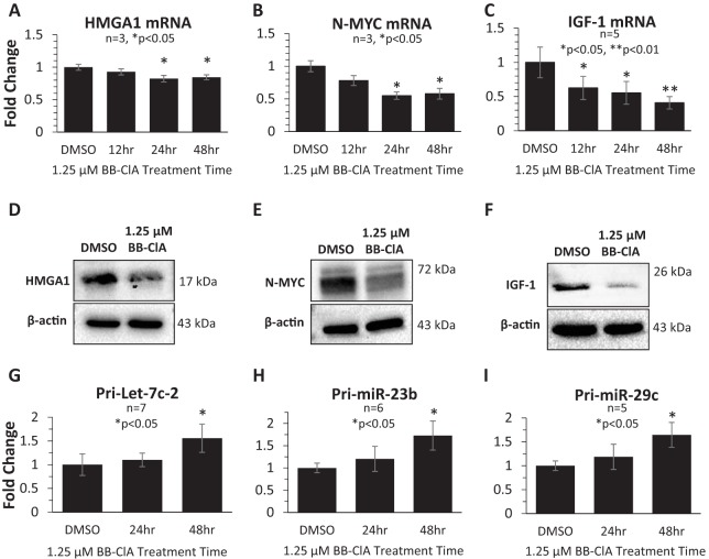
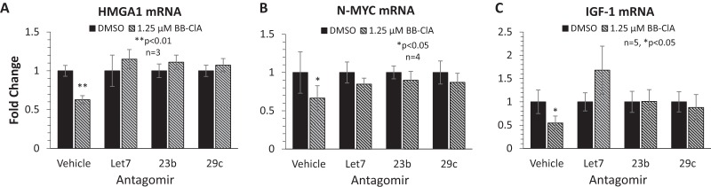
Peptidylarginine deiminase (PAD) enzymes convert histone arginine residues into citrulline to modulate chromatin organization and gene expression. Although PADs are expressed in anterior pituitary gland cells, their functional role and expression in pituitary adenomas are unknown. To begin to address these issues, we first examined normal human pituitaries and pituitary adenomas and found that PAD2, PAD4, and citrullinated histones are highest in prolactinomas and somatoprolactinomas. In the somatoprolactinoma-derived GH3 cell line, PADs citrullinate histone H3, which is attenuated by a pan-PAD inhibitor. RNA sequencing and chromatin immunoprecipitation (ChIP) studies show that the expression of microRNAs (miRNAs) let-7c-2, 23b, and 29c is suppressed by histone citrullination. Our studies demonstrate that these miRNAs directly target the mRNA of the oncogenes encoding HMGA, insulin-like growth factor 1 (IGF-1), and N-MYC, which are highly implicated in human prolactinoma/somatoprolactinoma pathogenesis. Our results are the first to define a direct role for PAD-catalyzed histone citrullination in miRNA expression, which may underlie the etiology of prolactinoma and somatoprolactinoma tumors through regulation of oncogene expression.

Keywords: citrullination; epigenetics; miRNA; oncogenes; prolactinoma.


Figures

FIG 1
FIG 1
Human prolactinomas and somatoprolactinomas express elevated levels of PADs and citrullinated histones. (A) Normal human pituitary sections (n = 8 female and n = 6 male) were examined by IHC by probing with anti-PAD2 and anti-PAD4 antibodies or with an equal mass of rabbit IgG as a negative control. Representative images for a 29-year-old female and 33-year-old male were taken with a Zeiss Axio Vert.A1 microscope using the 20× objective except for IgG, which was taken with a 10× objective. (B) Human prolactinoma (n = 11) and somatoprolactinoma (n = 6) sections were examined by IHC using anti-PAD2, anti-PAD4, and anti-H3Cit2,8,17 antibodies. Sections were probed with an equal mass of rabbit IgG as a negative control. Representative images were taken using the 40× objective except for IgG, which was taken with a 10× objective. The scale bars represent 100 μm for the 10× objective, 50 μm for the 20× objective, and 20 μm for the 40× objective.
FIG 2
FIG 2
PAD2 and PAD4 localize to the nucleus and citrullinate histones in the somatolactotrope-derived GH3 cell line. (A) GH3 cells were lysed and subjected to Western blotting using anti-PAD2 and anti-PAD4 antibodies or an anti-PAD4 antibody that was preabsorbed with the N-terminal 15 amino acids of the rat PAD4 isoform. Anti-β-actin was used as a loading control. To generate positive controls, GH3 cells were transiently transfected with a PAD2 or PAD4 expression plasmid for 24 h and subsequently lysed. (B) GH3 cells were fixed and examined by ICC using anti-PAD2 or anti-PAD4 antibodies or an equal mass of rabbit IgG as a negative control and stained with 4′,6-diamidino-2-phenylindole (DAPI; blue). Arrows indicate nuclear PAD staining. Images were taken with a Zeiss LSM 710 confocal microscope using a 40× objective. The scale bars represent 10 μm.
FIG 3
FIG 3
The pan-PAD inhibitor BB-ClA decreases basal histone citrullination to regulate the expression of pri-miRNAs in GH3 cells. (A) GH3 cells were treated with either DMSO or 1.25 μM BB-ClA. Following cell lysis, histones were purified and examined by Western blotting using anti-H3Cit2,8,17 antibody and anti-histone H3 antibody as a loading control. In vitro-citrullinated histones were used as a positive control. (B) Western blots were quantified with Bio-Rad Image Lab; means were separated via a two-tailed t test (n = 3; P < 0.01), and values are expressed as means ± SEM. (C) GH3 cells were treated with either DMSO or 1.25 μM BB-ClA. The RNA from three independent experiments was purified and sequenced on an Illumina platform. Bioinformatic analysis identified 81 upregulated genes, of which the top 12 genes in DMSO- versus BB-ClA-treated GH3 cells are listed. Genes with Padj values of <0.05 were considered significant.
FIG 4
FIG 4
Inhibiting histone citrullination or knocking down PAD2 increases the expression of primary microRNAs let-7c, miR-23b, and miR-29c. (A) GH3 cells were treated with either DMSO or 1.25 μM BB-ClA and then lysed, and RNA was subjected to qPCR with primers specific to pri-let-7c-2, pri-miR-23b, and pri-miR-29c. Results were quantified (n ≥ 4) and means were separated with a two-tailed t test. (B) Dispersed mouse pituitary cells were treated with either DMSO or 1.25 μM BB-ClA. Cells were lysed and purified RNA was subjected to qPCR with mouse primers for the indicated pri-miRNAs. Experiments were quantified (n ≥ 4) and means were separated with a one-tailed paired t test. (C to F) GH3 cells were transiently transfected with nontargeting, PAD2, and PAD4 siRNA constructs for 48 h. Following lysis and RNA purification, samples were examined by qPCR for expression of PAD2 and -4 (C), pri-let7c-2 (D), pri-miR-23b (E), and pri-miR-29c (F), with GAPDH as the endogenous reference control. Means were separated with either a one-tailed paired t test (C; n = 5) or a one-way repeated-measures analysis of variance (ANOVA) with a post hoc Dunnett's t test (D to F; n ≥ 4). (G) After DMSO or BB-ClA treatment, GH3 cells were lysed and purified small RNAs were subjected to qPCR with TaqMan probes specific for let-7c, miR-23b, and miR-29c. U6 snRNA was used as an endogenous reference control. Experiments were quantified and means were separated with a two-tailed t test (n = 4). All values provided are means ± SEM.
FIG 5
FIG 5
Citrullinated histones are directly associated with and repress primary miRNA expression in GH3 cells. For ChIP, GH3 protein-DNA complexes were immunoprecipitated with anti-H3Cit2,8,17, anti-histone H3 (positive control), or nonspecific IgG (negative control). Cross-links were reversed, and the DNA was purified and subjected to qPCR analysis with pri-miRNA specific primers. Means were separated with a one-tailed paired t test (n ≥ 3). All values provided are means ± SEM.
FIG 6
FIG 6
PAD inhibition significantly decreases oncogene mRNA and protein expression in GH3 cells. GH3 cells were treated every 12 h with 1.25 μM BB-ClA for 0, 12, 24, or 48 total hours. Following cell lysis, RNA was purified, reverse transcribed, and subjected to qPCR analysis using intron-spanning primers specific to HMGA1 (A), N-MYC (B), or IGF-1 (C). GAPDH was used as an endogenous reference control. Means were separated with a one-way repeated-measures ANOVA with a post hoc Dunnett's t test (n ≥ 3). Following the same paradigm, HMGA1 (D), N-MYC (E), and IGF-1 (F) were examined by Western blotting. pri-let7c-2 (G), pri-miR-23b (H), and pri-miR-29c (I) expression was examined by qPCR after 24 and 48 h of BB-ClA treatment to confirm that pri-miRNAs were increased when oncogene expression was suppressed. Means were separated with a one-way repeated-measures ANOVA with a post hoc Dunnett's t test (n ≥ 5). All values provided are means ± SEM.
FIG 7
FIG 7
Antagomirs to let7c, miR-23b, and miR-29c attenuate oncogene mRNA expression in the presence of PAD inhibition. GH3 cells were transiently transfected with vehicle, let7c, miR-23b, and miR-29c antagomirs for 48 h. Immediately following transfection, cells were treated every 12 h with DMSO or 1.25 μM BB-ClA for 48 total hours. RNA was purified, reverse transcribed, and subjected to qPCR analysis using intron-spanning primers specific to HMGA1 (A), N-MYC (B), or IGF-1 (C). GAPDH was used as an endogenous reference control. Means were separated with a one-tailed paired t test. All values provided are means ± SEM.
FIG 8
FIG 8
PAD inhibition attenuates GH3 proliferation. GH3 cells were treated every 12 h with DMSO or 1.25 μM BB-ClA for 24, 48, 72, or 96 total hours. (A) To quantify proliferation, the MTT tetrazolium reagent was added to each well. After 2 h, the resulting formazan solution was collected and the absorbance measured at 490 nm. Means were separated with one-tailed paired t tests (n = 3). (B and C) Cell growth (B) and viability (C) for DMSO and 1.25 μM BB-ClA treated GH3 cells were measured at 24, 48, 72, or 96 total hours using a Countess automated cell counter. Means were separated with one-tailed paired t tests (n ≥ 5). All values provided are means ± SEM.
FIG 9
FIG 9
A working model for PAD catalyzed histone citrullination in lactotrope and somatolactotrope proliferation.

Similar articles

Cited by

References

    1. Vossenaar ER, Zendman AJW, van Venrooij WJ, Pruijn GJM. 2003. PAD, a growing family of citrullinating enzymes: genes, features and involvement in disease. Bioessays News Rev Mol Cell Dev Biol 25:1106–1118. doi:10.1002/bies.10357. - DOI - PubMed
    1. Horibata S, Coonrod SA, Cherrington BD. 2012. Role for peptidylarginine deiminase enzymes in disease and female reproduction. J Reprod Dev 58:274–282. doi:10.1262/jrd.2011-040. - DOI - PubMed
    1. Cuthbert GL, Daujat S, Snowden AW, Erdjument-Bromage H, Hagiwara T, Yamada M, Schneider R, Gregory PD, Tempst P, Bannister AJ, Kouzarides T. 2004. Histone deimination antagonizes arginine methylation. Cell 118:545–553. doi:10.1016/j.cell.2004.08.020. - DOI - PubMed
    1. Wang Y, Wysocka J, Sayegh J, Lee Y-H, Perlin JR, Leonelli L, Sonbuchner LS, McDonald CH, Cook RG, Dou Y, Roeder RG, Clarke S, Stallcup MR, Allis CD, Coonrod SA. 2004. Human PAD4 regulates histone arginine methylation levels via demethylimination. Science 306:279–283. doi:10.1126/science.1101400. - DOI - PubMed
    1. Cherrington BD, Zhang X, McElwee JL, Morency E, Anguish LJ, Coonrod SA. 2012. Potential role for PAD2 in gene regulation in breast cancer cells. PLoS One 7:e41242. doi:10.1371/journal.pone.0041242. - DOI - PMC - PubMed

Publication types

MeSH terms