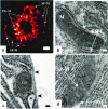
Mitochondrial junctions with cellular organelles: Ca2+ signalling perspective
- PMID: 29982949
- PMCID: PMC6060751
- DOI: 10.1007/s00424-018-2179-z
Mitochondrial junctions with cellular organelles: Ca2+ signalling perspective
Abstract
Cellular organelles form multiple junctional complexes with one another and the emerging research area dealing with such structures and their functions is undergoing explosive growth. A new research journal named "Contact" has been recently established to facilitate the development of this research field. The current consensus is to define an organellar junction by the maximal distance between the participating organelles; and the gap of 30 nm or less is considered appropriate for classifying such structures as junctions or membrane contact sites. Ideally, the organellar junction should have a functional significance, i.e. facilitate transfer of calcium, sterols, phospholipids, iron and possibly other substances between the organelles (Carrasco and Meyer in Annu Rev Biochem 80:973-1000, 2011; Csordas et al. in Trends Cell Biol 28:523-540, 2018; Phillips and Voeltz in Nat Rev Mol Cell Biol 17:69-82, 2016; Prinz in J Cell Biol 205:759-769, 2014). It is also important to note that the junction is not just a result of a random organelle collision but have active and specific formation, stabilisation and disassembly mechanisms. The nature of these mechanisms and their role in physiology/pathophysiology are the main focus of an emerging research field. In this review, we will briefly describe junctional complexes formed by cellular organelles and then focus on the junctional complexes that are formed by mitochondria with other organelles and the role of these complexes in regulating Ca2+ signalling.
Keywords: Ca2+ signalling; Endoplasmic reticulum; Membrane contact sites; Mitochondria; Organellar junctions; Reactive oxygen species.
Figures


Similar articles
-
Plant organellar calcium signalling: an emerging field.J Exp Bot. 2012 Feb;63(4):1525-42. doi: 10.1093/jxb/err394. Epub 2011 Dec 26. J Exp Bot. 2012. PMID: 22200666 Free PMC article. Review.
-
Endoplasmic reticulum/mitochondria calcium cross-talk.Novartis Found Symp. 2007;287:122-31; discussion 131-9. Novartis Found Symp. 2007. PMID: 18074635 Review.
-
Interactions between the endoplasmic reticulum, mitochondria, plasma membrane and other subcellular organelles.Int J Biochem Cell Biol. 2009 Oct;41(10):1805-16. doi: 10.1016/j.biocel.2009.02.017. Epub 2009 Mar 5. Int J Biochem Cell Biol. 2009. PMID: 19703651 Review.
-
Regulation of calcium and phosphoinositides at endoplasmic reticulum-membrane junctions.Biochem Soc Trans. 2016 Apr 15;44(2):467-73. doi: 10.1042/BST20150262. Biochem Soc Trans. 2016. PMID: 27068956 Free PMC article. Review.
-
The Role of Mitochondria in the Activation/Maintenance of SOCE: Membrane Contact Sites as Signaling Hubs Sustaining Store-Operated Ca2+ Entry.Adv Exp Med Biol. 2017;993:277-296. doi: 10.1007/978-3-319-57732-6_15. Adv Exp Med Biol. 2017. PMID: 28900920 Review.
Cited by
-
Pathological Mechanisms Underlying Myalgic Encephalomyelitis/Chronic Fatigue Syndrome.Diagnostics (Basel). 2019 Jul 20;9(3):80. doi: 10.3390/diagnostics9030080. Diagnostics (Basel). 2019. PMID: 31330791 Free PMC article. Review.
-
Ca2+ Signaling in Exocrine Cells.Cold Spring Harb Perspect Biol. 2020 May 1;12(5):a035279. doi: 10.1101/cshperspect.a035279. Cold Spring Harb Perspect Biol. 2020. PMID: 31636079 Free PMC article. Review.
-
A genetically encoded toolkit of functionalized nanobodies against fluorescent proteins for visualizing and manipulating intracellular signalling.BMC Biol. 2019 May 23;17(1):41. doi: 10.1186/s12915-019-0662-4. BMC Biol. 2019. PMID: 31122229 Free PMC article.
-
β2-adrenergic receptor regulates ER-mitochondria contacts.Sci Rep. 2021 Nov 2;11(1):21477. doi: 10.1038/s41598-021-00801-w. Sci Rep. 2021. PMID: 34728663 Free PMC article.
-
T lymphocytes from malignant hyperthermia-susceptible mice display aberrations in intracellular calcium signaling and mitochondrial function.Cell Calcium. 2021 Jan;93:102325. doi: 10.1016/j.ceca.2020.102325. Epub 2020 Dec 1. Cell Calcium. 2021. PMID: 33310301 Free PMC article.
References
-
- Appenzeller-Herzog C, Simmen T. ER-luminal thiol/selenol-mediated regulation of Ca2+ signalling. Biochem Soc Trans. 2016;44:452–459. - PubMed
-
- Barrow SL, Voronina SG, da Silva Xavier G, Chvanov MA, Longbottom RE, Gerasimenko OV, Petersen OH, Rutter GA, Tepikin AV. ATP depletion inhibits Ca2+ release, influx and extrusion in pancreatic acinar cells but not pathological Ca2+ responses induced by bile. Pflugers Arch. 2008;455:1025–1039. - PubMed
-
- Belousov VV, Fradkov AF, Lukyanov KA, Staroverov DB, Shakhbazov KS, Terskikh AV, Lukyanov S. Genetically encoded fluorescent indicator for intracellular hydrogen peroxide. Nat Methods. 2006;3:281–286. - PubMed
Publication types
MeSH terms
Substances
LinkOut - more resources
Full Text Sources
Other Literature Sources
Miscellaneous

