

Papillary thyroid carcinoma with pleomorphic tumor giant cells in a pregnant woman - a case report
- PMID: 29976183
- PMCID: PMC6034243
- DOI: 10.1186/s12902-018-0275-x
Papillary thyroid carcinoma with pleomorphic tumor giant cells in a pregnant woman - a case report
Abstract
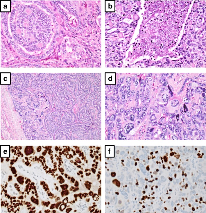
Background: Papillary thyroid carcinoma with pleomorphic tumor giant cells (PTC-PC) is characterized by the occurrence of bizarre, pleomorphic cells within a small area of a conventional PTC. The histologic distinction between PTC-PC and PTC's with a focal anaplastic thyroid cancer (ATC) component (denoted in the 2004 WHO classification as "papillary thyroid carcinoma with spindle and giant cell carcinoma", PTC-SGC) is debated, however the prognosis is thought to be different (excellent for PTC-PC, poor for PTC-SGC). Therefore, this diagnostic challenge is significant for any endocrine pathologist to recognize. Herein, we report the histological and clinical workup of a PTC-PC case, with particular focus on the molecular analyses that facilitated the establishment of the final diagnosis.
Case presentation: The patient was a pregnant, 28-year-old female presenting with a 30 mm conventional PTC, with focal areas with undifferentiated cells exhibiting exaggerated nuclear pleomorphism. No foci of extrathyroidal extension, angioinvasion or lymph node engagement were seen. Immunohistochemical analyses revealed the pleomorphic cells exhibiting retained differentiation. Molecular genetic analyses demonstrated a codon V600 missense mutation of the BRAF gene, but no TP53 or TERT promoter mutations. The absence of an aggressive phenotype in addition to the lack of mutations in two major ATC-related genes led to the diagnosis of a PTC-PC. Postoperative MRI showed no evidence of metastatic disease. Radioiodine ablation was performed seven months post-operatively, and a SPECT-CT imaging did not show signs of residual tissue. She is well and without signs of disease 16 months post-operatively.
Conclusions: PTC-PC is a differential diagnosis to PTC-SGC that mandates careful considerations. Taken together with previous publications, PTC-PC seems to be histologically similar to PTC-SGC, but clinically distinct. Even so, the distinction is not easily made given the different therapeutic consequences for each individual patient. This is the first report that includes molecular genetics to aid in finalizing the diagnosis. Exclusion of mutations in TP53 and the TERT promoter could be considered as an adjunct tool when assessing papillary thyroid cancer with focal pleomorphism.
Keywords: Anaplastic thyroid cancer; Molecular testing; Papillary thyroid cancer; Pathology; Pleomorphism.
Conflict of interest statement
Ethics approval and consent to participate
The local Karolinska Insitutet ethical committee has granted ethical approval, and written informed consent has been obtained.
Consent for publication
Consent to publish has been obtained from the person described in this case report.
Competing interests
The authors declare that they have no competing interests.
Publisher’s Note
Springer Nature remains neutral with regard to jurisdictional claims in published maps and institutional affiliations.
Figures
Similar articles
-
Robot-assisted Sistrunk's operation, total thyroidectomy, and neck dissection via a transaxillary and retroauricular (TARA) approach in papillary carcinoma arising in thyroglossal duct cyst and thyroid gland.Ann Surg Oncol. 2012 Dec;19(13):4259-61. doi: 10.1245/s10434-012-2674-y. Epub 2012 Oct 16. Ann Surg Oncol. 2012. PMID: 23070784
-
TERT Promoter Mutations and Tumor Persistence/Recurrence in Papillary Thyroid Cancer.Cancer Res Treat. 2016 Jul;48(3):942-7. doi: 10.4143/crt.2015.362. Epub 2015 Dec 28. Cancer Res Treat. 2016. PMID: 26727717 Free PMC article.
-
Clear Cell Variant of Papillary Thyroid Carcinoma With Associated Anaplastic Thyroid Carcinoma: Description of an Extraordinary Case.Int J Surg Pathol. 2019 Sep;27(6):658-663. doi: 10.1177/1066896919837678. Epub 2019 Apr 1. Int J Surg Pathol. 2019. PMID: 30931661
-
Evidence that one subset of anaplastic thyroid carcinomas are derived from papillary carcinomas due to BRAF and p53 mutations.Cancer. 2005 Jun 1;103(11):2261-8. doi: 10.1002/cncr.21073. Cancer. 2005. PMID: 15880523 Review.
-
Anaplastic Transformation of Papillary Thyroid Carcinoma Only Seen in Pleural Metastasis: A Case Report with Review of the Literature.Head Neck Pathol. 2017 Jun;11(2):162-167. doi: 10.1007/s12105-016-0751-4. Epub 2016 Aug 22. Head Neck Pathol. 2017. PMID: 27550513 Free PMC article. Review.
Cited by
-
miRNA-144-5p/ITGA3 Suppressed the Tumor-Promoting Behaviors of Thyroid Cancer Cells by Downregulating ITGA3.Comput Math Methods Med. 2021 Dec 13;2021:9181941. doi: 10.1155/2021/9181941. eCollection 2021. Comput Math Methods Med. 2021. PMID: 34938358 Free PMC article.
-
Inhibitory effects of miR‑26b‑5p on thyroid cancer.Mol Med Rep. 2019 Aug;20(2):1196-1202. doi: 10.3892/mmr.2019.10315. Epub 2019 May 31. Mol Med Rep. 2019. PMID: 31173209 Free PMC article.
-
Thyroid cancer and pregnancy: a systematic ten-year-review.Gland Surg. 2024 Jun 30;13(6):1097-1107. doi: 10.21037/gs-24-52. Epub 2024 Jun 20. Gland Surg. 2024. PMID: 39015727 Free PMC article. Review.
-
Efficacy and Safety of Fuzheng Yiqi Kang-Ai Decoction Combined with External Irradiation in the Treatment of Undifferentiated Thyroid Carcinoma and Its Influence on Antiangiogenesis.J Oncol. 2022 May 16;2022:3589924. doi: 10.1155/2022/3589924. eCollection 2022. J Oncol. 2022. PMID: 35615246 Free PMC article.
References
-
- RA DL, Lloyd RV, Heitz PU, Eng C. International Agency for Research on Cancer, World Health Organization. Pathology and genetics of tumours of endocrine organs. 3. Lyon: IARC Press; 2004.
-
- Volante M, Collini P, Nikiforov YE, Sakamoto A, Kakudo K, Katoh R, Lloyd RV, LiVolsi VA, Papotti M, Sobrinho-Simoes M, Bussolati G, Rosai J. Poorly differentiated thyroid carcinoma: the Turin proposal for the use of uniform diagnostic criteria and an algorithmic diagnostic approach. Am J Surg Pathol. 2007;31:1256–1264. doi: 10.1097/PAS.0b013e3180309e6a. - DOI - PubMed
-
- Lloyd RV, Osamura RY, Klöppel G, Rosai J. International Agency for Research on Cancer, World Health Organization. WHO classification of tumours of endocrine organs. World Health Organization classification of tumours. 4. Lyon: IARC Press; 2017.
-
- Adeniran AJ, Zhu Z, Gandhi M, Steward DL, Fidler JP, Giordano TJ, Biddinger PW, Nikiforov YE. Correlation between genetic alterations and microscopic features, clinical manifestations, and prognostic characteristics of thyroid papillary carcinomas. Am J Surg Pathol. 2006;30:216–222. doi: 10.1097/01.pas.0000176432.73455.1b. - DOI - PubMed
Publication types
MeSH terms
Grants and funding
LinkOut - more resources
Full Text Sources
Other Literature Sources
Medical
Research Materials
Miscellaneous
