

DDR1 and DDR2 in skin
- PMID: 29952722
- PMCID: PMC6363048
- DOI: 10.1080/19336918.2018.1485618
DDR1 and DDR2 in skin
Abstract
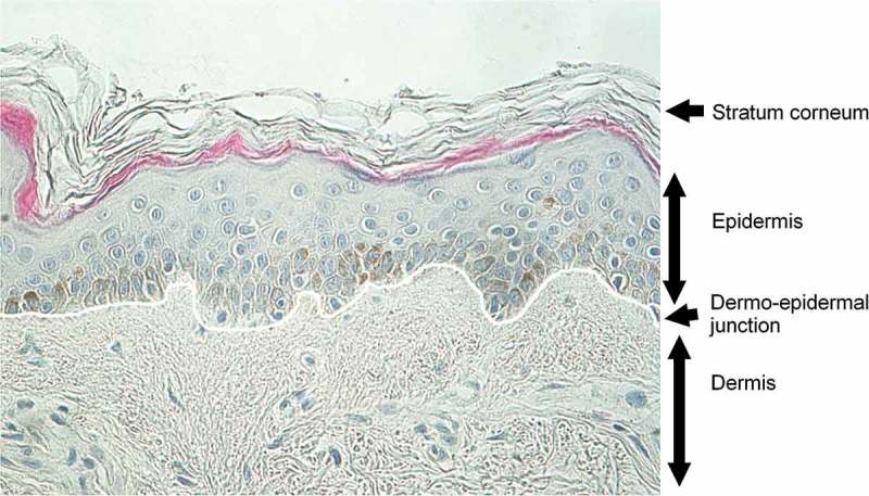
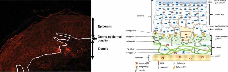
DDR1 and DDR2 are expressed in skin but their expression differs according to the skin compartment, epidermis, dermis, hypodermis and to the embryonic origin of the cells. In skin, it seems that during physiological processes such as wound healing or pathological processes such as tumorigenesis or systemic sclerosis development only one of the DDR is dysregulated. Furthermore, the altered DDR in pathological process is not necessarily the DDR implicated in basal homeostasis. Indeed, in epidermis, while DDR1 is the main DDR involved in melanocyte homeostasis, DDR2 seems to be the main DDR implicated in melanoma. On the contrary, in dermis, while DDR2 is necessary for normal wound healing, dysregulation of DDR1 is associated with abnormal wound healing leading to keloid. In conclusion, targeting DDR could be a therapeutic solution, however side effects have to be managed carefully.
Keywords: CCN3; Melanocyte; cadherin; cell adhesive proteins; fibroblast; integrins; keratinocyte.
Figures
Similar articles
-
The Journey of DDR1 and DDR2 Kinase Inhibitors as Rising Stars in the Fight Against Cancer.Int J Mol Sci. 2021 Jun 18;22(12):6535. doi: 10.3390/ijms22126535. Int J Mol Sci. 2021. PMID: 34207360 Free PMC article. Review.
-
DDR1 and DDR2 physical interaction leads to signaling interconnection but with possible distinct functions.Cell Adh Migr. 2018;12(4):324-334. doi: 10.1080/19336918.2018.1460012. Epub 2018 Jun 25. Cell Adh Migr. 2018. PMID: 29616590 Free PMC article.
-
Synthesis and biological evaluation of novel dasatinib analogues as potent DDR1 and DDR2 kinase inhibitors.Chem Biol Drug Des. 2017 Mar;89(3):420-427. doi: 10.1111/cbdd.12863. Epub 2016 Oct 17. Chem Biol Drug Des. 2017. PMID: 27589335
-
Discoidin domain receptors and their ligand, collagen, are temporally regulated in fetal rat fibroblasts in vitro.Plast Reconstr Surg. 2001 Mar;107(3):769-76. doi: 10.1097/00006534-200103000-00018. Plast Reconstr Surg. 2001. PMID: 11304604
-
Inhibitors of Discoidin Domain Receptor (DDR) Kinases for Cancer and Inflammation.Biomolecules. 2021 Nov 10;11(11):1671. doi: 10.3390/biom11111671. Biomolecules. 2021. PMID: 34827669 Free PMC article. Review.
Cited by
-
Role of the p53‑TRPM1/miR‑211‑MMP9 axis in UVB‑induced human melanocyte migration and its potential in repigmentation.Int J Mol Med. 2020 Apr;45(4):1017-1026. doi: 10.3892/ijmm.2020.4478. Epub 2020 Jan 27. Int J Mol Med. 2020. PMID: 31985026 Free PMC article.
-
Discoidin domain receptors orchestrate cancer progression: A focus on cancer therapies.Cancer Sci. 2021 Mar;112(3):962-969. doi: 10.1111/cas.14789. Epub 2021 Jan 27. Cancer Sci. 2021. PMID: 33377205 Free PMC article. Review.
-
The Journey of DDR1 and DDR2 Kinase Inhibitors as Rising Stars in the Fight Against Cancer.Int J Mol Sci. 2021 Jun 18;22(12):6535. doi: 10.3390/ijms22126535. Int J Mol Sci. 2021. PMID: 34207360 Free PMC article. Review.
-
ABL1/2 and DDR1 Drive MEKi Resistance in NRAS-Mutant Melanomas by Stabilizing RAF/MYC/ETS1 and Promoting RAF Homodimerization.Cancers (Basel). 2023 Feb 2;15(3):954. doi: 10.3390/cancers15030954. Cancers (Basel). 2023. PMID: 36765910 Free PMC article.
-
Extracellular Matrix Dynamics as an Emerging yet Understudied Hallmark of Aging and Longevity.Aging Dis. 2023 Jun 1;14(3):670-693. doi: 10.14336/AD.2022.1116. Aging Dis. 2023. PMID: 37191434 Free PMC article. Review.
References
-
- Makino K, Jinnin M, Aoi J, et al. Discoidin domain receptor 2–microRNA 196a–mediated negative feedback against excess Type I collagen expression is impaired in scleroderma dermal fibroblasts. J Invest Dermatol. 2013;133:110–119. - PubMed
-
- Alves F, Vogel W, Mossie K, et al. Distinct structural characteristics of discoidin I subfamily receptor tyrosine kinases and complementary expression in human cancer. Oncogene. 1995;10:609–618. - PubMed
-
- Reichert-Faria A, Jung JE, Moreschi Neto V, et al. Reduced immunohistochemical expression of Discoidin Domain Receptor 1 (DDR1) in vitiligo skin. J Eur Acad Dermatol Venereol. 2013;27:1057–1059. - PubMed
-
- Ricard AS, Pain C, Daubos A, et al. Study of CCN3 (NOV) and DDR1 in normal melanocytes and vitiligo skin. Exp Dermatol. 2012;21:411–416. - PubMed
-
- Di Marco E, Cutuli N, Guerra L, et al. Molecular cloning of trkE, a novel trk-related putative tyrosine kinase receptor isolated from normal human keratinocytes and widely expressed by normal human tissues. J Biol Chem. 1993;268:24290–24295. - PubMed
Publication types
MeSH terms
Substances
LinkOut - more resources
Full Text Sources
Other Literature Sources