Review
. 2018 Jul;20(7):745-754.
doi: 10.1038/s41556-018-0124-1. Epub 2018 Jun 27.

The multifaceted contributions of mitochondria to cellular metabolism

Affiliations
Review

The multifaceted contributions of mitochondria to cellular metabolism

Jessica B Spinelli et al. Nat Cell Biol. 2018 Jul.

Abstract

Although classically appreciated for their role as the powerhouse of the cell, the metabolic functions of mitochondria reach far beyond bioenergetics. In this Review, we discuss how mitochondria catabolize nutrients for energy, generate biosynthetic precursors for macromolecules, compartmentalize metabolites for the maintenance of redox homeostasis and function as hubs for metabolic waste management. We address the importance of these roles in both normal physiology and in disease.


Conflict of interest statement

Competing Interests: The authors declare no competing interests.

Figures

Figure 1:
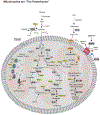
Figure 1:. Mitochondria are the powerhouse of the cell.
Mitochondria integrate fuel metabolism to generate energy in the form of ATP. Mitochondria oxidize pyruvate (derived from glucose or lactate), fatty acids, and amino acids to harness electrons onto the carriers NADH and FADH2. NADH and FADH2 transport these electrons to the electron transport chain, in which an electrochemical gradient is formed to facilitate ATP production through oxidative phosphorylation. Enzymes have the following abbreviations: LDH: lactate dehydrogenase, VDAC: Voltage-dependent anion channel, MPC: mitochondrial pyruvate carrier, PDC: pyruvate dehydrogenase complex, PC: pyruvate carboxylase, CS: citrate synthase, IDH2: isocitrate dehydrogenase 2, OGDH: α-ketoglutarate dehydrogenase, SDH: succinate dehydrogenase, MDH2: malate dehydrogenase 2, GLS: glutaminase, GDH: glutamate dehydrogenase, BCAT2: branched chain amino transferase 2, BCKDH: branched chain ketoacid dehydrogenase, PHD3: prolyl hydroxylase 3, AMPK: adenosine monophosphate kinase, ACC: Acetyl CoA Carboxylase, , ACS: acyl CoA synthetase, CPT1/2: carnitine palmitoyltransferase 1/2. Electrons and reducing equivalents are shown in yellow.
Figure 2.
Figure 2.. Mitochondria are biosynthetic hubs.
The mitochondria are a critical source of building blocks for biosynthetic pathways including nucleotide synthesis, fatty acid and cholesterol synthesis, amino acid synthesis, and glucose and heme synthesis. Compartmentalization is a key feature of biosynthetic pathways. While many of the enzymes listed are bi-directional, arrows are drawn to highlight the biosynthetic functions. Enzymes are circled in grey and brown with the following abbreviations: Nucleotide Synthesis: MTHFD1/2: methylenetetrahydrofolate dehydrogenase, SHMT1/2: serine hydroxymethyltransferase, DHODH: dihydroorotate dehydrogenase, FTDH: formate dehydrogenase. Fatty Acid and Cholesterol Synthesis: GLS: glutaminase, GDH: glutamate dehydrogenase, TA: transaminase, ACLY: ATP citrate lyase, ACC2: acetyl CoA carboxylase, PHD3: prolyl hydroxylase 3, MPC: mitochondrial pyruvate carrier. Amino Acid Synthesis: GDH: glutamate dehydrogenase, GS: glutamine synthetase, P5CS: Pyrroline-5-carboxylate synthase, PYCR1: Pyrroline-5-carboxylate reductase 1, OAT: ornithine aminotransferase, GOT2: glutamate oxaloacetate transaminase 2, GPT2: glutamate pyruvate transaminase 2, GC: glutamate carrier, AGC: aspartate-glutamate carrier, ORNT1: ornithine translocator. Glucose and Heme Synthesis: PCK1/2: phosphoenolpyruvate carboxykinase, MDH1/2: malate dehydrogenase, PC: pyruvate carboxylase, ALAS: aminolevulinate synthase, FECH: ferrochetolase, ABCB6: ATP binding cassette subfamily B member 6, FLVCR: feline leukemia virus subgroup C receptor 1.
Figure 3.
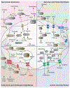
Figure 3.. Mitochondria balance redox equivalents.
In the absence of a direct mode for NAD transport, cells rely on compartmentalized flux of metabolites to support balance of reducing equivalents NAD/NADH, and NADP/NADPH. Generally, redox shuttles favor cytosolic NAD+ synthesis and mitochondrial NADH synthesis. Enzymes and transporters have with the following abbreviations: Malate-aspartate shuttle. GOT1/2: Glutamate oxaloacetate transaminase, MDH1/2: malate dehydrogenase, ME1: malic enzyme 1, Glu-Asp antiporter: glutamate-aspartate antiporter, Malate-α-KG Antiporter: malate-α-ketoglutarate antiporter. Malate-citrate shuttle. ACLY: ATP citrate lyase, MDH1/2: malate dehydrogenase, CS: citrate synthase. α-glycerophosphate shuttle. (m/c)GPDH: mitochondrial/cytosolic glycerol-3-phosphate dehydrogenase. Folate Shuttle (1C Metabolism): MTHFD1/2: methylenetetrahydrofolate dehydrogenase, SHMT1/2: serine hydroxymethyltransferase.
Figure 4.
Figure 4.. Mitochondria orchestrate waste management.
(A). Tumor cells increase nutrient consumption and metabolic fitness relative to healthy tissue, leading to accumulation of waste products in the tumor microenvironment. To manage metabolic waste, cancer cells engage recycling pathways for these metabolic by-products. (B). Ammonia. Production of and metabolic clearance of ammonia (NH3) in cell metabolism. NH3 is generated by amino acid and nucleotide catabolism. NH3 is assimilated in the mitochondria through GS (glutamine synthetase), GDH (glutamate dehydrogenase), and CPS1 (carbamoyl phosphate synthase 1). CPS1 initiates the urea cycle for production of the metabolic waste product urea. Urea can be re-catabolized by urease positive bacteria in the microbiome to regenerate NH3. AGC: aspartate-glutamate carrier, ORNT1: ornithine translocator (C). Hydrogen Sulfide. Production of and metabolic clearance of hydrogen sulfide (H2S) in cell metabolism. H2S is generated by the mammalian enzymes CBS (cystathionine β synthase), CSE (cystathionine γ lyase), 3-MST (3-mercaptopyruvate sulfurtransferase) and from the metabolic reactions in the microbiome. H2S is cleared by iterative oxidation catalyzed by sulfide quinone reductase (SQR), thiosulfate reductase (TR), and sulfite oxidase (SO). TR utilizes oxidized glutathione (GS) as a sink for electrons. Oxidations catalyzed by SQR and SO are linked to mitochondrial ETC and oxidative phosphorylation. (D). Reactive Oxygen Species. Reactions that generate and sequester ROS (reactive oxygen species). ROS are generated in the mitochondria through the ETC and NOX4 (NADPH oxidase). SOD2 (superoxide dismutase 2) converts superoxide into a the less reactive molecule hydrogen peroxide (H2O2). In the mitochondria, H2O2 is turned over by combined functions of periodxins (Prx) and thioredoxins (Trx). H2O2 also reacts with Fe+ (the Fenton Reaction) to generated OH. in the mitochondria. ROS inflict oxidative damage to proteins in the mitochondria and cytosol, and also function as potent mitogen signaling agents.

Comment in

Similar articles

Cited by

References

    1. Castresana J, Saraste M Evolution of Energetic Metabolism: the respiration-early hypothesis. Trends in biochemical sciences 20, 443–448 (1995). - PubMed
    1. Lyons TW, Reinhard CT & Planavsky NJ The rise of oxygen in Earth’s early ocean and atmosphere. Nature 506, 307–315, doi:10.1038/nature13068 (2014). - DOI - PubMed
    1. Sagan L On the Origin of Mitosing Cells. J.Theoret. Biol 14, 225–274 (1966). - PubMed
    1. Timmis JN, Ayliffe MA, Huang CY & Martin W Endosymbiotic gene transfer: organelle genomes forge eukaryotic chromosomes. Nature reviews. Genetics 5, 123–135, doi:10.1038/nrg1271 (2004). - DOI - PubMed
    1. Vyas S, Zaganjor E & Haigis MC Mitochondria and Cancer. Cell 166, 555–566, doi:10.1016/j.cell.2016.07.002 (2016). - DOI - PMC - PubMed

Publication types

Substances