. 2018 Aug 16;92(17):e00340-18.
doi: 10.1128/JVI.00340-18. Print 2018 Sep 1.

Human Cytomegalovirus Immediate Early 1 Protein Causes Loss of SOX2 from Neural Progenitor Cells by Trapping Unphosphorylated STAT3 in the Nucleus

Affiliations

Human Cytomegalovirus Immediate Early 1 Protein Causes Loss of SOX2 from Neural Progenitor Cells by Trapping Unphosphorylated STAT3 in the Nucleus

Cong-Cong Wu et al. J Virol. .

Abstract

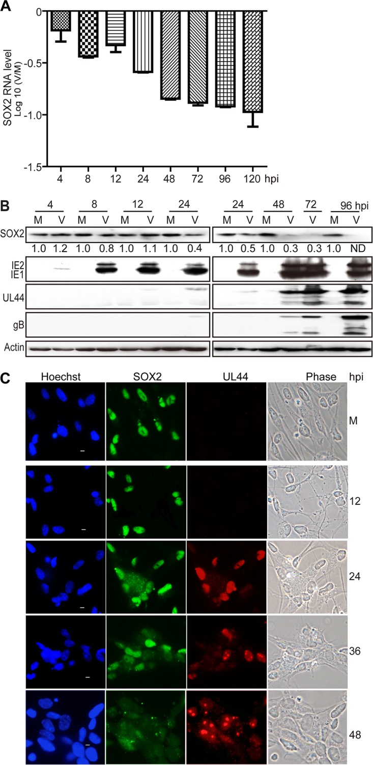
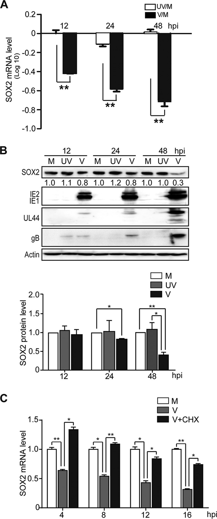
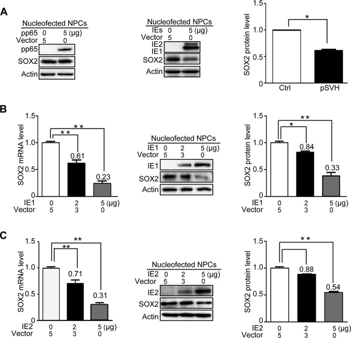
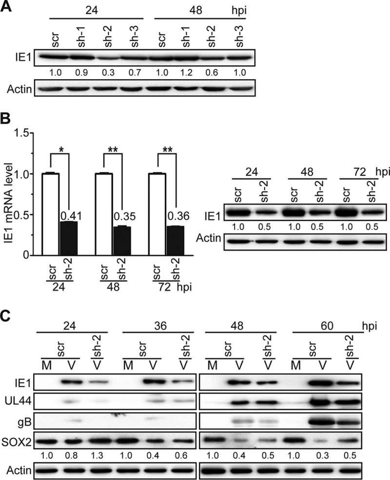
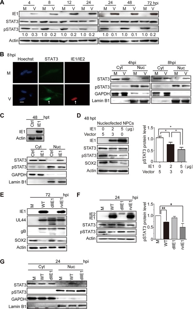
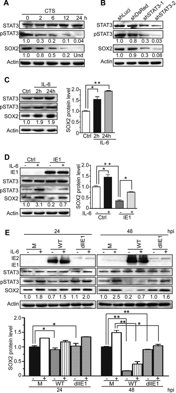
The mechanisms underlying neurodevelopmental damage caused by virus infections remain poorly defined. Congenital human cytomegalovirus (HCMV) infection is the leading cause of fetal brain development disorders. Previous work has linked HCMV infection to perturbations of neural cell fate, including premature differentiation of neural progenitor cells (NPCs). Here, we show that HCMV infection of NPCs results in loss of the SOX2 protein, a key pluripotency-associated transcription factor. SOX2 depletion maps to the HCMV major immediate early (IE) transcription unit and is individually mediated by the IE1 and IE2 proteins. IE1 causes SOX2 downregulation by promoting the nuclear accumulation and inhibiting the phosphorylation of STAT3, a transcriptional activator of SOX2 expression. Deranged signaling resulting in depletion of a critical stem cell protein is an unanticipated mechanism by which the viral major IE proteins may contribute to brain development disorders caused by congenital HCMV infection.IMPORTANCE Human cytomegalovirus (HCMV) infections are a leading cause of brain damage, hearing loss, and other neurological disabilities in children. We report that the HCMV proteins known as IE1 and IE2 target expression of human SOX2, a central pluripotency-associated transcription factor that governs neural progenitor cell (NPC) fate and is required for normal brain development. Both during HCMV infection and when expressed alone, IE1 causes the loss of SOX2 from NPCs. IE1 mediates SOX2 depletion by targeting STAT3, a critical upstream regulator of SOX2 expression. Our findings reveal an unanticipated mechanism by which a common virus may cause damage to the developing nervous system and suggest novel targets for medical intervention.

Keywords: HCMV; IE1; SOX2; STAT3; neural progenitor cells.


Figures

FIG 1
FIG 1
HCMV infection downregulates SOX2 at the mRNA and protein levels in NPCs. NPC monolayers were mock infected (M) or infected with HCMV (TNWT) at an MOI of 3 (V) and collected at the indicated times postinfection for mRNA or protein analyses. (A) SOX2 mRNA levels during HCMV infection of NPCs. The levels of SOX2 mRNA, normalized to GAPDH, were determined by qRT-PCR at 4 to 120 hpi. The results shown are averages and SD of data from three independent experiments, each conducted in triplicate. (B) SOX2 and viral protein levels during HCMV infection of NPCs. SOX2, IE1/IE2, UL44, and gB steady-state protein levels were determined by Western blotting at 4 to 96 hpi. Actin served as a loading control. The values listed below the SOX2 blots indicate the relative SOX2 protein levels compared to corresponding mock-infected controls following actin normalization. ND, not detectable. (C) Cellular distribution of SOX2 in relation to viral replication compartments during HCMV infection of NPCs. The distributions of SOX2 and UL44 were determined by indirect immunofluorescence assay at 12 to 48 hpi. NPCs grown on poly-d-lysine-coated coverslips were stained with antibodies against SOX2 (green) and UL44 (red), and nuclei were counterstained with Hoechst 33342 (blue). Phase-contrast images are also shown. Scale bars, 5 μm.
FIG 2
FIG 2
De novo synthesis of HCMV proteins is required for SOX2 downregulation in NPCs. Shown are comparisons of HCMV infections with active virus (V) and UV-inactivated virus (UV). NPCs were mock infected (M) or infected with active or UV-irradiated TNWT at an MOI of 3 and collected at the indicated times postinfection. (A) SOX2 mRNA levels normalized to GAPDH were determined by qRT-PCR. Log10 values of inactivated virus/mock-infected virus (UV/M) and active virus/mock-infected virus (V/M) ratios are shown. Data from three independent experiments were analyzed by one-way analysis of variance (ANOVA), and the results are presented as averages and SD. **, P ≤ 0.01. (B) Levels of SOX2 and representative viral proteins (IE1/IE2, UL44, and gB) were determined by Western blotting. Actin served as a loading control. The values listed below the SOX2 blots indicate the relative SOX2 protein levels compared to corresponding mock-infected controls following actin normalization. The data are from three independent experiments, and the results are presented as averages and SD. *, P ≤ 0.05; **, P ≤ 0.01. (C) Effect of protein synthesis inhibition by CHX treatment on SOX2 mRNA levels. NPCs were pretreated with CHX for 1 h prior to infection and then mock infected (M) or infected with HCMV (TNWT) at an MOI of 3 (V). Cells were collected at 4, 8, 12, and 16 hpi for analysis of SOX2 mRNA by qRT-PCR. Data from three independent experiments were analyzed by one-way ANOVA, and the results are presented as averages and SD. *, P ≤ 0.05; **, P ≤ 0.01.
FIG 3
FIG 3
HCMV major IE proteins downregulate SOX2 mRNA and protein in NPCs. HCMV pp65, IE1, IE2, or IE1 and IE2 combined were transiently expressed in NPCs following nucleofection. Samples were collected at 48 h posttransfection for mRNA (qRT-PCR) or protein (Western blotting) analysis. Actin served as a loading control. Shown is the relative level of SOX2 protein compared to corresponding controls following actin normalization. Data from three independent experiments were analyzed by one-way ANOVA, and the results are presented as averages and SD. *, P < 0.05; **, P < 0.01. (A) Effects of pp65 and major IE proteins (IE1 and IE2) on SOX2 protein levels. (Left blot) NPCs were transfected with 5 μg pcDNA3.0 (Vector) or pcDNA3-pp65. (Right blot) NPCs were transfected with 5 μg pcDNA3-pp65 as a control (Vector) or pSVH (IE1 and IE2). (B) Effects of IE1 on SOX2 mRNA and protein levels. NPCs were transfected with 5 μg pcDNA3-pp65 (Vector) or 2 to 5 μg pcDNA3-IE1. (C) Effects of IE2 on SOX2 mRNA and protein levels. NPCs were transfected with 5 μg pcDNA3-pp65 (Vector) or 2 to 5 μg pcDNA3-IE2.
FIG 4
FIG 4
IE1 knockdown attenuates HCMV-induced SOX2 downregulation in NPCs. (A) IE1-directed knockdown efficiencies of candidate shRNAs in HEL cells. HEL cells were transduced with lentiviruses expressing shRNA-IE1-1 (sh-1), shRNA-IE1-2 (sh-2), shRNA-IE1-3 (sh-3), or shRNA-scramble (scr). At 48 h postransduction, the cells were infected with HCMV (TNWT) at an MOI of 1 and collected at 24 or 48 hpi. IE1 protein levels were determined by Western blotting. Actin served as a loading control. (B) IE1-directed knockdown efficiency of sh-2 in HEL cells. HEL cells transduced with sh-2- or scr-expressing lentiviruses were infected with HCMV (TNWT) as for panel A. (Left) IE1 mRNA levels were determined by qRT-PCR at the indicated times postinfection; data from two independent experiments were analyzed by one-way ANOVA, and the results are presented as averages and SD. *, P ≤ 0.05; **, P ≤ 0.01. (Right) Protein levels were determined at the indicated times by Western blotting. (C) Effects of sh-2 on IE1 and SOX2 expression in NPCs. NPCs were transduced with sh-2- or scr-expressing lentiviruses, cultured for 48 h, reseeded at a density of 3 × 10⁶ cells/dish, and mock infected (M) or infected with TNWT at an MOI of 1 (V). Protein levels of IE1, UL44, gB, and SOX2 at the indicated times postinfection are shown. Actin served as a loading control.
FIG 5
FIG 5
HCMV infection or IE1 expression inhibits STAT3 tyrosine phosphorylation and promotes nuclear accumulation of unphosphorylated STAT3 in NPCs. (A) Inhibition of STAT3 tyrosine (Y705) phosphorylation by HCMV infection. NPCs were mock infected (M) or infected with TNWT at an MOI of 3 (V) and collected at the indicated times postinfection. The protein levels of IE1, pSTAT3, and total STAT3 were determined by Western blotting. Actin served as a loading control. (B) Nuclear trapping of STAT3 by HCMV infection. NPCs were mock infected (M) or -infected with TNWT at an MOI of 1 (V). (Left) For indirect immunofluorescence analysis, NPCs on coverslips collected at 8 hpi were stained with antibodies against STAT3 (green) or IE1/IE2 (red), and nuclei were counterstained with Hoechst 33342 (blue). Infected (IE1/IE2-positive) cells are indicated by arrows. Scale bar, 10 μm. (Right) For cellular-fractionation analysis, fractions enriched in cytoplasmic (Cyt) or nuclear (Nuc) proteins were prepared from cells collected at 4 or 8 hpi. Protein levels of pSTAT3 and total STAT3 in each fraction were determined by Western blotting. GAPDH and lamin B1 served as controls for the Cyt and Nuc fractions, respectively. (C to G) Inhibition of tyrosine phosphorylation and nuclear sequestration of unphosphorylated STAT3 by IE1. Fractions enriched in cytosolic or nuclear proteins or total cell extracts were prepared. (C) For transient-transfection analysis, NPCs were nucleofected with pcDNA3-IE1 or empty vector (Ctrl) and harvested at 48 h postnucleofection. Protein levels of IE1, pSTAT3, and total STAT3 were determined by Western blotting. GAPDH and lamin B1 served as controls for the Cyt and Nuc fractions, respectively. (D) To examine dose-dependent effects of IE1 on pSTAT3 levels, NPCs transfected with the indicated amounts of pcDNA-IE1 and empty vector (pcDNA3.0) were harvested 48 h postnucleofection. The protein levels of IE1, STAT3, pSTAT3, and SOX2 were determined by Western blotting. *, P ≤ 0.05. (E to G) For HCMV infection analysis, NPCs were mock infected (M) or infected with TNWT (WT), TNdlIE1 (dlIE1), or TNrvIE1 (rvIE1) viruses at an MOI of 10. The levels of the indicated viral and cellular proteins in whole-cell extracts at 72 hpi (E) and 24 hpi (F) or in the Cyt and Nuc fractions at 24 hpi (G) were determined by Western blotting. Actin, GAPDH, and lamin B1 served as controls for total extracts or Cyt and Nuc fractions, respectively. *, P ≤ 0.05; **, P ≤ 0.01.
FIG 6
FIG 6
SOX2 expression strictly depends on pSTAT3, and IE1 mediates SOX2 depletion by inhibiting STAT3 activation. (A) Inhibition of STAT3 correlates with suppression of SOX2 expression. NPCs were treated with the chemical inhibitor CTS for the indicated times. The protein levels of total STAT3, pSTAT3, and SOX2 were determined by Western blotting. Actin served as a loading control. Und, undetectable. (B) Silencing of STAT3 correlates with suppression of SOX2 expression. NPCs were transduced with the lentivirus Tet-pLKO-puro-shLuci (shLuci), Tet-pLKO-puro-shDsRed (shDsRed), Tet-pLKO-puro-shSTAT3-1 (shSTAT3-1), or Tet-pLKO-puro-shSTAT3-2 (shSTAT3-2) and treated with doxycycline for 48 h to induce shRNA expression. The protein levels of total STAT3, pSTAT3, and SOX2 were determined by Western blotting. Actin served as a loading control. (C) IL-6-mediated activation of STAT3 correlates with induction of SOX2 expression. NPCs were treated with IL-6 for 2 or 24 h. The protein levels of pSTAT3, total STAT3, and SOX2 were monitored by Western blotting. Actin served as a loading control. Data from three independent experiments were analyzed by one-way ANOVA, and the results are presented as averages and SD. *, P ≤ 0.05; **, P ≤ 0.01. (D) IL-6 counteracts IE1-dependent SOX2 downregulation in transiently transfected NPCs. NPCs were transfected with pcDNA3-IE1, treated with IL-6 for 4 h or left untreated, and collected at 48 h posttransfection. The protein levels of IE1, total STAT3, pSTAT3, and SOX2 were determined by Western blotting. Actin served as a loading control. Data from three independent experiments were analyzed by one-way ANOVA, and the results are presented as averages and SD. *, P ≤ 0.05; **, P ≤ 0.01. (E) IL-6 counteracts IE1-dependent SOX2 downregulation in HCMV-infected NPCs. NPCs were infected with TNWT or TNdlIE1 virus at an MOI of 10, treated with IL-6 for 4 h or left untreated, and collected at 48 hpi. The protein levels of IE1/IE2, total STAT3, pSTAT3, and SOX2 were determined by Western blotting. Actin served as a loading control. Data from three independent experiments were analyzed by one-way ANOVA, and the results are presented as averages and SD. *, P ≤ 0.05; **, P ≤ 0.01.

Similar articles

Cited by

References

    1. Bale JF., Jr 1984. Human cytomegalovirus infection and disorders of the nervous system. Arch Neurol 41:310–320. doi:10.1001/archneur.1984.04050150092023. - DOI - PubMed
    1. Stagno S, Pass RF, Cloud G, Britt WJ, Henderson RE, Walton PD, Veren DA, Page F, Alford CA. 1986. Primary cytomegalovirus infection in pregnancy. Incidence, transmission to fetus, and clinical outcome. JAMA 256:1904–1908. - PubMed
    1. Bale JF., Jr 2014. Congenital cytomegalovirus infection. Handb Clin Neurol 123:319–326. doi:10.1016/B978-0-444-53488-0.00015-8. - DOI - PubMed
    1. Conboy TJ, Pass RF, Stagno S, Britt WJ, Alford CA, McFarland CE, Boll TJ. 1986. Intellectual development in school-aged children with asymptomatic congenital cytomegalovirus infection. Pediatrics 77:801–806. - PubMed
    1. Pass RF, Stagno S, Myers GJ, Alford CA. 1980. Outcome of symptomatic congenital cytomegalovirus infection: results of long-term longitudinal follow-up. Pediatrics 66:758–762. - PubMed

Publication types

MeSH terms

LinkOut - more resources