. 2018 Aug 16;92(17):e01036-18.
doi: 10.1128/JVI.01036-18. Print 2018 Sep 1.

Attenuated Herpes Simplex Virus 1 (HSV-1) Expressing a Mutant Form of ICP6 Stimulates a Strong Immune Response That Protects Mice against HSV-1-Induced Corneal Disease

Affiliations

Attenuated Herpes Simplex Virus 1 (HSV-1) Expressing a Mutant Form of ICP6 Stimulates a Strong Immune Response That Protects Mice against HSV-1-Induced Corneal Disease

David J Davido et al. J Virol. .

Abstract

We previously isolated a herpes simplex virus 1 (HSV-1) mutant, KOS-NA, that carries two nonsynonymous mutations in UL39, resulting in L393P and R950H amino acid substitutions in infected cell protein 6 (ICP6). Our published data studying KOS-NA pathogenesis strongly suggest that one of these ICP6 substitutions expressed from KOS-NA, R950H, severely impaired acute viral replication in the eyes and trigeminal ganglia of mice after inoculation onto the cornea and consequently impaired establishment and reactivation from latency. Because of its significant neuroattenuation, we tested KOS-NA as a potential prophylactic vaccine against HSV-1 in a mouse model of corneal infection. KOS-NA stimulated stronger antibody and T cell responses than a replication-competent ICP0-null mutant and a replication-incompetent ICP8-null mutant optimized for immunogenicity. Immunizations with the ICP0-, ICP8-, and KOS-NA viruses all reduced replication of wild-type HSV-1 challenge virus in the corneal epithelium to similar extents. Low immunizing doses of KOS-NA and the ICP8- virus, but not the ICP0- virus, protected mice against eyelid disease (blepharitis). Notably, only KOS-NA protected almost completely against corneal disease (keratitis) and greatly reduced latent infection by challenge virus. Thus, vaccination of mice with KOS-NA prior to corneal challenge provides significant protection against HSV-1-mediated disease of the eye, even at a very low immunizing dose. These results suggest that KOS-NA may be the foundation of an effective prophylactic vaccine to prevent or limit HSV-1 ocular diseases.IMPORTANCE HSV-1 is a ubiquitous human pathogen that infects the majority of the world's population. Although most infections are asymptomatic, HSV-1 establishes lifelong latency in infected sensory neurons, from which it can reactivate to cause deadly encephalitis or potentially blinding eye disease. No clinically effective vaccine is available. In this study, we tested the protective potential of a neuroattenuated HSV-1 mutant (KOS-NA) as a vaccine in mice. We compared the effects of immunization with KOS-NA to those of two other attenuated viruses, a replication-competent (ICP0-) virus and a replication-incompetent (ICP8-) virus. Our data show that KOS-NA proved superior to the ICP0- and ICP8-null mutants in protecting mice from corneal disease and latent infection. With its significant neuroattenuation, severe impairment in establishing latency, and excellent protective effect, KOS-NA represents a significant discovery in the field of HSV-1 vaccine development.

Keywords: HSV-1; ICP6; cornea; herpes simplex virus; immunization; keratitis; mutant; ocular; vaccine.


Figures

FIG 1
FIG 1
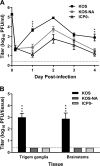
Virus titers in tear film and neural tissues after corneal inoculation. Groups of BALB/c mice were inoculated on their scarified corneas with 2 × 10⁵ PFU per eye of wild-type KOS, KOS-NA, or ICP0 (7134) virus. (A) Titers of virus collected on corneal swabs were determined 4 h and 1 to 4 days postinfection. **, P = 0.0083, and ***, P = 0.0009 for KOS-NA compared with KOS (P > 0.05 for KOS-NA compared with ICP0 virus). (B) Mice were euthanized on day 5 postinfection, and viral titers in TG and brainstems were determined. The values represent means and standard errors of the mean (SEM) of a total of 6 to 10 mice per group compiled from 2 independent experiments. ***, P < 0.0001 for KOS-NA or ICP0 virus compared with KOS. The dashed lines indicate limits of detection.
FIG 2
FIG 2
T cell responses to vaccination. Mononuclear cells were isolated from draining lymph nodes 6 days after immunization of BALB.B mice with a low (2 × 10⁴ PFU) dose of the indicated virus or control supernatant. Activated CD4 T cells in draining lymph nodes were quantified by stimulation with PMA and ionomycin, followed by intracellular staining for IFN-γ. (A) Percentages of CD4+ T cells that were IFN-γ+. (B) Total numbers of CD4+ IFN-γ-producing cells in draining lymph nodes. The results are the means of numbers from individual mice compiled from 3 independent experiments (total numbers of mice, 5 for control group and 6 to 9 for vaccine groups). *, P < 0.05 for control or ICP0 virus compared with KOS-NA. HSV-specific CD8 T cell responses were compared using gB498–505 peptide as the stimulus in an IFN-γ ELISpot assay. (C) Numbers of spot-forming cells (SFC) per million lymph node cells. (D) Total numbers of SFC in draining lymph nodes. The results are the means of numbers from individual mice compiled from 3 independent experiments (total numbers of mice, 7 for control group and 8 to 11 for vaccine groups). **, P < 0.001 for control supernatant or ICP0 virus compared with KOS-NA. P < 0.05 to 0.01 for ICP8 compared with KOS-NA. The error bars represent SEM.
FIG 3
FIG 3
Titers of HSV-specific antibody in immunized mice. Groups of BALB/c mice were immunized with high (5 × 10⁵ PFU), medium (1 × 10⁵ PFU), or low (2 × 10⁴ PFU) doses of the indicated viruses, and 1 group of mice was immunized with control supernatant of uninfected cells. Blood was collected 21 days postimmunization, and HSV-specific serum IgG was quantified by ELISA. The data represent the geometric mean titers for 12 mice per group plus SEM and are the combined results of 2 independent experiments with similar results. *, P < 0.01, and **, P < 0.001 for KOS-NA compared to the ICP0 virus. P < 0.05 for KOS-NA compared to the ICP8 virus. nd, not detected.
FIG 4
FIG 4
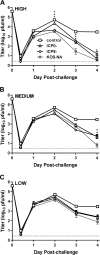
Eye titers of challenge virus shed from the corneal epithelium. Groups of 10 BALB/c mice immunized with the high (A), medium (B), or low (C) dose of virus vaccine or control supernatant as described in the legend to Fig. 3 were challenged with HSV-1 strain mP (4 × 10⁵ PFU/eye) 4 weeks postimmunization. The eyes of 6 mice per group were swabbed at the indicated times, and the titers of challenge virus in them were determined. The experiment was repeated once. The eye swab data represent the geometric means ± SEM of the combined results from the two independent experiments (total number of mice, 12 per group). The control group was the same for all three graphs. *, P = 0.0221; **, P = 0.0044; and ***, P = 0.0002 to 0.0001 for KOS-NA compared with the ICP0 virus. P = 0.0013 on day 3 at the high dose and P = 0.0019 on day 4 at the medium dose for KOS-NA compared with the ICP8 virus. The dashed lines indicate limits of detection.
FIG 5
FIG 5
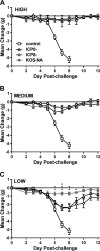
Body weight change after virus challenge. The groups of 10 BALB/c mice described in the legend to Fig. 4 were weighed prior to challenge and at the indicated times postchallenge. The data are the mean changes in weight ± SEM for each group and represent the combined results from two independent experiments (total number of mice, 20 per group). *, P = 0.0300 to 0.0171; **, P = 0.007 to 0.002; ***, P < 0.0001 for KOS-NA compared with the ICP0 virus. P = 0.0189 to 0.0005 at the low dose on days 6 through 10 for KOS-NA compared with the ICP8 virus.
FIG 6
FIG 6
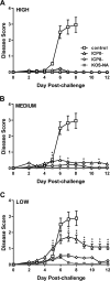
Protection of mice from blepharitis after corneal challenge. Groups of mice as described in the legend to Fig. 4 were scored daily for signs of eyelid disease. The values are the means ± SEM of 36 to 40 eyes per group and are the combined results of two independent experiments. *, P = 0.043 to 0.01, and **, P = 0.007 to <0.001 for KOS-NA relative to the ICP0 virus. Differences between KOS-NA and ICP8 virus were not statistically significant.
FIG 7
FIG 7
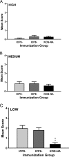
Protection of mice from keratitis after corneal challenge. The eyes of the same groups of mice described in the legend to Fig. 4 were examined at 14 days postchallenge for signs of keratitis. The values represent the mean keratitis scores and SEM of eyes from surviving mice (36 to 40 eyes per group). **, P < 0.001 for KOS-NA compared with ICP8 or ICP0 virus and are the combined results of two independent experiments.
FIG 8
FIG 8
Immunization with KOS-NA impairs the establishment of latency of the challenge virus. Groups of mice immunized with the indicated virus or medium were infected with challenge virus as described in the legend to Fig. 4. (A to C) One month postchallenge, TG were removed and DNA was extracted. The relative viral DNA content was assessed by real-time PCR using primers for the HSV-1 UL50 gene after normalization to the mouse cellular adipsin gene. The data represent the relative mean fold decrease in the latent genome in 5 to 14 TG of virus-immunized mice compared with 3 TG from mice immunized with control supernatant and are the results from one of two experiments performed. HSV-1 DNA in some TG from KOS-NA high- and KOS-NA medium-dose groups were below the limit of detection. The TG were assigned a cycle number at the limit of detection for statistical purposes. *, P < 0.05, and **, P < 0.01 by ANOVA for TG from KOS-NA compared with all other groups. Additional mice were immunized with 2 × 10⁴ PFU of the indicated viruses and challenged 1 month later as described in the legend to Fig. 4. (D) TG were removed 4 days postchallenge, and the challenge virus titer in disrupted tissue was determined. ***, P < 0.001 by ANOVA for KOS-NA compared with ICP0 virus. The data are the means and SEM of 7 to 8 mice (14 to 15 TG) compiled from 2 independent experiments. Also 4 days postchallenge, cervical lymph nodes were removed and used in an IFN-γ ELISpot assay for HSV-specific CD8+ T cells with gB498–505 as the stimulus. (E) Numbers of SFC per 1 × 10⁶ cells. *, P < 0.05 for control compared with KOS-NA. (F) Total numbers of SFC per mouse. *, P < 0.01 for control or ICP0- compared with KOS-NA. P < 0.05 for ICP8- compared with KOS-NA. The results are the means of the results for individual mice compiled from 2 independent experiments (total number of mice, 6 per group).
FIG 9
FIG 9
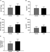
Neuroattenuated KOS-NA stimulates immune responses similar to those stimulated by the wild-type strain, KOS. Groups of mice were immunized s.c. with 2 × 10⁴ PFU (low dose) of KOS-NA or KOS, and immune responses were evaluated. Antigen-specific IFN-γ-producing cells in draining lymph nodes were enumerated by ELISpot assay 6 days postvaccination and by HSV-specific IgG in the serum 21 days postvaccination. (A and B) CD4+ IFN-γ-producing cells per 106 lymph node cells from individual BALB/c mice (A) and total CD4+ IFN-γ-producing cells responding to inactivated virus antigen (B) (11 mice per group). (C and D) CD8+ IFN-γ-producing cells specific for the gB498–505 epitope per 106 lymph node cells from individual BALB.B mice (C) and total CD8+ IFN-γ-producing cells (D) (10 mice per group). (E) Concentrations of HSV-specific serum IgG (8 BALB/c mice per group). The error bars represent SEM.
FIG 10
FIG 10
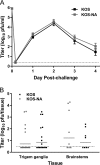
Mice immunized with KOS-NA or KOS are equivalently protected from challenge. Mice immunized with 2 × 10⁴ PFU of KOS-NA or KOS were challenged with 4 × 10⁵ PFU of HSV-1 strain mP, and protection from acute infection was assessed. (A) Titers of virus shed from the corneal epithelium over time postchallenge. (B) Virus titer in the TG and brainstems 4 days postchallenge. The data are the combined results of two independent experiments with 8 mice per group. The dashed lines indicate the limits of detection. The error bars represent SEM.

Similar articles

Cited by

References

    1. Roizman R, Knipe DM, Whitley RJ. 2007. Herpes simplex viruses, p 2501–2601. In Knipe DM, Howley PM (ed), Fields virology, vol 2 Lippincott Williams & Wilkins, New York, NY.
    1. Dana MR, Qian Y, Hamrah P. 2000. Twenty-five-year panorama of corneal immunology: emerging concepts in the immunopathogenesis of microbial keratitis, peripheral ulcerative keratitis, and corneal transplant rejection. Cornea 19:625–643. doi:10.1097/00003226-200009000-00008. - DOI - PubMed
    1. Liesegang TJ. 2001. Herpes simplex virus epidemiology and ocular importance. Cornea 20:1–13. doi:10.1097/00003226-200101000-00001. - DOI - PubMed
    1. Barron BA, Gee L, Hauck WW, Kurinij N, Dawson CR, Jones DB, Wilhelmus KR, Kaufman HE, Sugar J, Hyndiuk RA, Laibson PR, Stulting D, Asbell PA. 1994. Herpetic Eye Disease Study. A controlled trial of oral acyclovir for herpes simplex stromal keratitis. Ophthalmology 101:1871–1882. - PubMed
    1. Koelle DM, Corey L. 2003. Recent progress in herpes simplex virus immunobiology and vaccine research. Clin Microbiol Rev 16:96–113. doi:10.1128/CMR.16.1.96-113.2003. - DOI - PMC - PubMed

Publication types

MeSH terms

LinkOut - more resources