. 2018 Aug 15;29(17):2045-2054.
doi: 10.1091/mbc.E18-05-0282. Epub 2018 Jun 27.

Rab18 is not necessary for lipid droplet biogenesis or turnover in human mammary carcinoma cells

Affiliations

Rab18 is not necessary for lipid droplet biogenesis or turnover in human mammary carcinoma cells

Christina B K Jayson et al. Mol Biol Cell. .

Abstract

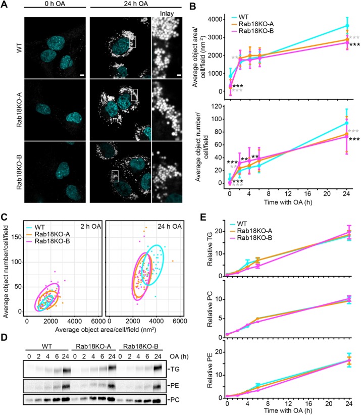
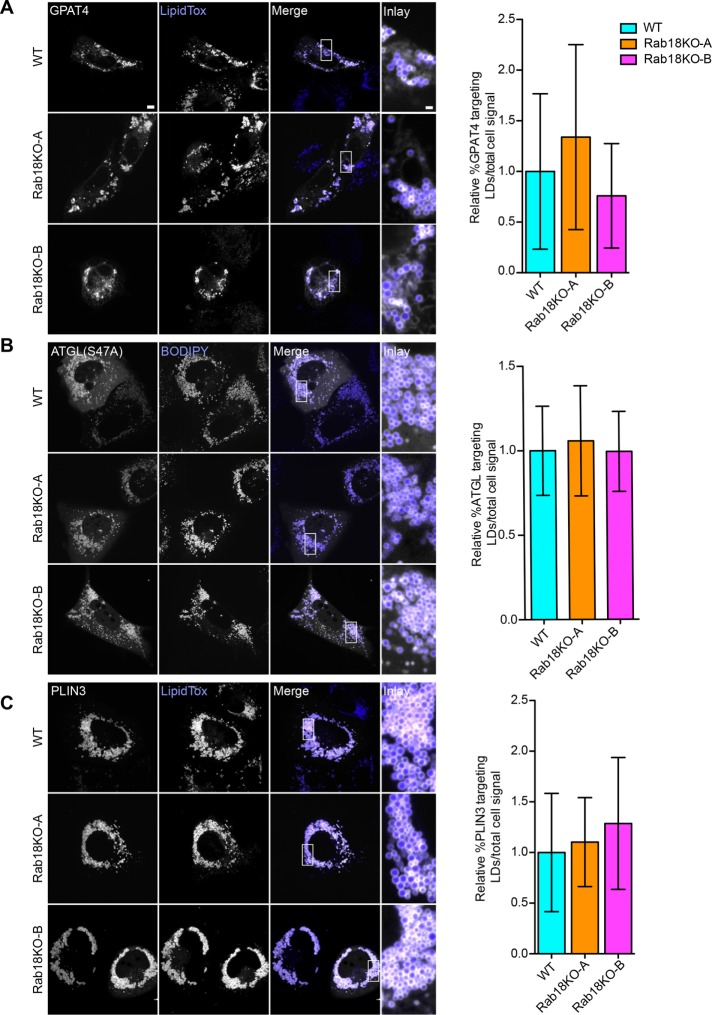
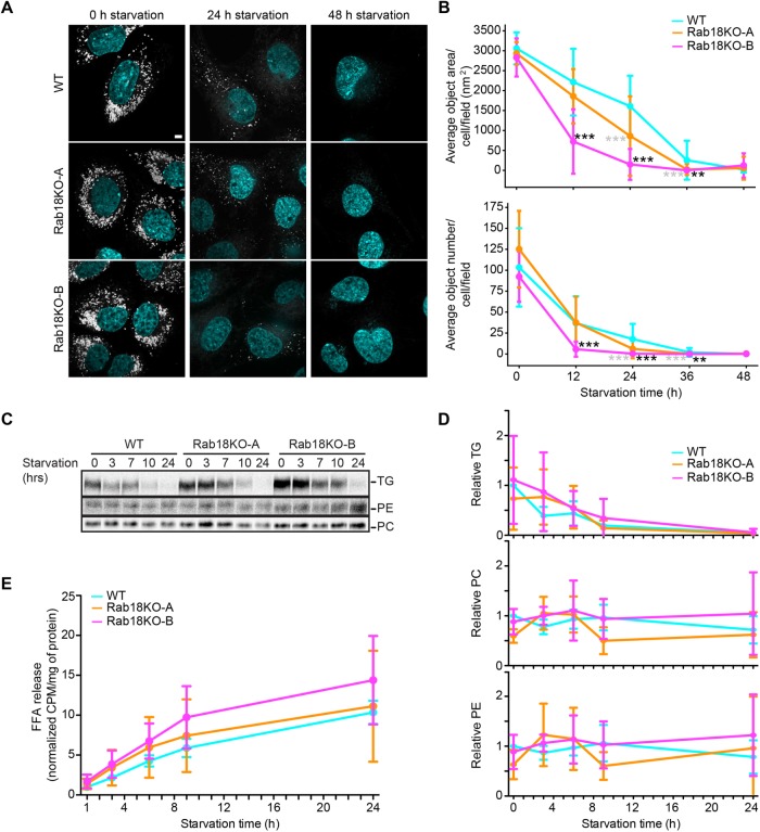
Rab GTPases recruit peripheral membrane proteins and can define organelle identity. Rab18 localizes to the endoplasmic reticulum (ER) but also to lipid droplets (LDs), where it has been implicated in effector protein recruitment and in defining LD identity. Here, we studied Rab18 localization and function in a human mammary carcinoma cell line. Rab18 localized to the ER and to LD membranes on LD induction, with the latter depending on the Rab18 activation state. In cells lacking Rab18, LDs were modestly reduced in size and numbers, but we found little evidence for Rab18 function in LD formation, LD turnover on cell starvation, or the targeting of several proteins to LDs. We conclude that Rab18 is not a general, necessary component of the protein machinery involved in LD biogenesis or turnover.


Figures

FIGURE 1:
FIGURE 1:
Rab18 localizes distinctly to LDs and the ER in SUM159 cells. (A) Overexpressed GFP-Rab18 localizes to LDs (LipidTox) (white arrowheads) and the ER (mCherry-ER3), and localization depends on GTP state (white arrows). SUM159 cells coexpressing mCherry-ER3 and GFP-tagged WT Rab18, GDP-bound Rab18(S22N) mutant, or GTP-bound Rab18(Q67L) mutant were incubated with OA for 0 or 18 h and imaged with spinning disk confocal. Scale bar 5 µm and for inlay 1 µm. (B) Rab18 localizes to the ER and LD structures. SUM159 cells co-expressing mCherry-ER3 and GFP-Rab18 were incubated with oleic acid for 18 h and imaged by SIM. Max projections of 1.25-µm stacks are shown. Scale bars, 1 µm. (C) Quantification of Rab18 signal distribution in SIM images. n = 5 fields. (D) Rab18 was detected in LD fractions and total cell lysates of SUM159 cells. LD fractions and cell lysates isolated from SUM159 cells after 18 h oleic acid were analyzed by mass spectrometry to detect proteins on LDs compared with total lysate. ND = not detected.
FIGURE 2:
FIGURE 2:
Rab18 deletion does not affect ER morphology. (A) Sequence analysis of Rab18 KO clones A and B. CRISPR/Cas9-mediated genome editing of the Rab18 locus introduces early stop codons at exons 4 (clone A) and 5 (clone B). (B) qPCR data reveal decreased Rab18 mRNA levels by 98 and 96% in Rab18KO-A and –B, respectively, compared with WT control. WT vs. Rab18KO-A* in gray, WT vs. Rab18KO-B* in black. (C) No Rab18 protein is detected in knockout clones by Western blot. Expression levels of Rab18 protein in WT and Rab18 KO cells were analyzed by Western blot with an antibody against endogenous Rab18. No detectable Rab18 protein was found in the Rab18KO-A or Rab18KO-B. (D) Rab18 peptide fragments were not detected by mass spectrometry in Rab18KO-A. WT SUM159 cell lysates and Rab18KO-A cell lysates were analyzed by mass spectrometry with sequence coverage of 68.4% for Rab18. (E) ER morphology in Rab18 KO clones is similar to WT cells. Cells were transfected with GFP-ERox to analyze general ER morphology. Separately, cells were fixed and probed with Reticulon 4 (Rtn4) antibody to visualize ER tubules. Scale bar 5 µm and for inlay 1 µm.
FIGURE 3:
FIGURE 3:
LD biogenesis is not affected in Rab18 KO cells. (A) LD morphology is similar in Rab18KO clones and WT cells with oleic acid incubation. Representative images of WT and Rab18 KO cells prestarved for 5 h before addition of oleic acid and after 24 h of oleic acid incubation. Scale bar 5 µm and for inlay 1 µm. (B) Rab18 KO average BODIPY object area and number are slightly smaller than WT after 24 h oleic acid incubation. WT and Rab18 KO cells were incubated with oleic acid for indicated time points, fixed, and imaged by high-throughput microscopy. Average BODIPY object area and number per cell per field (>five cells per field to obtain representative measurements) were quantified per condition. n > 27 fields/point. WT vs. Rab18KO-A* in gray; WT vs. Rab18KO-B* in black. (C) Rab18KO and WT average object area vs. number 90% confidence intervals overlap at 2 and 24 h oleic acid incubation. Average object number plotted against average object area per cell per field with 2 and 24 h oleic acid. Ellipses represent 90% confidence intervals. (D, E) Rab18KO clones have similar synthesis of TG, PE, and PC as WT cells with oleic acid incubation. Incorporation of [14C] oleate into triglycerides (TGs), phophatidylethanolamine (PE), and phosphatidycholine (PC) were measure over time. Lipids were extracted from cells and separated by TLC. Representative autoradiographs of three replicates per genotype are shown (D). (E) Quantified TG, PC, and PE levels are similar between Rab18KO clones and WT cells over time. TLC plates were developed and incorporation of [14C] oleate into TGs, PE, and PC was quantified using Fiji. Data presented are normalized CPM to mg/ml protein and relative to WT at t = 0 h. n = 3.
FIGURE 4:
FIGURE 4:
Protein targeting to LDs is similar in Rab18KO and WT. (A–C) Representative confocal images of WT, Rab18KO-A, and Rab18KO-B cells transfected with GPAT4-GFP (A), catalytically inactive mCherry-ATGL(S47A) (B), and GFP-PLIN3 (C) were incubated with oleic acid for 24 h. Targeting to LDs stained with LipidTox (A, C) or BODIPY (B) compared with total cell signal quantified in (A) n ≥ 39 cells, (B) n ≥ 36 cells, and (C) n ≥ 43 cells. Scale bar 5 µm and for inlay 1 µm.
FIGURE 5:
FIGURE 5:
Rab18 deletion does not affect TG turnover. (A) LDs are degraded similarly in Rab18KO clones and WT cells with starvation. Representative images of WT, Rab18KO-A, and Rab18KO-B cells after 12 h OA loading (t = 0) and 24 or 48 h of starvation. LDs stained with BODIPY 493/503 and nuclei with Hoechst. Scale bar 5 µm and inlay 1 µm. (B) Rab18KO average BODIPY object area and number are similar to WT after 48 h starvation. WT and Rab18KO cells were incubated with oleic acid for indicated time points as in A. Cells were fixed and imaged by high-throughput microscopy. Average BODIPY object area and number per cell per field (>5 cells per field) was quantified per condition. n > 7 fields. WT vs. Rab18KO-A * in gray; WT vs. Rab18KO-B * in black. (C) Rab18KO clones have similar synthesis of TG and PE and less PC than WT cells with starvation. WT and Rab18KO cells were incubated with [14C] oleic acid for 18 h, followed by starvation for increasing time. Total lipids were extracted at each time point and separated by TLC to detect radiolabeled TG, PE, and PC levels. Representative autoradiographs of three replicates per genotype. (D) Quantified TG and PE levels are similar, and PC levels are less between Rab18KO clones and WT cells over time with starvation. TG, PC, and PE signals were quantified from TLC plates using Fiji. Data presented are normalized CPM to mg/ml protein and relative to WT at t = 0 h. n = 3 biological replicates. (E) Rab18KO clones have decreased free fatty acid release over time with starvation. The [14C]-labeled free fatty acid release measured over time with starvation after WT; Rab18KO-A, and Rab18KO-B cells were incubated with [14C] oleic acid for 18 h. n = 3 biological replicates.

Similar articles

Cited by

References

    1. Barr FA. (2013). Rab GTPases and membrane identity: causal or inconsequential? J Cell Biol , 191–199. - PMC - PubMed
    1. Bulankina AV, Deggerich A, Wenzel D, Mutenda K, Wittmann JG, Rudolph MG, Burger KNJ, Höning S. (2009). TIP47 functions in the biogenesis of lipid droplets. J Cell Biol , 641–655. - PMC - PubMed
    1. Bussell R. (2005). Helix periodicity, topology, and dynamics of membrane-associated-Synuclein. Protein Sci , 862–872. - PMC - PubMed
    1. Carpanini SM, McKie L, Thomson D, Wright AK, Gordon SL, Roche SL, Handley MT, Morrison H, Brownstein D, Wishart TM, et al. (2014). A novel mouse model of Warburg Micro syndrome reveals roles for RAB18 in eye development and organisation of the neuronal cytoskeleton. Dis Model Mech , 711–722. - PMC - PubMed
    1. Carpenter AE, Jones TR, Lamprecht MR, Clarke C, Kang IH, Friman O, Guertin DA, Chang JH, Lindquist RA, Moffat J, Golland P, Sabatini DM. (2006). CellProfiler: image analysis software for identifying and quantifying cell phenotypes. Genome Biol , R100. - PMC - PubMed

Publication types