. 2018 Jun 1;9(42):26817-26833.
doi: 10.18632/oncotarget.25510.

Nucleolin facilitates nuclear retention of an ultraconserved region containing TRA2β4 and accelerates colon cancer cell growth

Affiliations

Nucleolin facilitates nuclear retention of an ultraconserved region containing TRA2β4 and accelerates colon cancer cell growth

Yuzuru Satake et al. Oncotarget. .

Abstract

Transcribed-ultraconserved regions (T-UCRs), which contain conserved sequences with 100% identity across human, rat and mouse species, are a novel category of functional RNAs. The human transformer 2β gene (TRA2B) encodes a UCR that spans exon 2 (276 bp) and its neighboring introns. Among five spliced RNA variants (TRA2β1-5) transcribed from the TRA2B gene, only TRA2β4 contains the conserved exon 2. TRA2β4 is overexpressed in colon cancer cells and accelerates cell growth by blocking the transcription of CDKN1A. However, the mechanisms underlying the overexpression of TRA2β4 in colon cancer cells are unknown. Using biotinylated RNA pull-down assays followed by liquid chromatography-mass spectrometric analysis, we identified nucleolin as a TRA2β4-binding protein. Knockdown of nucleolin reduced the nuclear retention of TRA2β4 and accelerated its degradation in the cytoplasm, whereas nucleolin overexpression increased TRA2β4 levels and its mitogenic activity. Nucleolin directly bound to TRA2β4 exon 2 via the glycine/arginine-rich (GAR) domain. Overexpression of GAR-deficient nucleolin failed to increase TRA2β4 expression and growth of colon cancer cells. RNA fluorescence in situ hybridization showed that TRA2β4 co-localized with nucleolin in nuclei but not with the mutant lacking GAR. Our results suggest that specific interactions between nucleolin and UCR-containing TRA2β4 may be associated with abnormal growth of colon cancer cells.

Keywords: TRA2β4; cell growth; colon cancer; nucleolin; transcribed-UCR.


Conflict of interest statement

CONFLICTS OF INTEREST The authors declare that they have no conflict of interest.

Figures

Figure 1
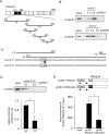
Figure 1. TRA2β4 interacts with nucleolin
(A) Schematic diagram of the human TRA2B gene. Exons are indicated by open boxes and Arabic numbers. Filled boxes denote the ultraconserved exon 2. Two major splice variants TRA2β1 and TRA2β4 are generated from the TRA2B gene and the use of each exon is shown. Black arrows show the specific primers used to detect each of the transcripts. PTCs; premature stop codons. (B) After biotinylated RNA pull-down assays using biotinylated exon 2 probes, the purified proteins were resolved by SDS-PAGE and visualized by silver staining. According to the analysis of the separated ~100 kDa protein by mass spectrometry, nucleolin (NP_005372) was one of RNA-binding proteins in the precipitated materials. (C) Cell lysates from HCT116 were subjected to a RIP assay using an anti-nucleolin antibody. Immunoprecipitated RNAs were quantified by qPCR. Data are shown as enrichment relative to GAPDH. Values are means ± s.d. (n = 6). *Significantly different by unpaired Student's t-test (p < 0.05).
Figure 2
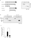
Figure 2. Identification of nucleolin-binding sites in TRA2β4
(A) Schema of the fragments of TRA2β4 that were used for in vitro binding assays. (B) A biotinylated RNA pull-down assay was carried out using lysates prepared from HCT116 cells and the biotinylated RNA fragments. (C) The RNA sequence in exon 2 F3 to F5. The introduction of two-point mutations (mt; 485-GGGG-488 to 485-GGAA-488) in TRA2β4 exon 2. (D) A biotinylated RNA pull-down assay was carried out using biotinylated RNA fragments with (mt) or without (wt) mutations. (E) The constructs coding FLAG-tagged full length TRA2β4 (pCMV- TRA2β4) or the mutated TRA2β4 (pCMV- TRA2β4 mt) were generated. These plasmids were transfected into HCT116 cells and then RIP assays were carried out with an anti-nucleolin antibody. The exogenous TRA2β4 in IP materials was detected using the primers shown as arrows. Each value represents the mean ± s.d. from 3 independent experiments. *Significantly different by unpaired Student's t-test (p < 0.05).
Figure 3
Figure 3. Characterization of TRA2β4 recognition domain in nucleolin
(A) The scheme of constructs encoding full-length nucleolin (NCL FL), lacking the GAR (NCL ΔGAR), lacking the C-terminus (NCL ΔC), lacking the N-terminal domain (NT) (NCL ΔN), and RNA-binding domains (RBDs) only (NCL RBD). Each plasmid was transfected into HCT116 cells for 48 hours, and then whole-cell lysates were subjected to Western blotting using an anti-FLAG antibody. (B) A biotinylated RNA pull-down assay was carried out using biotinylated RNA fragment F2 and lysates prepared from HCT116 cells that were transfected with each plasmid. The bound nucleolin was detected by Western blotting using anti-FLAG antibody. (C) The plasmids bearing full-length or GAR-deficient nucleolin were transfected into HCT116 cells and then a RIP assay was carried out with an anti-FLAG antibody. TRA2β4 in IP materials was detected by qPCR. Each value represents the mean ± s.d. from 3 independent experiments. *Significantly different by unpaired Student's t-test (p < 0.05).
Figure 4
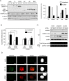
Figure 4. Effect of nucleolin on TRA2β4 stability
(A) HCT116 cells were treated with siRNAs for 48 h as follows: 10 nM control, NCL#1 (targeting the coding region of nucleolin), or NCL #2 (targeting 3′-UTR). Nucleolin levels were analyzed by Western blotting and qPCR. (B) The levels of TRA2β4 in control or nucleolin-silenced cells were measured by qPCR. (C) HCT116 cells were transfected with control, NCL #1, or NCL #2 siRNAs for 48 h and then incubated in the presence of 2 μg/mL actinomycin D for the indicated times. TRA2β1 and TRA2β4 levels were measured by qPCR and plotted on a logarithmic scale to calculate the time required for each RNA to reach one-half of its initial abundance (50%, dashed line). (D) After downregulation of endogenous nucleolin using an siRNA targeting its 3′-UTR (NCL#2), nucleolin expression was rescued by transfection with plasmids encoding a FLAG-tagged coding region of nucleolin with or without GAR. (E) The levels of TRA2β4 in nucleolin-rescued cells were measured by qPCR. (F) TRA2β4 levels in nucleolin-rescued cells after incubation with 2 μg/mL actinomycin D for the indicated times were measured by qPCR. The results were plotted on a logarithmic scale to calculate the time required for each RNA to reach one-half of its initial abundance (50%, dashed line).
Figure 5
Figure 5. Nucleolin is essential for nuclear localization of TRA2β4
(A) After transfection with the truncated nucleolin, the subcellular localization was measured by Western blotting. FLAG signals were quantified by densitometry. (B) After NMD was inhibited with cycloheximide treatment (100 μg/mL), the amounts of TRA2β4 and TRA2β1 mRNAs in the cytoplasmic or nuclear fraction were analyzed by qPCR. (C) Purities of the extracted nuclear and cytoplasmic fractions were confirmed by Western blotting using antibodies for cytosolic (α-tubulin) or nuclear proteins (hnRNP C1/C2) and by RT-PCR using primers targeting GAPDH pre-mRNA as a nuclear marker. (D) After a 24-h transfection of full-length or ΔGAR nucleolin, subcellular localization of TRA2β4 (green) and nucleolin (red) was examined by RNA-fluorescence in situ hybridization (RNA-FISH) using a probe that specifically hybridized to exon 2 (313-588 nt) and anti-FLAG antibody. Nucleoli were counterstained with TO-PRO-3. Scale bars, 5 μM.
Figure 6
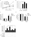
Figure 6. Association between nucleolin and TRA2β4 is essential for cell growth and susceptibility to an anticancer drug
(A) HCT116 cells (1 × 10⁴ cells) were seeded in 35-mm dishes and transfected with the indicated siRNA. Subsequently, growing cells were harvested and counted at the indicated times. Values are means ± s.d. from 4 independent experiments. *Significantly different by unpaired Student's t-test compared with control siRNA-treated cells (p < 0.05). (B) After transfection with control (ctrl) or NCL siRNA #2 for 12 h, the cells were treated with plasmids encoding a FLAG-tagged full-length or ΔGAR nucleolin. After a 48-h transfection, these cells were harvested and counted. Values are means ± s.d. from 4 independent experiments. *Significantly different by unpaired Student's t-test (p < 0.05). (C, D) HCT116 cells were treated with the indicated siRNAs and plasmids for 24 h, and then cells were exposed to 10 μM etoposide for 24 h. Subsequently, growing cells were harvested and counted or used for measurement of caspase 3/7 activities. Values are means ± s.d. from 4 independent experiments. *Significantly different by unpaired Student's t-test (p < 0.05). (E) After HCT116 cells were treated with the indicated siRNAs and plasmids for 24 h, the cells were exposed to 10 μM etoposide for 24 h. Subsequently, growing cells were harvested and caspase 3/7 activities were measured. Values are means ± s.d. from 3 independent experiments. *Significantly different by unpaired Student's t-test (p < 0.05).
Figure 7
Figure 7. Nucleolin regulates TRA2β4 expression in colon and breast cancer cells
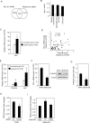
(A) Microarray analysis showed that NCL siRNA #1-treated cells differentially expressed 2,493 genes (≥ 1.5-fold), compared with control siRNA-treated cells. TRA2β4-silenced cells altered the expression of 3,044 genes (≥ 1.5-fold). A total of 630 genes were commonly changed in the same direction between TRA2β4- and nucleolin-silenced cells. (B) Commonly regulated 630 genes were subjected to Ingenuity Pathway Analysis (QIAGEN Bioinformatics) to identify biologically relevant functions. (C) Using colon adenocarcinoma TissueScan Tissue qPCR arrays (HCRT103, OriGene), NCL and ACTB mRNAs were measured by qPCR. ACTB mRNA was used as an endogenous quality control. *Significantly different by paired Student's t-test (p < 0.05). (D) The correlation between TRA2β4 and NCL expression in a colon cancer cDNA array was analyzed by determining the Pearson's correlation coefficient. (E) Expression levels of TRA2β4, NCL, and ACTB mRNAs were measured by qPCR using lung cancer cDNA libraries (CSRT101, OriGene). ACTB mRNA was used as an endogenous quality control. Values are means ± s.d. *Significantly different by unpaired Student's t-test (p < 0.05). (F) Both protein and mRNA levels of nucleolin in lung carcinoma (A549) and lung epithelial cells (BEAS-2B) were analyzed by qPCR or Western blotting. Values are means ± s.d. from 3 independent experiments. *Significantly different by unpaired Student's t-test (p < 0.05). (G) The levels of TRA2β4 expression were measured by qPCR in A549 and BEAS-2B cells. Values are means ± s.d. from 4 independent experiments. *Significantly different by unpaired Student's t-test (p < 0.05). (H) After A549 cells were treated with 10 nM of the indicated siRNA for 48 h, changes in the expression levels of TRA2β4 were measured by qPCR. GAPDH mRNA was used as an endogenous quality control. Values are means ± s.d. from 4 independent experiments. *Significantly different by unpaired Student's t-test compared with control (ctrl) siRNA-treated cells (p < 0.05). (I) After BEAS-2B cells were transfected with mock or plasmids encoding nucleolin with/without GAR for 48 h, changes in the expression level of TRA2β4 were measured by qPCR. GAPDH mRNA was used as an endogenous quality control. Values are means ± s.d. from 3 independent experiments. *Significantly different by unpaired Student's t-test compared with mock-transfected cells (p < 0.05).

Similar articles

Cited by

References

    1. Dauwalder B, Amaya-Manzanares F, Mattox W. A human homologue of the Drosophila sex determination factor transformer-2 has conserved splicing regulatory functions. Proc Natl Acad Sci U S A. 1996;93:9004–9. https://doi.org/10.1073/pnas.93.17.9004. - DOI - PMC - PubMed
    1. Tsuda K, Someya T, Kuwasako K, Takahashi M, He F, Unzai S, Inoue M, Harada T, Watanabe S, Terada T, Kobayashi N, Shirouzu M, Kigawa T, et al. Structural basis for the dual RNA-recognition modes of human Tra2-β RRM. Nucleic Acids Res. 2011;39:1538–53. https://doi.org/10.1093/nar/gkq854. - DOI - PMC - PubMed
    1. Watermann DO, Tang Y, Zur Hausen A, Jäger M, Stamm S, Stickeler E. Splicing factor Tra2-β1 is specifically induced in breast cancer and regulates alternative splicing of the CD44 gene. Cancer Res. 2006;66:4774–80. https://doi.org/10.1158/0008-5472.CAN-04-3294. - DOI - PubMed
    1. Gabriel B, Zur Hausen A, Bouda J, Boudova L, Koprivova M, Hirschfeld M, Jager M, Stickeler E. Significance of nuclear hTra2-beta1 expression in cervical cancer. Acta Obstet Gynecol Scand. 2009;88:216–21. https://doi.org/10.1080/00016340802503021. - DOI - PubMed
    1. Kuwano Y, Nishida K, Kajita K, Satake Y, Akaike Y, Fujita K, Kano S, Masuda K, Rokutan K. Transformer 2β and miR-204 regulate apoptosis through competitive binding to 3′ UTR of BCL2 mRNA. Cell Death Differ. 2015;22:815–25. https://doi.org/10.1038/cdd.2014.176. - DOI - PMC - PubMed

LinkOut - more resources