. 2018 Jun 21;3(12):e120121.
doi: 10.1172/jci.insight.120121.

γδ T cells: an immunotherapeutic approach for HIV cure strategies

Affiliations

γδ T cells: an immunotherapeutic approach for HIV cure strategies

Carolina Garrido et al. JCI Insight. .

Abstract

Current strategies aimed to cure HIV infection are based on combined efforts to reactivate the virus from latency and improve immune effector cell function to clear infected cells. These strategies are primarily focused on CD8+ T cells and approaches are challenging due to insufficient HIV antigen production from infected cells and poor HIV-specific CD8+ T cells. γδ T cells represent a unique subset of effector T cells that can traffic to tissues, and selectively target cancer or virally infected cells without requiring MHC presentation. We analyzed whether γδ T cells represent a complementary/alternative immunotherapeutic approach towards HIV cure strategies. γδ T cells from HIV-infected virologically suppressed donors were expanded with bisphosphonate pamidronate (PAM) and cells were used in autologous cellular systems ex vivo. These cells (a) are potent cytotoxic effectors able to efficiently inhibit HIV replication ex vivo, (b) degranulate in the presence of autologous infected CD4+ T cells, and (c) specifically clear latently infected cells after latency reversal with vorinostat. This is the first proof of concept to our knowledge showing that γδ T cells target and clear autologous HIV reservoirs upon latency reversal. Our results open potentially new insights into the immunotherapeutic use of γδ T cells for current interventions in HIV eradication strategies.

Keywords: AIDS/HIV; Immunotherapy.


Conflict of interest statement

Conflict of interest: DMM holds public stock in Gilead Sciences, outside of the content of this work.

Figures

Figure 1
Figure 1. Expansion of Vδ2 cells after 6 days of culture.
(A) Greater Vδ2 cell frequency in uninfected donors. PBMCs of uninfected (n = 10) and HIV-infected donors (n = 13) were stained for CD3 and Vδ2 and analyzed by flow cytometry. As expected, uninfected individuals showed a statistically higher percentage of Vδ2 cells compared with HIV-infected donors. Data represent mean ± SEM (Mann-Whitney U test, P < 0.001). (B) Representative histograms showing Vδ2 cell expansion. PBMCs from uninfected (left histogram) or ART-suppressed HIV-infected donors (right panel) were incubated for 6 days using HMBPP + IL-2 (blue), pamidronate (PAM) + IL-2 (orange), or IL-2 alone (green). (C) Vδ2 cells from HIV-infected individuals expand in response to PAM and IL-2. Vδ2 cell fold change relative to basal cell numbers is represented. HIV-infected donors’ response to HMBPP was lower, not statistically significant after FDR adjustment, compared with uninfected individuals (FDR P = 0.11). Response to PAM and IL-2 was similar between uninfected and HIV-infected donors (FDR P = 0.29). Response to HMBPP and PAM in uninfected donors was comparable (FDR P = 0.22), while response to HMBPP in HIV-infected donors was statistically lower (FDR P = 0.04). Uninfected donors (n = 9) are represented with gray circles and HIV-infected donors (n = 11) with pink squares. Uninfected and HIV-infected donors were compared using Mann-Whitney U test. HMBPP, PAM, and IL-2 conditions in uninfected donors and in HIV-infected donors were compared using Wilcoxon’s signed-rank test.
Figure 2
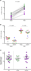
Figure 2. Expansion of Vδ2 cells from ART-suppressed HIV-infected donors in response to pamidronate (PAM).
(A) PAM exposure significantly increased Vδ2 cell frequency. Vδ2 cells from suppressed HIV+ donors (n = 21) significantly expanded in response to PAM. Patients treated in the acute infection (n = 9) are represented with green triangles and patients treated in chronic infection (n = 12) are represented with purple squares. (B) Decreased Vδ2 cell numbers in donors treated in the chronic phase of the infection. HIV-infected donors treated in the chronic phase of the infection (n = 12) showed significantly reduced number of Vδ2 cells (FDR P = 0.007) compared with those treated in the acute phase of the infection (n = 9). After PAM expansion, Vδ2 cells remained significantly lower in donors treated in chronic infection. Mean ± SEM. Mann-Whitney U test, FDR P = 0.02. (C) Comparable expansion capacity between patients who initiated ART in the acute or chronic phase of HIV infection. Fold change of Vδ2 cell expansion with PAM was similar in patients treated in the acute and chronic infection. Mean ± SEM. Mann-Whitney U test.
Figure 3
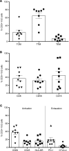
Figure 3. Phenotype of pamidronate-expanded Vδ2 cells in ART-suppressed HIV-infected donors.
Phenotype of Vδ2 cells was analyzed by flow cytometry in 8 HIV-infected individuals after expansion. Mean ± SEM is represented. (A) Memory populations defined as central memory (TCM: CD45CD27+CCR7+), transitional memory (TTM: CD45CD27+CCR7), or effector memory (TEM: CD45CD27CCR7). (B) Expression of cytotoxic markers CD8, CD56, and CD16. (C) Expression of activation markers CD69, CD25, and HLA-DR, and exhaustion markers PD-1 and CTLA-4.
Figure 4
Figure 4. Vδ2 T cells inhibit active HIV replication.
(A) Ex vivo–isolated Vδ2 T cells reduce HIV p24 production. HIV p24 production from autologous superinfected CD4+ T cells was significantly reduced in the presence of Vδ2 (n = 8 for the 1:1 effector/target ratio). Bars represent average viral production normalized to the condition where only superinfected CD4+ cells were cultured. Ratios expressed as effector/target cells. (B) Pamidronate-expanded (PAM-expanded) Vδ2 T cells retain their capacity to inhibit viral replication. After 14 days of exposure to PAM, γδ T cells were cocultured with autologous superinfected CD4+ cells. Vδ2 T cells (n = 10 for the 1:1 effector/target ratio) significantly reduced HIV p24 production. (C) Comparable inhibition capacity between basal and PAM-expanded V2 T cells. Data from basal HIV inhibition assays were compared to their respective inhibition capacity after PAM exposure. Inhibition capacity was similar in Vδ2 cells (n = 7 for the 1:1 effector/target ratio). Mean ± SEM is represented. Mann-Whitney U test. *P < 0.05, **P < 0.005, ***P < 0.0005.
Figure 5
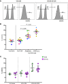
Figure 5. Vδ2 cells degranulate in the presence of autologous HIV-infected CD4+ T cells.
(A) Flow cytometry plots showing an example of CD107a detection in cocultures of expanded Vδ2 cells with autologous CD4+ cells (left) and with autologous JR-CSF–superinfected CD4+ cells (right). (B) Greater CD107a production in the presence of HIV-infected cells. CD107a production was statistically higher when Vδ2 cells were cocultured with HIV-superinfected CD4+ cells compared with cocultures of autologous isolated CD4+ cells (FDR P = 0.006). CD107a production was the highest when Vδ2 cells were cocultured with PHA-activated, HIV-superinfected CD4+ cells (FDR P = 0.02) but without statistical differences compared with cells infected using polybrene. Mean ± SEM is represented. P = 0.08, Wilcoxon’s matched-pairs signed-rank test. (C) Comparable degranulation capacity of Vδ2 cells between donors treated in acute and chronic HIV infection. CD107a production was not statistically different between acute and chronic patients. Both groups of patients showed statistically higher CD107a expression in cocultures of Vδ2 cells and superinfected CD4+ target cells than in cultures of Vδ2 cells cocultured with ex vivo–isolated CD4+ cells. Effector/target ratio (1:1). Mann-Whitney U test.
Figure 6
Figure 6. γδ T cells clear latently infected cells after latency reversal with vorinostat (VOR).
Isolated resting CD4+ (r-CD4+) cells from ART-suppressed HIV-infected donors were reactivated with 0.5 μM VOR. After washing, r-CD4+ cells were cultured alone or with γδ T cells, which were removed from the culture after 24 hours. The same number of replicate cultures of 1 × 10⁶ from each condition were then cultured in parallel for 19 days. VOR efficiently reactivated latent HIV in 6 (represented in the graph) of the 8 patients analyzed in the condition where r-CD4+ cells were cultured alone. Frequency of HIV recovery (number of positive wells for HIV p24 measured by ELISA) decreased significantly (P = 0.03, Wilcoxon’s signed-rank test) in cultures of r-CD4+ cells in the presence of γδ T cells, demonstrating that γδ T cells can recognize and clear latently HIV-infected cells upon latency reversal.

Similar articles

Cited by

References

    1. Archin NM, Sung JM, Garrido C, Soriano-Sarabia N, Margolis DM. Eradicating HIV-1 infection: seeking to clear a persistent pathogen. Nat Rev Microbiol. 2014;12(11):750–764. doi: 10.1038/nrmicro3352. - DOI - PMC - PubMed
    1. Chun TW, et al. Quantification of latent tissue reservoirs and total body viral load in HIV-1 infection. Nature. 1997;387(6629):183–188. doi: 10.1038/387183a0. - DOI - PubMed
    1. Wong JK, et al. Recovery of replication-competent HIV despite prolonged suppression of plasma viremia. Science. 1997;278(5341):1291–1295. doi: 10.1126/science.278.5341.1291. - DOI - PubMed
    1. Finzi D, et al. Latent infection of CD4+ T cells provides a mechanism for lifelong persistence of HIV-1, even in patients on effective combination therapy. Nat Med. 1999;5(5):512–517. doi: 10.1038/8394. - DOI - PubMed
    1. Siliciano JD, Siliciano RF. Recent developments in the effort to cure HIV infection: going beyond N = 1. J Clin Invest. 2016;126(2):409–414. doi: 10.1172/JCI86047. - DOI - PMC - PubMed

Publication types

MeSH terms

Substances