

Dengue fever with encephalitis: a rare phenomenon
- PMID: 29909394
- PMCID: PMC6011480
- DOI: 10.1136/bcr-2018-225463
Dengue fever with encephalitis: a rare phenomenon
Abstract
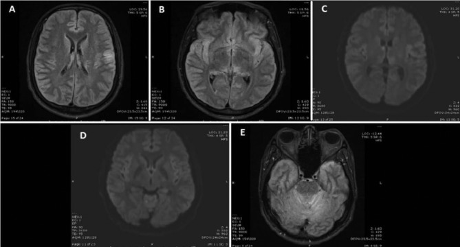
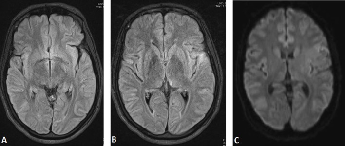
The clinical profile and presentation of patients with dengue fever may differ from asymptomatic infection to the dreadful complications like dengue shock syndrome. However, neurological complications are very rare. Dengue encephalitis occurs by a direct involvement of central nervous system by the dengue virus which is an extremely rare complication. A 33-year-old man presented with fever, vomiting and severe headache. He had one episode of generalised tonic-clonic seizure followed by an altered sensorium on the day of admission to the hospital. The diagnosis of dengue fever was confirmed by dengue serology (IgM) and (NS1) antigen assay. MRI brain was suggestive of encephalitis. Thus, the patient was treated symptomatically and discharged in stable condition with minimal neurological deficit.
Keywords: general practice / family medicine; infection (neurology); meningitis; tropical mkedicine (infectious disease).
© BMJ Publishing Group Ltd (unless otherwise stated in the text of the article) 2018. All rights reserved. No commercial use is permitted unless otherwise expressly granted.
Conflict of interest statement
Competing interests: None declared.
Figures
Similar articles
-
A rare case of dengue encephalitis.BMJ Case Rep. 2013 Feb 13;2013:bcr2012008229. doi: 10.1136/bcr-2012-008229. BMJ Case Rep. 2013. PMID: 23413293 Free PMC article.
-
Fatal dengue encephalitis.Southeast Asian J Trop Med Public Health. 2005 Jan;36(1):200-2. Southeast Asian J Trop Med Public Health. 2005. PMID: 15906668
-
Dengue fever presenting with acute cerebellitis: a case report.BMC Res Notes. 2014 Mar 5;7:125. doi: 10.1186/1756-0500-7-125. BMC Res Notes. 2014. PMID: 24598036 Free PMC article.
-
Cerebral vasculitis and lateral rectus palsy - two rare central nervous system complications of dengue fever: two case reports and review of the literature.J Med Case Rep. 2018 Apr 19;12(1):100. doi: 10.1186/s13256-018-1627-x. J Med Case Rep. 2018. PMID: 29669602 Free PMC article. Review.
-
Neurological complications of dengue virus infection.Lancet Neurol. 2013 Sep;12(9):906-919. doi: 10.1016/S1474-4422(13)70150-9. Lancet Neurol. 2013. PMID: 23948177 Review.
Cited by
-
Diagnostic challenges in a patient with dengue shock syndrome presenting with acute meningoencephalitis.IDCases. 2024 Apr 15;36:e01964. doi: 10.1016/j.idcr.2024.e01964. eCollection 2024. IDCases. 2024. PMID: 38646600 Free PMC article.
-
Dengue meningoencephalitis in a child presenting with focal seizures.Int J Pediatr Adolesc Med. 2020 Sep;7(3):153-154. doi: 10.1016/j.ijpam.2020.01.007. Epub 2020 Jan 31. Int J Pediatr Adolesc Med. 2020. PMID: 33094145 Free PMC article.
-
Acute appendicitis during the recovery phase of dengue hemorrhagic fever: two case reports.J Med Case Rep. 2022 Jun 5;16(1):219. doi: 10.1186/s13256-022-03443-2. J Med Case Rep. 2022. PMID: 35659758 Free PMC article.
References
-
- Centers for Disease Control and Prevention. Dengue: Epidemiology. 2014. https://www.cdc.gov/dengue/epidemiology/index.html (accessed 30th Mar 2018).
-
- Koley TK, Jain S, Sharma H, et al. . Dengue encephalitis. J Assoc Physicians India 2003;51:422–3. - PubMed
-
- Kamble R, Peruvamba JN, Kovoor J, et al. . Bilateral thalamic involvement in dengue infection. Neurol India 2007;55:418–9. - PubMed
Publication types
MeSH terms
Substances
LinkOut - more resources
Full Text Sources
Other Literature Sources
Medical