Review
. 2018 Mar;97(S 01):S3-S47.
doi: 10.1055/s-0043-121594. Epub 2018 Mar 22.

Immunotherapy - The New Era of Oncology

[Article in English, German]
Affiliations
Review

Immunotherapy - The New Era of Oncology

[Article in English, German]
Benjamin Kansy et al. Laryngorhinootologie. 2018 Mar.

Abstract

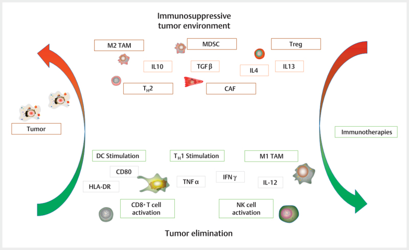
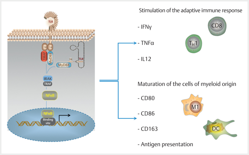
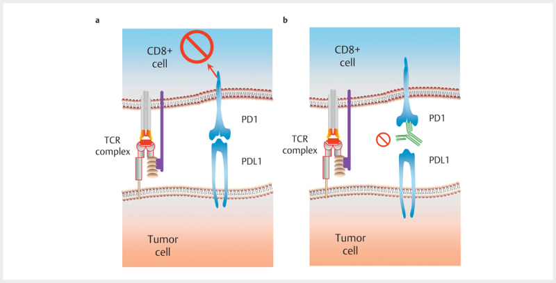
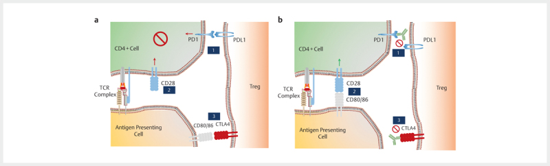
In the field of immunotherapy, essential progress was achieved over the past years partially demonstrating long lasting therapeutic responses in different tumor entities. A better understanding of the interactions between the tumor and the immune system as well as the integration of immunotherapeutic approaches into clinical routine were the foundations for this development. The different approaches intervene on multiple levels of the immune response and directly or indirectly mount the patient's own immune defense against tumor cells. Immunotherapeutic approaches are represented by cytokine therapies, vaccinations, the use of oncolytic viruses and monoclonal antibody therapies as well as adoptive cell transfer strategies.

In den letzten Jahren wurden bedeutende Fortschritte auf dem Gebiet der Immuntherapie erreicht mit teils langanhaltendem Therapieansprechen bei unterschiedlichsten Tumorentitäten. Die Basis hierfür bildet ein verbessertes Verständnis der Interaktion zwischen Tumor und Immunsystem und der damit verbundenen Integration immuntherapeutischer Ansätze in die klinische Routine. Die hierbei eingesetzten immuntherapeutischen Strategien greifen auf unterschiedlichen Ebenen der Immunantwort ein, fördern direkt oder indirekt die Zerstörung der Tumorzellen durch die körpereigene Abwehr und reichen von Zytokintherapien über Vakzinierungen und den Einsatz onkolytischer Viren bis hin zu monoklonalen Antikörpertherapien und dem adoptiven Zelltransfer.


Conflict of interest statement

Die Autoren geben an, dass kein Interessenkonflikt besteht.

Figures

Fig. 1
Fig. 1
MHC molecules. MHC molecules are expressed on nucleated, endogenous cells (MHC I) and antigen-presenting cells (MHC II). MHC I molecules consist of an α and a β subunit. The α subunit contains three domains, α1 and α2 are responsible for antigen presentation and α3 secures anchoring in the cell membrane. β2 microglobulin is the fourth soluble domain of MHC I molecules. The MHC II molecules consist of 2 subunits both anchored in the cell membrane. One subunit (α or β) consists of 2 domains each, α1 and α2 or β1 and β2, respectively; α1 and β1 domains are responsible for antigen presentation. (MHC: main histocompatibility complex)
Fig. 2
Fig. 2
Antigen presentation and antigen identification. CD8+ T cells identify antigens that are presented on MHC I molecules. CD4+ T cells recognize antigens of antigen presenting cells on MHC II molecules. MHC I molecules are charged with peptide fragments from the cellular cytosol that permanently develop from degradation products of proteasomes within the cell. This mechanism controls if the cell produces endogenous or exogenous proteins. MHC II molecules are exclusively expressed on antigen presenting cells and present foreign, extracellular peptide fragments that entered into the cell via phagocytosis or endocytosis in order to activate receptors of antigen-specific cells. In addition to activation via the TCR/MHC complex (signal 1) the T cells require a co-stimulating signal (CD80, signal 2) to be activated. (MHC: main histocompatibility complex; TCR: T cell receptor).
Fig. 3
Fig. 3
Immunoediting. a Elimination phase. In the elimination phase, the adaptive and innate immune system identify and destroy tumor cells. b Equilibrium phase. In the equilibrium phase, the immune system maintains the control over tumor cells, complete elimination does no longer take place. c Escape phase. In the escape phase, the immune system loses control over the tumor cells leading to tumor progress. The transition from the elimination phase via the equilibrium phase to the escape phase is called immunoediting. (DC: dendritic cells; T: T cells; G: granulocytes; M: macrophages).
Fig. 4
Fig. 4
The influence of the cytokine environment on the differentiation of naïve CD4+ T cells. CD4+ T cells are influenced by different cytokines for differentiation into T H 1 and T H 2 as well as T H 17 and Treg cells. Especially TGFβ induces the development of Treg. Treg itself influence the tumor environment by means of TGFβ and IL10. Additionally, the development of T cells is differentiated into T H 1, T H 2, and T H 17 cells. IL12 stimulates a differentiation into T H 1 cells that are associated with an anticancer response in the tumor environment. IL4 is responsible for induction of T H 2 cells that play a pro-tumor role in the tumor environment by means of IL4, IL10, and IL13 secretion. The role of T H 17 cells in the tumor environment is still controversially discussed, they are stimulated by IL6 and IL23 and TGFβ. (green: mainly anticancer effect; red: mainly pro-tumor effect; IL: interleukin; TGF: transforming growth factor; IFN: interferon; TNF: tumor necrosis factor; T-bet: T-box transcription factor; GATA3: GATA transcription factor; FoxP3: F box protein 3 transcription factor).
Fig. 5
Fig. 5
Myeloid suppressor cells and antigen presenting cells in the tumor environment. MDSC are stimulated by pro-inflammatory signals and suppress the immune response in the tumor environment. They induce the development of Treg and T H 2 cells and promote the differentiation to M2 phenotypes. Mature antigen presenting cells can stimulate the anticancer immune response by supporting CD8+ T cells, CD4+ T H 1 cells and NK cells by means of antigen presenting and co-stimulation. (green: mainly anticancer effect; red: mainly pro-tumor effect; IL: interleukin; TGF: transforming growth factor; IFN: interferon; TNF: tumor necrosis factor; M1: M1 phenotype; NK: natural killer cells; TLR: toll-like receptor stimulation; MMP: matrix metalloproteinase; VEGF: vascular endothelial growth factor; NO: nitric oxide).
Fig. 6
Fig. 6
Monocytes in the tumor environment. In the tumor environment, monocytes differentiate into M1 and M2 macrophages depending on the predominant influences. M1 macrophages stimulate the T H 1 response via IL12 and are associated with an anticancer effect. M2 macrophages stimulate a T H 2 response and have a pro-tumor effect by developing IL10, VEGF, and arginase in the tumor environment. (green: mainly anticancer effect; red: mainly pro-tumor effect; IL: interleukin; IFN: interferon; TNF: tumor necrosis factor, M1: M1 phenotype; CXCL: chemokine; VEGF: vascular endothelial growth factor; EGF: epidermal growth factor).
Fig. 7
Fig. 7
Overview about pro-tumor and anticancer influences in the tumor environment. Based on the tumor, a pro-tumor environment develops stimulating cell types that themselves promote tumor development. The different approaches of immunotherapy support the development of an anticancer environment with the intention of eliminating the tumor. (green: mainly anticancer effect; red: mainly pro-tumor effect; IL: interleukin; IFN: interferon; TNF: tumor necrosis factor; M1: M1 phenotype; M2: M2 phenotype; MDSC: myeloid suppressor cell; Treg: regulatory T cell; NK: natural killer cell; DC: dendritic cell; CD: cluster of differentiation; HLA-DR: human leukocyte antigen DR, CAF: cancer-associated fibroblast; TGF: transforming growth factor).
Fig. 8
Fig. 8
Toll-like receptors. Toll-like receptors may be bound on the cell surface as well as with the cells. Their activation leads to a signal cascade that mainly initiates a production of IFNγ, TNFα, and IL12 via MyD88 and NFKB. Additionally, cell differentiation is stimulated. (NFKB: transcription factor; MyD88: myeloid differentiation protein 88; TIR: toll/IL-1 R homology domain, TIRAP: adaptor molecule; IRAK: interleukin-1 receptor-associated kinase; TRAF: TNF receptor-associated factor; IFN: interferon; TNF: tumor necrosis factor).
Fig. 9
Fig. 9
Scheme of the monoclonal antibody. Monoclonal antibodies are classified according to their composition and the human percentage.
Fig. 10
Fig. 10
Effect of the checkpoint molecules and the checkpoint blockade. a The CD8+ T cell is inhibited via the binding of the PD1 receptor by PDL1 expressed by the tumor cells. The receptor binding leads to T cell anergy by inhibiting the proliferation and the T cell function. b The inhibiting effect is eliminated by the checkpoint blockade. (TCR: T cell receptor complex; PD1: programmed death receptor 1; PDL1: PD1 ligand).
Fig. 11
Fig. 11
Effect of checkpoint molecules and checkpoint blockade. a The CD4+ T cells is inhibited on several levels. Binding of the PD1 receptor by PDL1 leads to T cell anergy (1). The missing co-stimulatory signal by the antigen presenting cell also causes inhibition (2). CTLA4 expressed by the Treg competitively binds the co-stimulatory ligands of the antigen presenting cell (3) and thus impedes the co-stimulatory signal. b The inhibiting effect of the checkpoint blockade is eliminated in (1). The co-stimulatory ligand (CD80/86) is again available for the co-stimulatory signal (2) because the competitive binding partner CTLA4 is also blocked by an antibody (3). (TCR: T cell receptor complex; PD1: programmed death receptor 1; PDL1: PD1 ligand; CTLA4: cytotoxic T lymphocyte-associated protein; CD28: costimulatory receptor; CD80/86: co-stimulatory ligand).
Fig. 12
Fig. 12
Adoptive cell transfer. (1) Lymphocytes are taken from the patient either from the peripheral blood or the tumor itself. (2) Then the lymphocytes are isolated and modified if necessary. (3) Selection of the lymphocytes with the highest specificity is performed. (4) The selected lymphocytes are expanded and then re-infused (5).
Abb. 1
Abb. 1
MHC-Moleküle. MHC-Moleküle sind auf kernhaltigen, körpereigenen Zellen (MHC I) und Antigen-präsentierenden Zellen (MHC II) exprimiert. MHC I-Moleküle bestehen aus einer α- und einer β-Untereinheit. Die α-Untereinheit enthält drei Domänen, von denen die Domänen α 1 und α 2 die Antigenpräsentation übernehmen, und die α 3 Domäne die Verankerung in der Zellmembran sichert. Das β 2 -Mikroglobulin stellt die vierte lösliche Domäne der MHC I-Moleküle dar. MHC II-Moleküle bestehen aus 2 Untereinheiten, die beide in der Zellmembran verankert sind. Eine Untereinheit (α- oder β) besteht aus je 2 Domänen, α 1 und α 2 bzw. β 1 und β 2 , wobei die α 1 und die β 1 Domäne die Aufgabe der Antigenpräsentation übernehmen. MHC: Haupthistokompatibilitätskomplex.
Abb. 2
Abb. 2
Antigenpräsentation und Antigen-Erkennung. CD8+T-Zellen erkennen Antigene, die auf MHC I-Molekülen präsentiert werden. CD4+T-Zellen erkennen Antigene von Antigenpräsentierenden Zellen auf MHC II-Molekülen. MHC I-Moleküle werden mit Peptidfragmenten aus dem Zytosol der Zelle beladen, die permanent aus Abbauprodukten der Proteasomen im Zellinneren entstehen. Dies dient zur Kontrolle, ob die Zelle körpereigene oder körperfremde Proteine produziert. MHC-II Moleküle werden ausschließlich auf Antigenpräsentierenden Zellen exprimiert und präsentieren fremde, extrazelluläre Peptidfragmente, die über Phagozytose oder Endozytose in die Zelle aufgenommen wurden, um Rezeptoren Antigen-spezifischer Zellen zu aktivieren. Zusätzlich zur Aktivierung über den TZR/MHC-Komplex (Signal 1) benötigen die T-Zellen ein ko-stimulatorisches Signal (CD80, Signal 2) um aktiviert zu werden. MHC: Haupthistokompatibilitätskomplex; TZR: T-Zell-Rezeptor.
Abb. 3
Abb. 3
Immunoediting. a Eliminierungsphase: In der Eliminierungsphase erkennen und zerstören das adaptive und das native Immunsystem Tumorzellen. b Gleichgewichtsphase: In der Gleichgewichtsphase hält das Immunsystem die Tumorzellen unter Kontrolle, eine vollständige Elimination findet nicht mehr statt. c Entziehungsphase: In der Entziehungsphase verliert das Immunsystem die Kontrolle über die Tumorzellen mit darauffolgendem Tumorprogress. Der Übergang von Eliminierungsphase über die Gleichgewichtsphase zur Entziehungsphase wird als Immunoediting bezeichnet. DC: Dendritische Zelle; T: T-Zellen; G: Granulozyten; M: Makrophagen.
Abb. 4
Abb. 4
Der Einfluss des Zytokinmilieus auf die Differenzierung naiver CD4+ T-Zellen. CD4+ T-Zellen werden durch unterschiedliche Zytokine zur Differenzierung in T H 1 und T H 2 sowie T H 17 und Treg Zellen beeinflusst. Dabei induziert insbesondere TGFβ die Entwicklung von Treg. Treg selbst beeinflussen das Tumormilieu mittels TGFβ und IL10. Zusätzlich unterscheidet man bei der Entwicklung von T-Zellen die Differenzierung in T H 1, T H 2 und T H 17 Zellen. IL12 stimuliert eine Differenzierung in T H 1-Zellen, die mit einer antitumoralen Antwort im Tumormilieu assoziiert sind. IL4 ist für die Induktion von T H 2-Zellen wichtig, die eine protumorale Rolle im Tumormilieu mittels IL4, IL10 und IL13 Sekretion übernehmen. Die Rolle der T H 17-Zellen im Tumormilieu ist noch umstritten, sie werden durch IL6 und IL23 und TGFβ stimuliert. Grün: vorwiegend antitumorale Wirkung; Rot: vorwiegend protumorale Wirkung; IL: Interleukin; TGF: Transformierender Wachstumsfaktor; IFN: Interferon; TNF: Tumornekrosefaktor; T-bet: T-box Transkriptionsfaktor; GATA3: GATA Transkriptionsfaktor; FoxP3: F box protein 3-Transkriptionsfaktor.
Abb. 5
Abb. 5
Myeloide Suppressorzellen und Antigenpräsentierende Zellen im Tumormilieu. MDSC werden durch pro-inflammatorische Signale stimuliert und supprimieren die Immunantwort im Tumormilieu. Sie induzieren die Bildung von Treg und T H 2-Zellen und fördern die Differenzierung zu M2-Phänotypen. Reife Antigenpräsentierende Zellen können die antitumorale Immunantwort stimulieren, indem sie durch Antigenpräsentierende und Ko-Stimulation CD8+ T-Zellen, CD4+ T H 1-Zellen und NK-Zellen unterstützen. Grün: vorwiegend antitumorale Wirkung; Rot: vorwiegend protumorale Wirkung; IL: Interleukin; TGF: Transformierender Wachstumsfaktor; IFN: Interferon; TNF: Tumornekrosefaktor; M1: M1 Phänotyp; NK: Natürliche Killerzelle; TLR: Toll-like-Rezeptor-Stimulation; MMP: Matrixmetalloprotease; VEGF: vaskulärer endothelialer Wachstumsfaktor; NO: Stickoxid.
Abb. 6
Abb. 6
Monozyten im Tumormilieu. Monozyten differenzieren im Tumormilieu in M1- und M2-Makrophagen in Abhängigkeit der vorherrschenden Einflüsse. M1-Makrophagen stimulieren über IL12 die T H 1-Antwort und sind mit einer antitumoralen Wirkung assoziiert. M2-Makrophagen stimulieren eine T H 2-Antwort und wirken durch die Bildung von IL10, VEGF und Arginase protumoral im Tumormilieu. Grün: vorwiegend antitumorale Wirkung; Rot: vorwiegend protumorale Wirkung; IL: Interleukin; IFN: Interferon; TNF: Tumornekrosefaktor; M1: M1 Phänotyp; CXCL: Chemokin; VEGF: vaskulärer endothelialer Wachstumsfaktor; EGF: epidermaler Wachstumsfaktor.
Abb. 7
Abb. 7
Übersicht über pro- und antitumorale Einflüsse im Tumormilieu. Durch den Einfluss des Tumors wird ein protumorales Tumormilieu geschaffen und damit werden Zelltypen stimuliert, die ihrerseits die Tumorentwicklung begünstigen. Die verschiedenen Ansätze der Immuntherapien unterstützen die Bildung eines antitumoralen Milieus mit dem Ziel der Tumorelimination. Grün: vorwiegend antitumorale Wirkung; Rot: vorwiegend protumorale Wirkung; IL: Interleukin; IFN: Interferon; TNF: Tumornekrosefaktor; M1: M1-Phänotyp; M2: M2-Phänotyp; MDSC: Myeloide Suppressorzelle; Treg: regulatorische T-Zelle; NK: Natürliche Killerzelle; DC: Dendritische Zelle; CD: Cluster of Differentiation; HLA-DR: Humane Leukozytenantigen-DR; CAF: Krebs-assoziierter Fibroblast; TGF: Transformierender Wachstumsfaktor.
Abb. 12
Abb. 12
Adoptiver Zelltransfer. (1) Lymphozyten werden dem Patienten entnommen, entweder aus dem peripheren Blut, oder aus dem Tumor selbst. (2) Anschließend werden die Lymphozyten isoliert und gegebenenfalls modifiziert. (3) Dann erfolgt die Selektion der Lymphozyten mit der höchsten Spezifität. (4) Die selektierten Lymphozyten werden vermehrt und anschließend reinfundiert (5).
Abb. 8
Abb. 8
Toll-like-Rezeptoren. Toll-like-Rezeptoren können sowohl auf der Zelloberfläche als auch intrazellulär gebunden werden. Ihre Aktivierung hat eine Signalkaskade zur Folge, die hauptsächlich über MyD88 und NF K B eine Produktion von IFNγ, TNFα und IL12 initiiert. Zusätzlich wird eine Zelldifferenzierung stimuliert. NF K B: Transkriptionsfaktor; MyD88: Myeloides Differenzierungsprotein 88; TIR: Toll/IL-1 R Homologie Domäne; TIRAP: Adaptermolekül; IRAK: Interleukin-1 Rezeptor assoziierte Kinase; TRAF: TNF Rezeptor-assoziierter Faktor; IFN: Interferon; TNF: Tumornekrosefaktor.
Abb. 9
Abb. 9
Aufbau der monoklonalen Antikörper. Monoklonale Antikörper werden abhängig von ihrer Zusammensetzung und dem daraus resultierenden humanen Anteil unterschieden.
Abb. 10
Abb. 10
Wirkung der Checkpoint-Moleküle und der Checkpoint-Blockade. a Die CD8+ T-Zelle wird über die Bindung des PD1-Rezeptors durch das von den Tumorzellen exprimierte PDL1 inhibiert. Die Bindung des Rezeptors führt zur T-Zell-Anergie durch Hemmung der Proliferation und der T-Zell Funktion. b Durch die Checkpoint-Blockade wird die inhibierende Wirkung aufgehoben. TZR: T-Zell-Rezeptor-Komplex; PD1: Programmed Death-Rezeptor 1; PDL1: PD1 Ligand.
Abb. 11
Abb. 11
Wirkung der Checkpoint-Moleküle und der Checkpoint-Blockade. a Die CD4+ T-Zelle wird auf mehreren Ebenen inhibiert: Die Bindung des PD1 Rezeptors durch PDL1 führt zur T-Zell-Anergie (1). Das fehlende ko-stimulatorische Signal durch die Antigen-präsentierende Zelle bewirkt ebenso eine Inhibition (2). Das von der Treg exprimierte CTLA4 bindet den ko-stimulatorischen Liganden der Antigenpräsentierenden Zelle kompetitiv (3) und verhindert so das ko-stimulatorische Signal. b Durch die Checkpoint-Blockade wird die inhibierende Wirkung bei (1) aufgehoben. Ebenso steht der ko-stimulatorische Ligand (CD80/86) wieder für das ko-stimulatorische Signal zur Verfügung (2), da der kompetitive Bindungspartner CTLA4 ebenso durch einen Antikörper blockiert ist (3). TZR: T-Zell-Rezeptor-Komplex; PD1: Programmed Death-Rezeptor 1; PDL1: PD1 Ligand; CTLA4: Zytotoxische T-Lymphozyten-assoziiertes Protein; CD28: Ko-stimulatorischer Rezeptor; CD80/86 Ko-stimulatorischer Ligand.

Similar articles

References

    1. Coley W B., II Contribution to the Knowledge of Sarcoma. Annals of surgery. 1891;14:199–220. - PMC - PubMed
    1. Hodi F S, O'Day S J, McDermott D F et al.Improved survival with ipilimumab in patients with metastatic melanoma. N Engl J Med. 2010;363:711–723. - PMC - PubMed
    1. Topalian S L, Hodi F S, Brahmer J R et al.Safety, activity, and immune correlates of anti-PD-1 antibody in cancer. N Engl J Med. 2012;366:2443–2454. - PMC - PubMed
    1. Couzin-Frankel J. Breakthrough of the year 2013. Cancer immunotherapy. Science (New York, NY) 2013;342:1432–1433. - PubMed
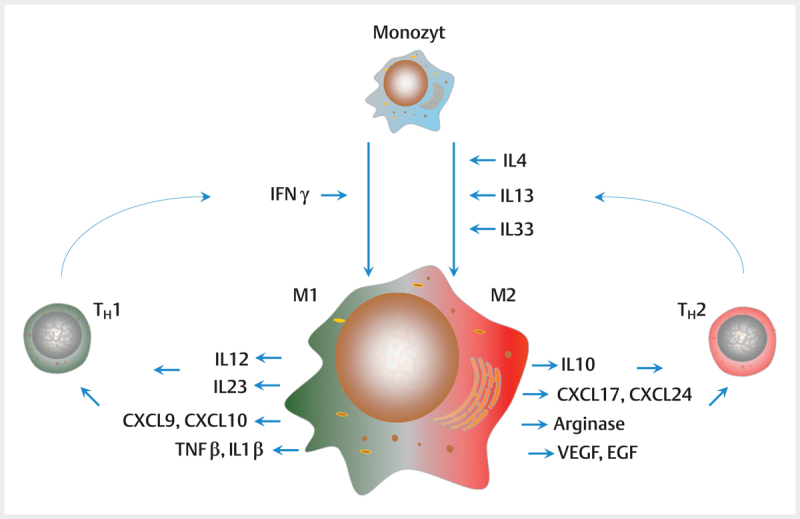
    1. Mantovani A, Sozzani S, Locati M et al.Macrophage polarization: tumor-associated macrophages as a paradigm for polarized M2 mononuclear phagocytes. Trends in immunology. 2002;23:549–555. - PubMed