. 2018 May 29:9:1091.
doi: 10.3389/fmicb.2018.01091. eCollection 2018.

Autophagy Benefits the Replication of Egg Drop Syndrome Virus in Duck Embryo Fibroblasts

Affiliations

Autophagy Benefits the Replication of Egg Drop Syndrome Virus in Duck Embryo Fibroblasts

Xueping Wang et al. Front Microbiol. .

Abstract

Egg drop syndrome virus (EDSV) is an economically important pathogen with a broad host range, and it causes disease that leads to markedly decreased egg production. Although EDSV is known to induce apoptosis in duck embryo fibroblasts (DEFs), the interaction between EDSV and its host needs to be further researched. Here, we provide the first evidence that EDSV infection triggers autophagy in DEFs through increases in autophagosome-like double-membrane vesicles, the conversion of LC3-I to LC3-II, and LC3 colocalization with viral hexon proteins. Conversely, P62/SQSTM1 degradation, LC3-II turnover, and colocalization of LAMP and LC3 confirmed that EDSV infection triggers complete autophagy. Furthermore, we demonstrated that inhibition of autophagy by chloroquine (CQ) and 3-methyladenine (3MA) or RNA interference targeting ATG-7 decreased the yield of EDSV progeny. In contrast, induction of autophagy by rapamycin increased the EDSV progeny yield. In addition, we preliminarily demonstrated that the class I phosphoinositide 3-kinase (PI3K)/Akt/mTOR pathway contributes to autophagic induction following EDSV infection. Altogether, these finding lead us to conclude that EDSV infection induces autophagy, which benefits its own replication in host cells. These findings provide novel insights into EDSV-host interactions.

Keywords: DEF cells; autophagic flux; autophagy; egg drop syndrome disease virus; virus replication.


Figures
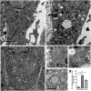
FIGURE 1
FIGURE 1
Egg drop syndrome virus (EDSV) infection triggers autophagosome-like vesicles formation in duck embryo fibroblasts (DEFs). (a) TEM observation. DEFs were mock-treated for 36 hpi, (b) infected with EDSV at an MOI of 1 for 36 hpi or (c) treated with rapamycin for 36 hpi and then processed for observation. Scale bar: 2 μm. (d,e) High magnification images of the typical autophagosome-like vesicles in EDSV-infected and (f) rapamycin-treated cells are shows. Scale bar: 0.5 μm. (g) The number of autophagosome-like vesicles per cell profile in mock- and EDSV-infected DEFs and rapamycin-treated DEFs were quantified. The results are shown as the mean ± SEM (n = 3). P < 0.05; ∗∗P < 0.01; ∗∗∗P < 0.001.
FIGURE 2
FIGURE 2
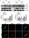
Modification of LC3 proteins during EDSV infection. (A) The significant conversion of LC3-l to LC3-ll was detected in mock-or EDSV-infected DEFs using an anti-LC3B antibody, and the expression of hexon from EDSV was also detected using anti-hexon antibodies. (B) The optical densities of each protein band were measured by densitometric scanning, and the optical density ratio of LC3-ll/LC3-l was calculated. The results are shown as the mean ± SEM (n = 3). P < 0.05; ∗∗P < 0.01; ∗∗∗P < 0.001. (C) Confocal immunofluorescence microscopy images of mock-infected control and EDSV-infected DEFs. Mock- and EDSV-infected cells were fixed at 36 hpi and stained with antibodies against LC3 and EDSV hexon and then merged. Scale bar: 10 μm. Cell nuclei were counterstained with Hoechst. Fluorescence images were examined under a confocal laser scanning microscope. Scale bar = 10 μm.
FIGURE 3
FIGURE 3
Egg drop syndrome virus infection enhances p62 degradation in DEFs. (A) A representative image of a western blot analysis of relative levels of p62 in mock- or EDSV-infected DEFs at indicated time points. (B) The optical densities of each protein band were measured by densitometric scanning, and the optical density ratio of p62/β-actin was calculated. The results are shown as the mean ± SEM (n = 3). P < 0.05; ∗∗P < 0.01; ∗∗∗P < 0.001; #Means no significant difference, P > 0.05.
FIGURE 4
FIGURE 4
Egg drop syndrome virus infection enhances autophagic flux. (A) Western blot analyses of the effect of CQ treatment on the expression of both LC3-ll and p62 proteins in DEFs. Cell lysates were subjected to western blot analyses using antibodies against LC3, p62, and β-actin. Representative profiles are shown, and similar results were obtained in three independent experiments. (B,C) Analysis of changes in the relative amounts of both LC3-ll and p62 in rapamycin- pretreated and EDSV-infected DEFs in the absence and presence of CQ. (D) Representative images of colocalization of LC3 with the endosomal/lysosomal marker LAMP1 in mock- and EDSV-infected DEFs. Cell nuclei were counterstained with Hoechst. Fluorescence images were examined under a confocal laser scanning microscope. The results are shown as the mean ± SEM (n = 3). P < 0.05; ∗∗P < 0.01; ∗∗∗P < 0.001. Scale bar = 10 μm.
FIGURE 5
FIGURE 5
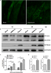
Western blot analysis of the ability of UV-inactivated EDSV to induce the conversion of LC3-l to LC3-ll in DEFs. (A) Cells infected with replication -competent EDSV or with UV-inactivated EDSV (UV-EDSV) at an MOI of 1. (B) Densitometric analysis of relative levels of LC3-II and LC3-I in EDSV- or UV-EDSV -infected DEFs. The results are shown as the mean ± SEM (n = 3). P < 0.05; ∗∗P < 0.01; ∗∗∗P < 0.001. (C) Confocal microscopy analysis of the ability of UV-EDSV to induce autophagy in DEFs. Scale bar = 15 μm.
FIGURE 6
FIGURE 6
Autophagy induction enhanced the replication of EDSV. (A) A representative image from confocal immunofluorescence microscopy analysis of LC3 expression in EDSV-infected DEFs or DEFs treated with 100 nM rapamycin prior to EDSV infection. Scale bar = 10 μm. (B) Western blot analyses the cells infected with DMSO (mock), cells treated with rapamycin for 4 h and then infected with EDSV at an MOI of 1 (Rapa + v), cells treated with DMSO for 4 h and then infected with EDSV at an MOI of 1 (DMSO + v), which were harvested at 24 and 48 hpi. (C) Densitometric analysis of both LC3-ll and β-actin proteins in mock, Rapa + v, and DMSO + v groups. (D) Quantification of the yields of virus progeny in EDSV-infected DEFs pretreated with DMSO (DMSO+V) and in EDSV-infected cells pretreated with rapamycin (Rapa+v) were determined according to the TCID50/mL. The results are shown as the mean ± SEM (n = 3). P < 0.05; ∗∗P < 0.01; ∗∗∗P < 0.001.
FIGURE 7
FIGURE 7
Inhibition of autophagy reduces EDSV replication in EDSV cells. (A) Western blot analysis of the effect of 3MA (5 mM) on the expression of both LC3 and hexon proteins in DEFs. Cells infected with DMSO (mock), cells infected with EDSV at an MOI of 1 (DMSO+V) and cells treated with 3MA for 4 h and then infected with EDSV an MOI of 1 (3MA+V) were harvested at 24 and 48 hpi. Cell samples were processed for western blot analysis, using antibodies against LC3, hexon, and β-actin, at 24 and 48 hpi. (C) Western blot analysis of the effect of ATG7 silencing on the expression of both LC3 and hexon proteins in DEFs. DEFs were transfected with either ATG7 siRNA (ATG7) or non-targeting siRNA (Scramble). Cell samples were processed for western blot analysis using antibodies against ATG7, LC3, hexon, and β-actin at 48 hpi. (B,D) Quantification of the yields of progeny virus in EDSV-infected DEFs were determined according to the TCID50/mL. The results are shown as the mean ± SEM (n = 3). P < 0.05; ∗∗P < 0.01; ∗∗∗P < 0.001.
FIGURE 8
FIGURE 8
Egg drop syndrome virus induced autophagy in DEFs via the P13k/Akt/mTOR pathway. (A) DEFs infected with EDSV was harvested at the indicated time points post-infection and analyzed for the expression of p-P13k, P13k, p-mTOR, mTOR p-Akt, and Akt by western blot assay. (C) The levels of Akt, p-Akt, p-mTOR, and mTOR in DEFs treated with 0.1% DMSO (control group), EDSV, EDSV + 740Y-P (25 μM), 740Y-P (25 μM) were measured by western blot assay. (B,D) The optical densities of each protein band were measured by densitometric scanning, and the optical density ratios of p-P13k/P13k, p-mTOR/mTOR, and p-Akt/Akt were calculated. The results are shown as the mean ± SEM (n = 3). P < 0.05; ∗∗P < 0.01; ∗∗∗P < 0.001, “#” means no significant difference, P > 0.05.
FIGURE 9
FIGURE 9
Hexon triggered autophagy in DEFs via the P13k/Akt/mTOR pathway. (A) Western blot analysis of the effect of 740Y-P (25 mM) on the expression of p-PI3k, PI3k, p-Akt, Akt, p-mTOR, mTOR, LC3, P62, and hexon proteins in DEFs. Cell lysates were subjected to western blot analyses using antibodies against p-PI3k, PI3k, LC3, p62, hexon, and β-actin. Representative profiles are shown, and similar results were obtained in three independent experiments. (B) The optical densities of each protein band were measured by densitometric scanning, and the optical density ratios of LC3II/I, P62/β-actin, hexon/β-actin, p-P13k/P13k, p-mTOR/mTOR, and p-Akt/Akt were calculated. The results are shown as the mean ± SEM (n = 3). P < 0.05; ∗∗P < 0.01; ∗∗∗P < 0.001, “#” means no significant difference, P > 0.05.
FIGURE 10
FIGURE 10
Pharmacological alterations had no effects on cell viability. DEF viability was determined by WST-8 cell proliferation assay after treatments with rapamycin (Rapa), chloroquine (CQ), 3-methyladenine (3MA) or 740Y-P or transfection with siATG7 for 48 h. Data are presented as the mean ± SEM of three independent experiments. “#” means no significant difference, P > 0.05.

Similar articles

Cited by

References

    1. Baxendale W. (1978). Egg drop syndrome 76. Vet. Rec. 102 285–286. 10.1136/vr.102.13.285 - DOI - PubMed
    1. Bidin Z., Lojkic I., Mikec M., Pokric B. (2007). Naturally occurring egg drop syndrome infection in turkeys. Acta Vet. Brno 76 415–421. 10.2754/avb200776030415 - DOI
    1. Boya P., Gonzalez-Polo R. A., Casares N., Perfettini J. L., Dessen P., Larochette N., et al. (2005). Inhibition of macroautophagy triggers apoptosis. Mol. Cell. Biol. 25 1025–1040. 10.1128/mcb.25.3.1025-1040.2005 - DOI - PMC - PubMed
    1. Brugh M., Beard C. W., Villegas P. (1984). Experimental infection of laying chickens with adenovirus 127 and with a related virus isolated from ducks. Avian Dis. 28 168–178. 10.2307/1590139 - DOI - PubMed
    1. Cao Z. X., Yang Y. T., Yu S., Li Y. Z., Wang W. W., Huang J., et al. (2017). Pogostone induces autophagy and apoptosis involving PI3K/Akt/mTOR axis in human colorectal carcinoma HCT116 cells. J. Ethnopharmacol. 202 20–27. 10.1016/j.jep.2016.07.028 - DOI - PubMed

LinkOut - more resources