

Prion protein stabilizes amyloid-β (Aβ) oligomers and enhances Aβ neurotoxicity in a Drosophila model of Alzheimer's disease
- PMID: 29887525
- PMCID: PMC6109938
- DOI: 10.1074/jbc.RA118.003319
Prion protein stabilizes amyloid-β (Aβ) oligomers and enhances Aβ neurotoxicity in a Drosophila model of Alzheimer's disease
Abstract
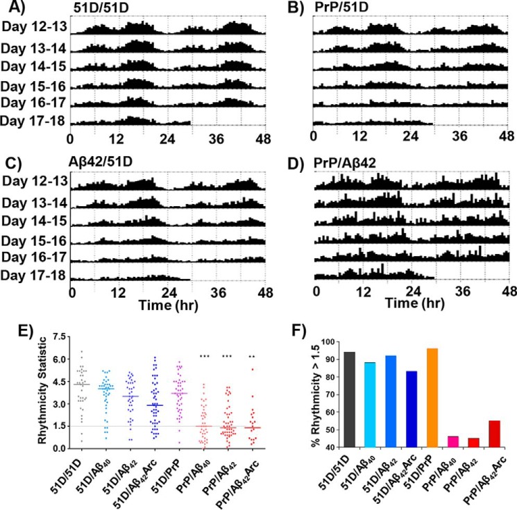
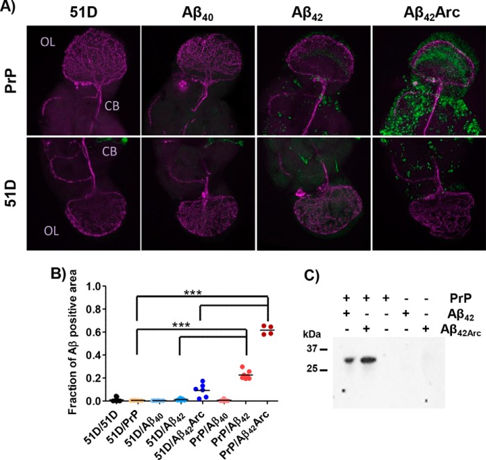
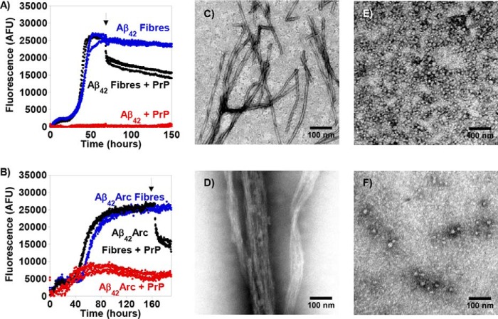
The cellular prion protein (PrPC) can act as a cell-surface receptor for β-amyloid (Aβ) peptide; however, a role for PrPC in the pathogenesis of Alzheimer's disease (AD) is contested. Here, we expressed a range of Aβ isoforms and PrPC in the Drosophila brain. We found that co-expression of Aβ and PrPC significantly reduces the lifespan, disrupts circadian rhythms, and increases Aβ deposition in the fly brain. In contrast, under the same conditions, expression of Aβ or PrPC individually did not lead to these phenotypic changes. In vitro studies revealed that substoichiometric amounts of PrPC trap Aβ as oligomeric assemblies and fragment-preformed Aβ fibers. The ability of membrane-anchored PrPC to trap Aβ as cytotoxic oligomers at the membrane surface and fragment inert Aβ fibers suggests a mechanism by which PrPC exacerbates Aβ deposition and pathogenic phenotypes in the fly, supporting a role for PrPC in AD. This study provides a second animal model linking PrPC expression with Aβ toxicity and supports a role for PrPC in AD pathogenesis. Blocking the interaction of Aβ and PrPC represents a potential therapeutic strategy.
Keywords: Alzheimer's disease; Drosophila; amyloid-beta (AB); animal model; circadian rhythm; fibril; neurodegenerative disease; oligomer; prion; protein misfolding.
© 2018 Younan et al.
Conflict of interest statement
The authors declare that they have no conflicts of interest with the contents of this article
Figures

Similar articles
-
The cellular prion protein traps Alzheimer's Aβ in an oligomeric form and disassembles amyloid fibers.FASEB J. 2013 May;27(5):1847-58. doi: 10.1096/fj.12-222588. Epub 2013 Jan 18. FASEB J. 2013. PMID: 23335053 Free PMC article.
-
Structural details of amyloid β oligomers in complex with human prion protein as revealed by solid-state MAS NMR spectroscopy.J Biol Chem. 2021 Jan-Jun;296:100499. doi: 10.1016/j.jbc.2021.100499. Epub 2021 Mar 3. J Biol Chem. 2021. PMID: 33667547 Free PMC article.
-
The anti-prion RNA aptamer R12 disrupts the Alzheimer's disease-related complex between prion and amyloid β.FEBS J. 2019 Jun;286(12):2355-2365. doi: 10.1111/febs.14819. Epub 2019 Apr 8. FEBS J. 2019. PMID: 30916478
-
Binding between Prion Protein and Aβ Oligomers Contributes to the Pathogenesis of Alzheimer's Disease.Virol Sin. 2019 Oct;34(5):475-488. doi: 10.1007/s12250-019-00124-1. Epub 2019 May 15. Virol Sin. 2019. PMID: 31093882 Free PMC article. Review.
-
Binding Sites for Amyloid-β Oligomers and Synaptic Toxicity.Cold Spring Harb Perspect Med. 2017 May 1;7(5):a024075. doi: 10.1101/cshperspect.a024075. Cold Spring Harb Perspect Med. 2017. PMID: 27940601 Free PMC article. Review.
Cited by
-
Soluble Prion Peptide 107-120 Protects Neuroblastoma SH-SY5Y Cells against Oligomers Associated with Alzheimer's Disease.Int J Mol Sci. 2020 Oct 1;21(19):7273. doi: 10.3390/ijms21197273. Int J Mol Sci. 2020. PMID: 33019683 Free PMC article.
-
The Toxicity and Polymorphism of β-Amyloid Oligomers.Int J Mol Sci. 2020 Jun 24;21(12):4477. doi: 10.3390/ijms21124477. Int J Mol Sci. 2020. PMID: 32599696 Free PMC article. Review.
-
Inducing prion protein shedding as a neuroprotective and regenerative approach in pathological conditions of the brain: from theory to facts.Neural Regen Res. 2023 Sep;18(9):1869-1875. doi: 10.4103/1673-5374.366496. Neural Regen Res. 2023. PMID: 36926701 Free PMC article. Review.
-
Alzheimer's Disease and Its Potential Alternative Therapeutics.J Alzheimers Dis Parkinsonism. 2019;9(5):477. doi: 10.4172/2161-0460.1000477. Epub 2019 Sep 13. J Alzheimers Dis Parkinsonism. 2019. PMID: 31588368 Free PMC article.
-
Tau Exon 10 Inclusion by PrPC through Downregulating GSK3β Activity.Int J Mol Sci. 2021 May 20;22(10):5370. doi: 10.3390/ijms22105370. Int J Mol Sci. 2021. PMID: 34065232 Free PMC article.
References
-
- Lambert M. P., Barlow A. K., Chromy B. A., Edwards C., Freed R., Liosatos M., Morgan T. E., Rozovsky I., Trommer B., Viola K. L., Wals P., Zhang C., Finch C. E., Krafft G. A., and Klein W. L. (1998) Diffusible, nonfibrillar ligands derived from Aβ1–42 are potent central nervous system neurotoxins. Proc. Natl. Acad. Sci. U.S.A. 95, 6448–6453 10.1073/pnas.95.11.6448 - DOI - PMC - PubMed
Publication types
MeSH terms
Substances
Grants and funding
- G0700990/MRC_/Medical Research Council/United Kingdom
- BB/M023877/1/BB_/Biotechnology and Biological Sciences Research Council/United Kingdom
- BB/D005027/1/BB_/Biotechnology and Biological Sciences Research Council/United Kingdom
- WT_/Wellcome Trust/United Kingdom
- 093241/Z/10/Z/WT_/Wellcome Trust/United Kingdom
LinkOut - more resources
Full Text Sources
Other Literature Sources
Medical
Molecular Biology Databases
Research Materials
