. 2018 Sep 1;441(1):52-66.
doi: 10.1016/j.ydbio.2018.06.006. Epub 2018 Jun 7.

CDK-11-Cyclin L is required for gametogenesis and fertility in C. elegans

Affiliations

CDK-11-Cyclin L is required for gametogenesis and fertility in C. elegans

Christopher W Williams et al. Dev Biol. .

Abstract

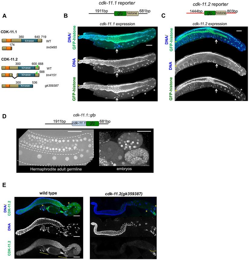
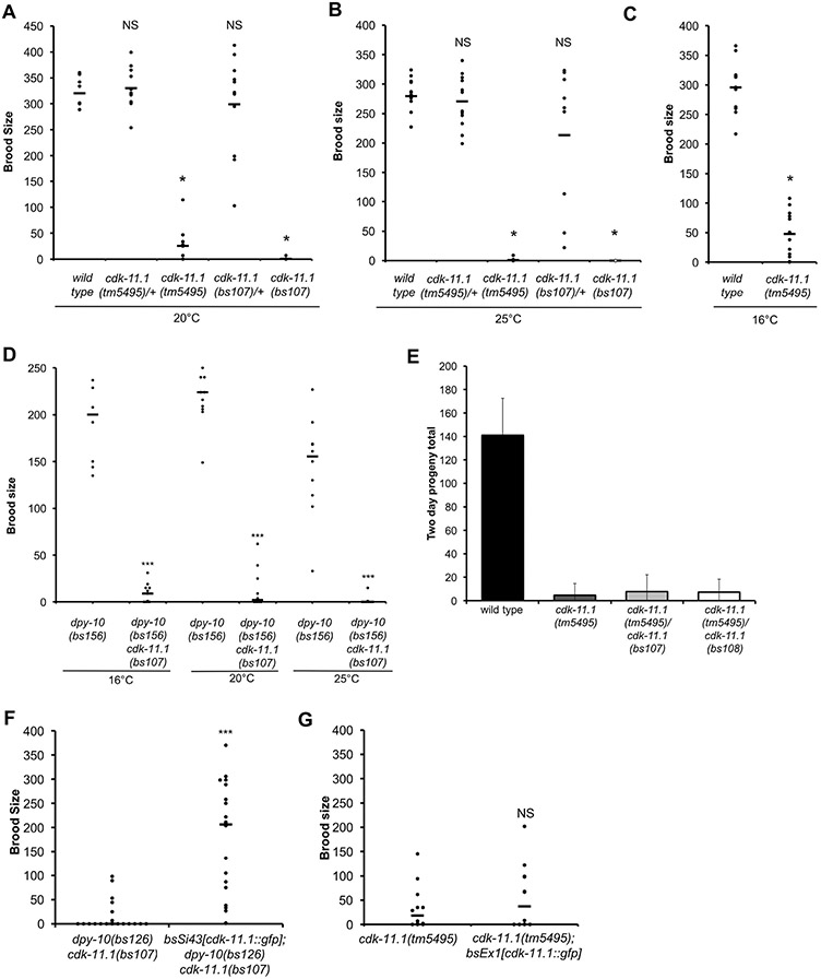
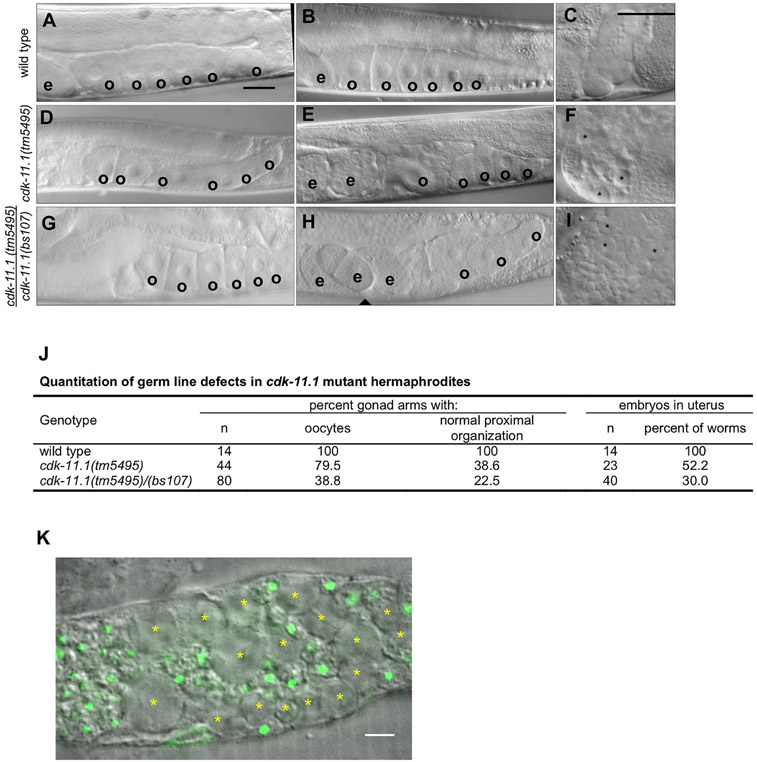
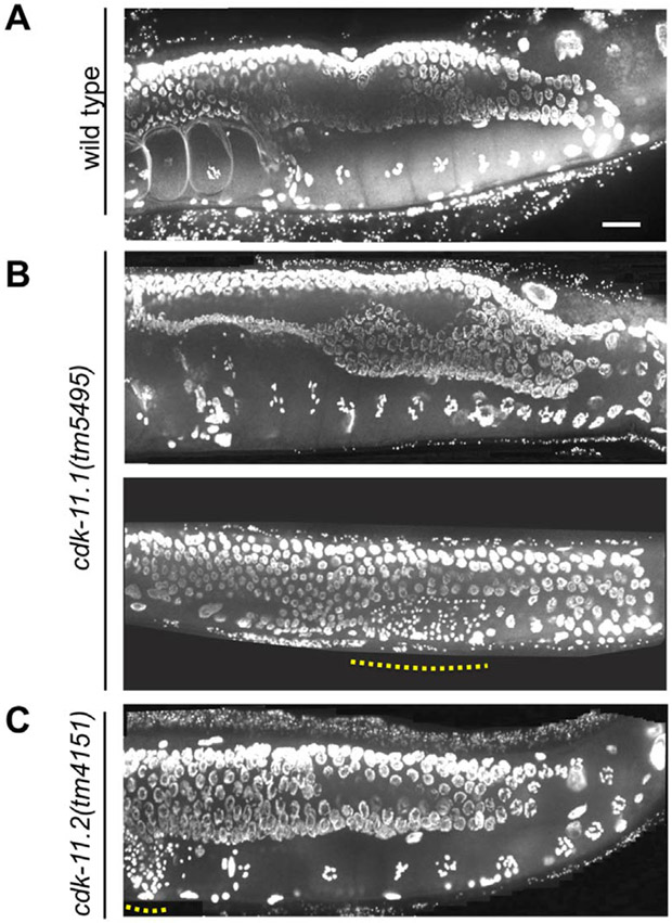
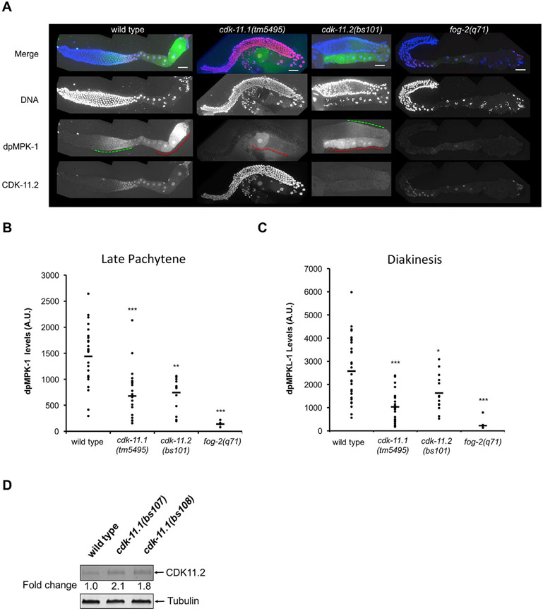
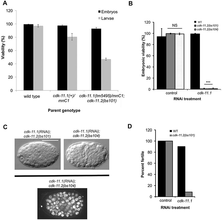
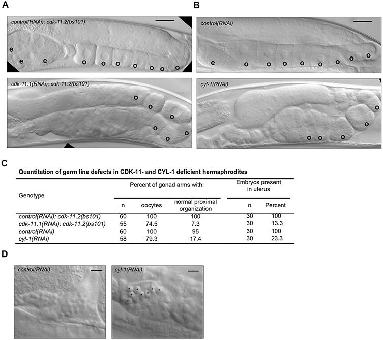
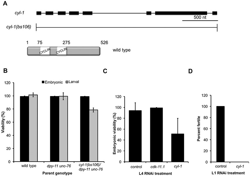
CDK11, a member of the cyclin-dependent kinase family, has been implicated in a diverse array of functions including transcription, RNA processing, sister chromatid cohesion, spindle assembly, centriole duplication and apoptosis. Despite its involvement in many essential functions, little is known about the requirements for CDK11 and its partner Cyclin L in a developing multicellular organism. Here we investigate the function of CDK11 and Cyclin L during development of the nematode Caenorhabditis elegans. Worms express two CDK11 proteins encoded by distinct loci: CDK-11.1 is essential for normal male and female fertility and is broadly expressed in the nuclei of somatic and germ line cells, while CDK-11.2 is nonessential and is enriched in hermaphrodite germ line nuclei beginning in mid pachytene. Hermaphrodites lacking CDK-11.1 develop normally but possess fewer mature sperm and oocytes and do not fully activate the RAS-ERK pathway that is required for oocyte production in response to environmental cues. Most of the sperm and eggs that are produced in cdk-11.1 null animals appear to complete development normally but fail to engage in sperm-oocyte signaling suggesting that CDK-11.1 is needed at multiple points in gametogenesis. Finally, we find that CDK-11.1 and CDK-11.2 function redundantly during embryonic and postembryonic development and likely do so in association with Cyclin L. Our results thus define multiple requirements for CDK-11-Cyclin L during animal development.

Keywords: C. elegans; CDK11; Cyclin L; Gametogenesis; RAS-ERK.


Figures

Fig. 1.
Fig. 1.
CDK-11.1 and CDK-11.2 are broadly expressed nuclear proteins with distinct germ line expression patterns. (A) Domain architecture of wild-type and mutant CDK-11.1 and CDK-11.2 proteins. Both proteins possess a C-terminal kinase domain and a N-terminal arginine- and glutamic acid-rich RE domain (orange box). Additionally, each protein contains a putative nuclear localization sequence within the RE domain (not shown). The position of the epitope recognized by the anti-CDK-11.2 antibody is indicated. (B) The structure of the cdk-11.1 reporter is shown at the top: the 5’ and 3’ flanking regions of cdk-11.1 were used to drive the expression of GFP::histone. GFP::histone is expressed throughout the entire germ line and in the sheath cells of the somatic gonad (white asterisks). The distal tip is marked by a yellow asterisk and the proximally located spermatheca by a white arrow. (C) The structure of the cdk-11.2 reporter is shown at the top: the 5’ and 3’ flanking regions of cdk-11.2 were used to drive the expression of GFP::histone. Expression of GFP::histone is observed throughout the germ line and somatic gonad. (D) The structure of the transgene expressing a CDK-11.1::GFP fusion protein. The fusion protein accumulates in nuclei throughout the germ line and in embryos. (E) Wild-type and cdk-11.2(gk359387) germ lines stained with anti-CDK-11.2 antibody and DAPI. CDK-11.2 is detected in the proximal germ line and somatic sheath cells (yellow asterisks). This staining is absent in the cdk-11.2(gk359387) mutant that lacks the epitope recognized by the antibody (see panel 1A). Note that the anti-CDK-11.2 antibody nonspecifically recognizes sperm (yellow dotted line). Scale bars are 25 μm in all images.
Fig. 2.
Fig. 2.
Loss of CDK-11.1 results in low fecundity. (A-C) Brood sizes of wild-type and cdk-11.1 mutant hermaphrodites at various temperatures. (D) Brood sizes of dpy-10(bs156), dpy-10(bs126) cdk-11.1(bs107), and dpy-10(bs141) cdk-11.1(bs108) hermaphrodites at various temperatures. (E) Complementation tests showing that the null alleles cdk-11.1(bs107) and cdk-11.1(bs108) fail to complement the cdk-11.1(tm5495) mutation for the fecundity defect. (F) The integrated cdk-11.1::gfp transgene (bsSi.43) rescues the fecundity defect of cdk-11.1(bs107) hermaphrodites. (G) The same cdk-11.1::gfp transgene does not rescue the fecundity defect when expressed from an extrachromosomal array (bsEx1). (ns = not significant, * p < 0.01, *** p < 0.001).
Fig. 3.
Fig. 3.
CDK-11.1 is required for normal gametogenesis. Hermaphrodites were synchronized at the L1 stage and grown for 3 days at 20 °C before analysis. (A and B) DIC images of wild-type gonads which invariably display a single row of cuboidal oocytes (o) in the proximal end of the germ line, and embryos (e) in the uterus. (C) An image of a wild-type spermatheca. Contained within are a population of sperm with uniform size. (D and E) DIC images of cdk-11.1(tm5495) hermaphrodite gonads. Although some appear normal (panel D) others display fewer oocytes that are less uniform in size and with a disorganized arrangement in the proximal germ line (panel E). (F) An image of a cdk-11.1(tm5495) spermatheca containing some normal looking sperm and some larger cells (asterisks). (G and H) DIC images of cdk-11.1(tm5495)/(bs107) hermaphrodite gonads. Some cdk-11.1(tm5495)/(bs107) trans-heterozygotes display normal-looking oocytes (panel G) while others possess fewer oocytes whose shape, size, and arrangement vary (panel H). (I) An image of a cdk-11.1(tm5495)/(bs107) spermatheca containing normal-sized sperm and some abnormally large cells (asterisks). (J) Quantitation of oocyte defects in various strains. (K) DAPI-stained spermatheca from a cdk-11.1(tm5495) hermaphrodite. Asterisks mark residual bodies which lack DNA (green). Scale bars are 25 μm (panels A–I) and 5 μm (panel K).
Fig. 4.
Fig. 4.
Germ-line defects in cdk-11.1(tm5495) hermaphrodites are restricted to the proximal end of the gonad. Animals were plated at 20 °C at the L1 stage, grown for 3 days, then fixed and stained with DAPI. (A) wild type (B) cdk-11.1(tm5495). Dotted line highlights region containing sperm nuclei. (C) cdk-11.2(tm4151). Scale bar is 25 μm.
Fig. 5.
Fig. 5.
CDK-11.1 is required for full activation of the RAS-ERK pathway. (A) Dissected germ lines stained for DNA (blue), activated ERK (dpMPK-1, green), and CDK-11.2 (red). In wild-type germ lines, dpMPK-1 levels peak in late pachytene (green dashed line) and again in diakinesis (red dashed line), The fog-2(q71) germ lines serve as a negative control where very little or no dpMPK-1 staining is observed. cdk-11.1(tm5495) mutant hermaphrodites display decreased levels of dpMPK-1 in both late pachytene and diakinesis, while cdk-11.2(bs101) animals exhibit a similar reduction in the level of dpMPK-1 in late pachytene but a milder reduction during diakinesis. Note that cdk-11.1(tm5495) mutants show an increase in the level of CDK-11.2 throughout the entire germ line when compared to wild-type germ lines. Scale bars are 25 μm in all images. B and C) Quantitation of dpMPK-1 levels (plotted as arbitrary units, A.U.) during late pachytene and diakinesis in wild-type, cdk-11.1(tm5495), cdk-11.2(bs101) and fog-2(q71) germ lines. Black bars represent the mean. A one-tailed t-test was used to determine if mutants exhibit a significantly different dpMPK-1 level relative to the wild type (*p < 0.05,**p < 0.0001, ***p < 0.00001). The number of gonads scored (late pachytene/diakinesis) = 29/31 for wild type, 22/23 for cdk-11.1(tm5495), 14/13 for cdk-11.2(bs101), and 8/10 for fog-2(q71). (D) Quantitative immunoblot demonstrating that loss of CDK-11.1 leads to an increase in expression of CDK-11.2. Whole worm lysates were prepared and the equivalent of thirty adult animals were loaded in each lane.
Fig. 6.
Fig. 6.
CDK-11.1 and CDK-11.2 function in a partially redundant manner. (A) The progeny of hermaphrodites of the indicated genotype were scored for embryonic and larval lethality. Note that cdk-11.1(tm5495)/mnC1; cdk-11.2(bs101) mothers produce about 25% more inviable larvae than +/mnC1 controls. (B) L4 hermaphrodites of the indicated genotypes were fed dsRNA targeting cdk-11.1 or the nonessential gene smd-1 as a negative control. Loss of embryonic viability was only observed when CDK-11.1 was depleted in animals harboring a cdk-11.2 null allele. (C) DIC Images of cdk-11.1(RNAi); cdk-11.2(null) embryos displaying the terminal phenotype (top) and an image of a DAPI-stained cdk-11.1(RNAi); cdk-11.2(bs104) embryo (bottom). Embryos arrest with approximately 130 cells but do not show any signs of gross aneuploidy, typical of mutants with cell division failures. Scale bar is 25 μm. (D) L1 hermaphrodites of the indicated genotypes were fed dsRNA targeting smd-1 or cdk-11.1. Loss of fertility was only observed when CDK-11.1 was depleted in animals harboring a cdk-11.2 null allele. The cdk-11.1(RNAi); cdk-11.2(null) animals display a nearly complete loss of fertility.
Fig. 7.
Fig. 7.
Germ line defects in cdk-11 null and cyl-1(RNAi) mutants. (A) DIC images of cdk-11.2(bs101) hermaphrodites treated with smd-1(RNAi) (top) as a negative control and cdk-11.1 (RNAi) (bottom), beginning at the L1 stage. cdk-11.2(bs101) hermaphrodites treated with smd-1 (RNAi) look normal with an ordered row of oocytes within the proximal end of the gonad. The cdk-11.1(RNAi); cdk-11.2(bs101) animals have irregularly sized and shaped oocytes which sometimes appear to form precociously. (B) Treatment of WT animals with cyl-1 RNAi from the L1 stage produces a disordered proximal germ line phenotype similar to that of cdk-11.1(RNAi); cdk-11.2(bs101) hermaphrodites. cyl-1(RNAi) animals exhibit premature cellularization in the loop region of the gonad and oocytes of abnormal shape, size and arrangement in the most proximal region. The oocytes also appear compacted, likely due to failed ovulation. (C) Quantitation of DIC phenotypes. (D) DIC images of spermathecae from smd-1(RNAi)-treated and cyl-1(RNAi)-treated animals. Note that the spermathecae of cyl-1(RNAi) animals possess unusually large cells (possibly residual bodies). Scale bars are 25 μm (panel A–B) and 5 μm (panel D).
Fig. 8.
Fig. 8.
Loss of CYL-1 phenocopies the various defects of cdk-11 mutants. (A) Schematic of the cyl-1 gene and the null allele cyl-1(bs106). The product of the cyl-1 gene is a Cyclin L protein containing a single CYCLIN box composed of two cyclin folds. (B) The progeny of hermaphrodites of the indicated genotype were scored for embryonic and larval lethality. Note that the homozygous cyl-1(bs106) progeny of a heterozygous mother die during larval development. (C) Percent embryonic viability of the progeny of wild-type hermaphrodites subjected to various RNAi treatments beginning at the L4 stage. smd-1(RNAi) serves as a negative control. (D) Percent of wild-type hermaphrodites that develop to fertile adults when exposed to smd-1(RNAi) or cyl-1(RNAi) beginning at the L1 stage.

Similar articles

Cited by

References

    1. Ariza ME, Broome-Powell M, Lahti JM, Kidd VJ, Nelson MA, 1999. Fas-induced apoptosis in human malignant melanoma cell lines is associated with the activation of thep34(cdc2)-related PITSLRE protein kinases. J. Biol. Chem 274, 28505–28513. - PubMed
    1. Arribere JA, Bell RT, Fu BXH, Artiles KL, Hartman PS, Fire AZ, 2014. Efficient marker-free recovery of custom genetic modifications with CRISPR/Cas9 in Caenorhabditis elegans. Genetics 198, 837–846. 10.1534/genetics.114.169730. - DOI - PMC - PubMed
    1. Arur S, Ohmachi M, Nayak S, Hayes M, Miranda A, Hay A, Golden A, Schedl T, 2009. Multiple ERK substrates execute single biological processes in Caenorhabditis elegans germ-line development. Proc. Natl. Acad. Sci. USA 106, 4776–4781. 10.1073/pnas.0812285106. - DOI - PMC - PubMed
    1. Barna M, Pusic A, Zollo O, Costa M, Kondrashov N, Rego E, Rao PH, Ruggero D, 2008. Suppression of Myc oncogenic activity by ribosomal protein haploinsufficiency. Nature 456, 971–975. 10.1038/nature07449. - DOI - PMC - PubMed
    1. Berke JD, Sgambato V, Zhu PP, Lavoie B, Vincent M, Krause M, Hyman SE, 2001. Dopamine and glutamate induce distinct striatal splice forms of Ania-6, an RNA polymerase II-associated cyclin. Neuron 32, 277–287. - PubMed

Publication types

LinkOut - more resources