

Isolation and Identification of Innate Lymphoid Cells (ILCs) for Immunotoxicity Testing
- PMID: 29882149
- PMCID: PMC6025753
- DOI: 10.1007/978-1-4939-8549-4_21
Isolation and Identification of Innate Lymphoid Cells (ILCs) for Immunotoxicity Testing
Abstract
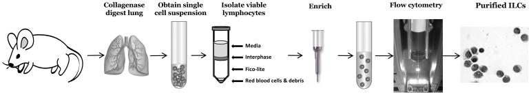
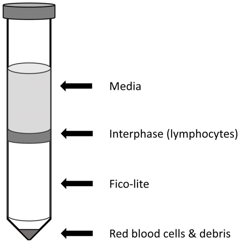
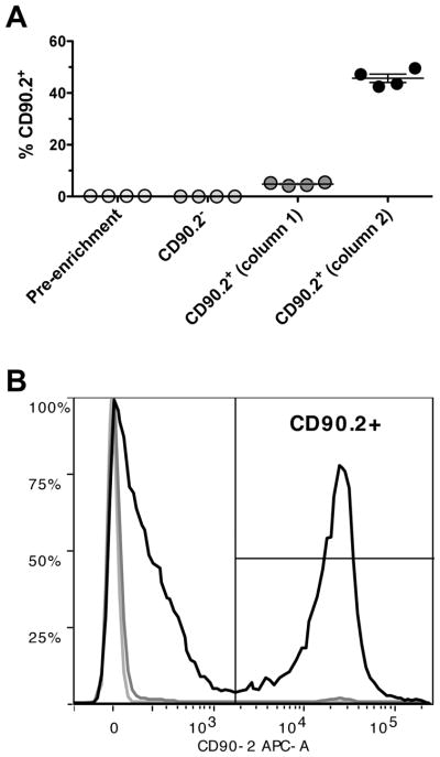
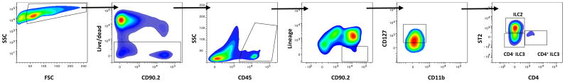
Innate lymphoid cells (ILCs) comprise a family of innate immune cells that orchestrate mucosal immune responses: initiating, sustaining, and even curbing immune responses. ILCs are relatively rare (≤1% of lymphocytes in mucosal tissues), lack classical cell-surface markers, and can be divided into three subsets (type 1-3 ILCs) based on differences in cytokine production, phenotype, and developmental pathway. Because ILCs can only be identified by combinations of cell-surface markers and cytokine production, multicolor flow cytometry is the most reliable method to purify, characterize, and assess the functionality of ILCs. Here, we describe the methods for cell preparation, flow cytometric analysis, and purification of murine ILCs from the lung.
Keywords: Flow cytometry; ILC2; ILC3; Innate lymphoid cells; Mouse; Respiratory tract.
Figures
Similar articles
-
Isolation and characterization of mouse innate lymphoid cells.Curr Protoc Immunol. 2014 Aug 1;106:3.25.1-3.25.13. doi: 10.1002/0471142735.im0325s106. Curr Protoc Immunol. 2014. PMID: 25081911 Review.
-
Rapid isolation of mouse ILCs from murine intestinal tissues.Methods Enzymol. 2020;631:305-327. doi: 10.1016/bs.mie.2019.10.001. Epub 2019 Nov 12. Methods Enzymol. 2020. PMID: 31948554
-
Purification and Adoptive Transfer of Group 3 Gut Innate Lymphoid Cells.Methods Mol Biol. 2016;1422:189-96. doi: 10.1007/978-1-4939-3603-8_18. Methods Mol Biol. 2016. PMID: 27246034
-
Single-cell RNA-seq identifies a PD-1hi ILC progenitor and defines its development pathway.Nature. 2016 Nov 3;539(7627):102-106. doi: 10.1038/nature20105. Epub 2016 Sep 29. Nature. 2016. PMID: 27749818
-
Human innate lymphoid cells (ILCs): Toward a uniform immune-phenotyping.Cytometry B Clin Cytom. 2018 May;94(3):392-399. doi: 10.1002/cyto.b.21614. Epub 2018 Jan 31. Cytometry B Clin Cytom. 2018. PMID: 29244250 Review.
Cited by
-
Detection, Isolation, and Functional Studies of Mouse Pulmonary Group 2 Innate Lymphoid Cells.Methods Mol Biol. 2022;2506:167-186. doi: 10.1007/978-1-0716-2364-0_12. Methods Mol Biol. 2022. PMID: 35771471 Free PMC article.
-
Emerging Roles of Interleukin-33-responsive Kidney Group 2 Innate Lymphoid Cells in Acute Kidney Injury.Int J Mol Sci. 2020 Feb 24;21(4):1544. doi: 10.3390/ijms21041544. Int J Mol Sci. 2020. PMID: 32102434 Free PMC article. Review.
-
Differential Regulation of Innate Lymphoid Cells in Human and Murine Oral Squamous Cell Carcinoma.Int J Mol Sci. 2023 Jan 13;24(2):1627. doi: 10.3390/ijms24021627. Int J Mol Sci. 2023. PMID: 36675138 Free PMC article.
-
Flow cytometric analysis of innate lymphoid cells: challenges and solutions.Front Immunol. 2023 Sep 22;14:1198310. doi: 10.3389/fimmu.2023.1198310. eCollection 2023. Front Immunol. 2023. PMID: 37809100 Free PMC article. Review.
-
Fate-mapping mice: new tools and technology for immune discovery.Trends Immunol. 2022 Mar;43(3):195-209. doi: 10.1016/j.it.2022.01.004. Epub 2022 Jan 31. Trends Immunol. 2022. PMID: 35094945 Free PMC article. Review.
References
MeSH terms
Grants and funding
LinkOut - more resources
Full Text Sources
Other Literature Sources
