. 2017 Jul 8;9(37):24653-24671.
doi: 10.18632/oncotarget.19065. eCollection 2018 May 15.

Collagen type 1 promotes survival of human breast cancer cells by overexpressing Kv10.1 potassium and Orai1 calcium channels through DDR1-dependent pathway

Affiliations

Collagen type 1 promotes survival of human breast cancer cells by overexpressing Kv10.1 potassium and Orai1 calcium channels through DDR1-dependent pathway

Mehdi Badaoui et al. Oncotarget. .

Abstract

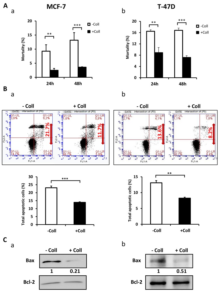
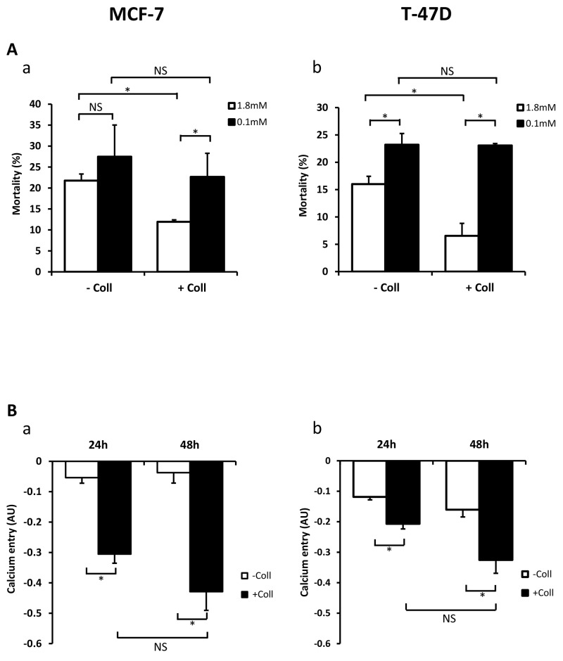
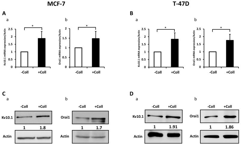
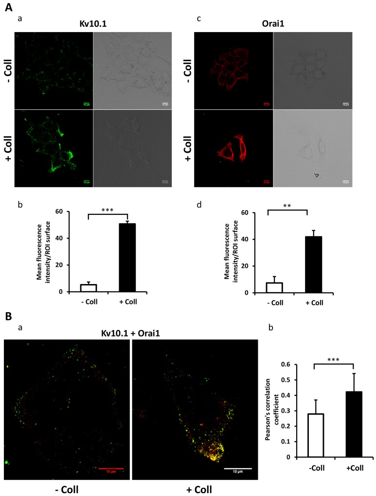
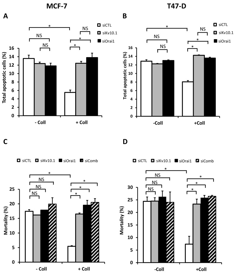
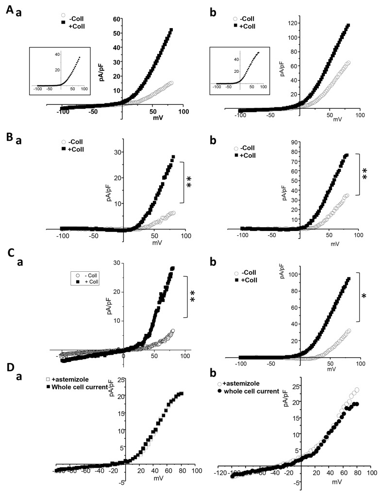
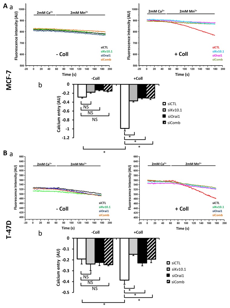
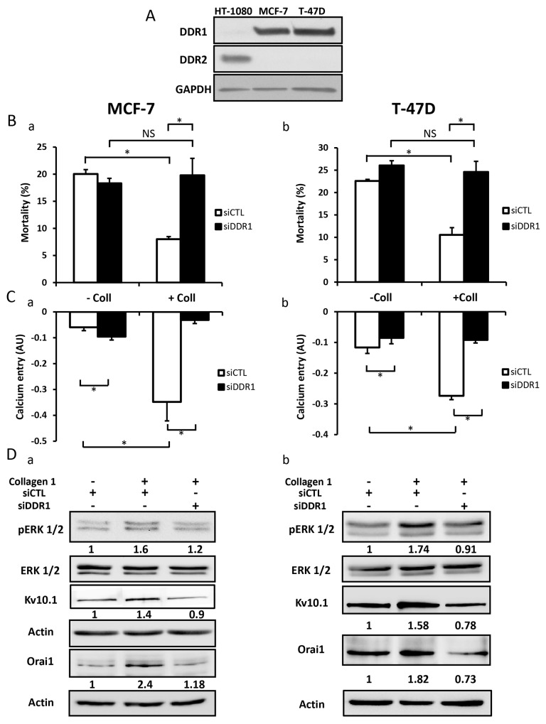
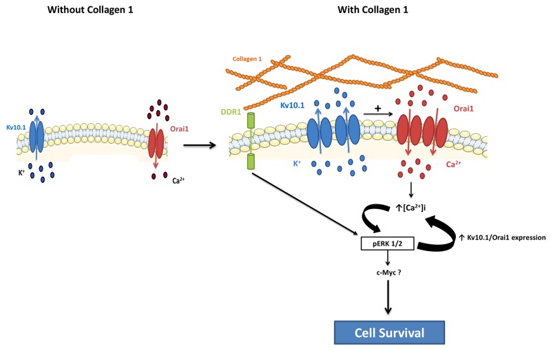
Collagen type 1 is among the tumor microenvironment (TM) factors, that regulates proliferation, survival, migration and invasion. Ion channels are key players in interactions between tumor cells and TM. Kv10.1 has been shown to play an essential role in breast cancer cell proliferation and migration by permitting Ca2+ influx notably via Orai1. Here, we show that human breast cancer (BC) cells growing, in culture media completely devoid of the serum and seeded on collagen 1 coating, exhibited less apoptotic rate and a decrease in Bax expression when compared to those grown on plastic. The survival conferred by collagen 1 was completely abolished by removing extracellular Ca2+ from the culture medium. In addition, Ca2+ entry was increased in collagen 1 condition along with increased Kv10.1 and Orai1 expressions. Moreover, collagen 1 was able to increase co-localization of Kv10.1 and Orai1 on the plasma membrane. Interestingly, silencing of Kv10.1 and Orai1 reduced survival and Ca2+influx without any additive effect. This calcium-dependent survival is accompanied by the activation of ERK1/2, and its pharmacological inhibition completely abolished the increase in Kv10.1 and Orai1 expressions, activities, and the cell survival induced by collagen 1. Moreover, both Kv10.1 and Orai1 knockdown reduced ERK1/2 activation but not Akt. Finally, DDR1 silencing but not β1-integrin reduced the collagen induced survival, ERK1/2 phosphorylation and the expression of Kv10.1 and Orai1. Together these data show that the Kv10.1/Orai1 complex is involved in BC cell survival and this is dependent on collagen 1/DDR1 pathway. Therefore, they represent a checkpoint of tumor progression induced by the tumor microenvironment.

Keywords: Kv10.1; Orai1; breast cancer; survival; tumour microenvironment.


Conflict of interest statement

CONFLICTS OF INTEREST The authors declare that they have no interest of any kind affecting this study.

Figures

Figure 1
Figure 1. Collagen 1 induced MCF-7 and T-47D cells survival under serum starvation
(A) Effect of collagen 1 on BC cell survival. The cell mortality was measured 24 h and 48 h post-starvation by Trypan Blue in MCF-7 (a) and T-47D (b) cells. (B) Measurement of rate of apoptosis in plastic and collagen 1 conditions 48 h post-starvation by Annexin V staining in MCF-7 (a) and T-47D (b) cells. (C) Representative western blot showing Bax and Bcl-2 expression in MCF-7 (a) and T-47D (b) cells 48 h post-starvation. Results were normalised as a percentage of the control condition (without collagen 1). Values are reported as mean ± SEM of triplicate experiments. **p < 0.01, ***p < 0.001. Student’s t-test. – Coll: without collagen 1, + Coll: with collagen 1.
Figure 2
Figure 2. Collagen 1 promotes calcium influx and regulates cell survival by a calcium-dependent mechanism
(A) Impact of decreasing extracellular Ca2+concentration on cell mortality of MCF-7 (a) and (b) T-47D cells. Mortality was measured 48 h post-starvation, with (+ Coll) or without collagen 1 (- Coll), after incubation with medium containing low (0.1 mM) or physiological Ca2+ concentrations (1.8 mM). Values are reported as mean ± SEM of triplicate experiments, *p<0.05, NS: not significant. ANOVA followed by Holm-Sidak post hoc tests. (B) Effect of collagen 1 on basal Ca2+ entry in the same batch of MCF-7 (a) and T-47D (b) cells using Mn2+ quenching experiments. Mean slope values are reported as mean ± SEM of triplicate experiments, *p<0.05, ANOVA followed by Holm-Sidak post hoc tests, NS: not significant.
Figure 3
Figure 3. Collagen 1 increases Kv10.1 and Orai1 expression in both MCF-7 and T-47D cells
(A) qRT-PCR expression of Kv10.1 (a) and Orai1 (b) in MCF-7 cells 48 h post-starvation in the presence of collagen 1. (B) qRT-PCR expression of Kv10.1 (a) and Orai1 (b) in T-47D cells 48 h post-starvation in the presence of collagen 1. (C) Representative western blots showing the expression of Kv10.1 (a) and Orai1 (b) in MCF-7 cells 48 h post-starvation in the presence of collagen 1. (D) Representative western blots showing the expression of Kv10.1 (a) and Orai1 (b) in T-47 cells 48 h post-starvation in collagen 1 condition. Results were normalized as a percentage of the control condition (- Coll). Values are reported as mean ± SEM of triplicate experiments. *p <0.05, Student’s t-test.
Figure 4
Figure 4. Collagen 1 promotes the co-localization of Kv10.1 and Orai 1 at plasma membrane in BC cells
(A) Localization of Kv10.1 (a) and Orai1 (c) proteins at the plasma membrane 48 h post-starvation in MCF-7 cells seeded (+ Coll) or not (- Coll) on collagen 1, visualized by confocal microscopy. Quantification of the Kv10.1 (b) and Orai1 (d) fluorescence intensity in the two conditions. (B) Merge of Kv10.1 and Orai1 (yellow) at the plasma membrane 48 h post-starvation visualized by confocal microscopy (a), and Pearson’s correlation coefficient (b). Values are reported as mean ± SEM of triplicate experiments. **p < 0.01, ***p < 0.001. Student’s t-test.
Figure 5
Figure 5. Kv10.1 and Orai1 are involved in collagen-dependent survival of BC cell lines
Effect of Kv10.1 or Orai1 silencing on the apoptotic MCF-7 (A) and T-47D cells (B) rate. Cells were starved for 48 h and the apoptosis assay was carried out by annexin V/IP staining, values are reported as mean ± SEM of triplicate experiments, *p<0.05, ANOVA followed by Holm-Sidak post hoc tests. (C-D) Effect of Kv10.1, Orai1 and Kv10.1 + Orai1 (siComb) silencing on MCF-7 (C) and T-47D (D) cell mortality. Cells were starved for 48 h and the mortality was measured by Trypan Blue assay, values are reported as mean ± SEM of triplicate experiments, *p<0.05, ANOVA followed by Holm-Sidak post hoc tests.
Figure 6
Figure 6. Collagen increases Kv10.1 functional channel activity in BC cells
(A) Whole cell currents recorded in cells treated or not with collagen 1. Cells were starved for 48 h and the patch-clamp measurements were performed. 500 msec voltage ramps from -100 to +80 mV from a holding potential of -40 mV were applied to record Kv10.1 channel activity in MCF-7 (a) and T-47D (b) cell lines. The collagen-activated outward current is shown in the small squares. (B) Astemizole-sensitive current traces in MCF-7 (a) and T-47D (b) cells seeded or not on collagen 1 obtained from the subtraction of the current remained after astemizole perfusion from the whole cell current. (C) siKv10.1 sensitive current in MCF-7 (a) and T-47D (b) cells treated or not with collagen, obtained from the subtraction of the whole cell current recorded in cells transfected with siKv10.1 from the average whole cell current recorded in cells transfected with si-control. Values are reported as mean. *p <0.05, **p< 0.01. Mann-withney test. (D) Effect of the perfusion of astemizole (5 μM) on the outward current recorded in siKv10.1-tranfected MCF-7 (a) and T-47D (b) cells seeded on collagen 1. Astemizole failed to affect the amplitude of the remaining outward current in siKv10.1-transfected cells.
Figure 7
Figure 7. Kv10.1 regulates collagen 1-calcium entry through Orai1
(A) Effect of Kv10.1 and Orai1 and Kv10.1 + Orai1 (siComb) silencing on Ca2+ entry in MCF-7 cells, by using Mn2+ quenching experiments (a). Mean slope values are reported as mean ± SEM of triplicate experiments performed on 5 different number of cell passage (b), *p<0.05, ANOVA followed by Holm-Sidak post hoc tests. (B) Effect of Kv10.1, Orai1 and kv10.1 + Orai1 (siComb) silencing on Ca2+ entry in T-47D cells, by using Mn2+ quenching experiments (a). Mean slope values are reported as mean ± SEM of triplicate experiments performed on 3 different number of cell passage (b), *p<0.05, ANOVA followed by Holm-Sidak post hoc tests.
Figure 8
Figure 8. Collagen 1 increases Kv10.1 and Orai1 expression through ERK1/2 pathway
(A) Representative western blots showing ERK1/2 phosphorylation in MCF-7 (a) and T-47D (b) cells 48 h post-starvation in presence (+ Coll) and absence (- Coll) of collagen 1. (B-C) Representative western blots showing the effect of PD98059 (40 μM) on ERK1/2 activation, and on Kv10.1 and Orai1 expression in MCF-7 (B) and T-47D (C) cells.Values were normalized as a percentage of the control condition (- Coll). (D-E) Representative western blots showing the effect of Kv10.1 and Orai1 silencing on ERK1/2 phosphorylation in MCF-7 (D) and T-47D (E) cells in collagen 1 conditions. – Coll: without collagen 1, + Coll: with collagen 1.
Figure 9
Figure 9. DDR1 is expressed in MCF-7 and T-47D cells and promoted collagen-dependent survival
(A) Representative western blot showing DDR1 and DDR2 expression in MCF-7 and T-47D cells. (B) Effect of DDR1 silencing on cell mortality of MCF-7 (a) and T-47D cells (b). Cells were starved for 48 h and the mortality was measured by Trypan Blue assay, values are reported as mean ± SEM of triplicate experiments performed on 3 different number of cell passage, *p<0.05, ANOVA followed by Holm-Sidak post hoc tests. (C) Effect of DDR1 silencing on Ca2+ entry in MCF-7 (a) and T-47D (b) cells. Mean slope values are reported as mean ± SEM of triplicate experiments performed on 4 different number of cell passage, *p<0.05, ANOVA followed by Holm-Sidak post hoc tests. (D) Representative western blot showing the effect of DDR1 silencing on ERK1/2 phosphorylation, Kv10.1 and Orai1 expression in MCF-7 (a) and T-47D (b) cells seeded on collagen 1.
Figure 10
Figure 10. Schematic representation of potential involvement of the collagen 1 in breast cancer cell survival

Similar articles

Cited by

References

    1. Liotta LA, Kohn EC. The microenvironment of the tumour-host interface. Nature. 2001;411:375–9. - PubMed
    1. Hynes RO. The extracellular matrix: not just pretty fibrils. Science. 2009;326:1216–9. - PMC - PubMed
    1. Di Lullo GA, Sweeney SM, Körkkö J, Ala-Kokko L, San Antonio JD. Mapping the ligand-binding sites and disease-associated mutations on the most abundant protein in the human, type I collagen. J Biol Chem. 2002;277:4223–31. - PubMed
    1. Saby C, Buache E, Brassart-Pasco S, El Btaouri H, Courageot MP, Van Gulick L, Garnotel R, Jeannesson P, Morjani H. Type I collagen aging impairs discoidin domain receptor 2-mediated tumor cell growth suppression. Oncotarget. 2016;7:24908–27. doi: 10.18632/oncotarget.8795. - DOI - PMC - PubMed
    1. Maquoi E, Assent D, Detilleux J, Pequeux C, Foidart JM, Noel A. MT1-MMP protects breast carcinoma cells against type I collagen-induced apoptosis. Oncogene. 2012;31:480–93. - PubMed