

Directed evolution and biophysical characterization of a full-length, soluble, human caveolin-1 variant
- PMID: 29857161
- PMCID: PMC6269214
- DOI: 10.1016/j.bbapap.2018.05.014
Directed evolution and biophysical characterization of a full-length, soluble, human caveolin-1 variant
Abstract
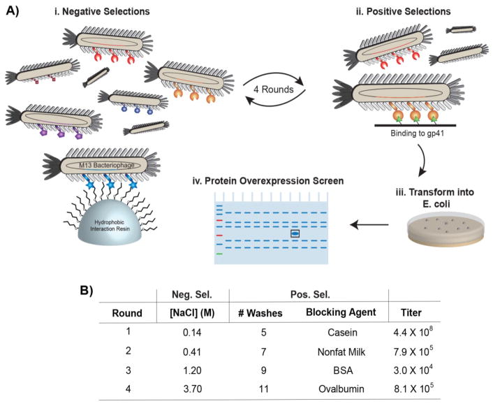
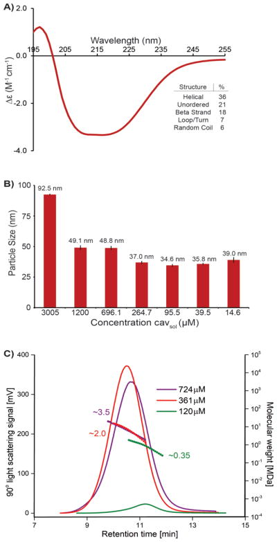
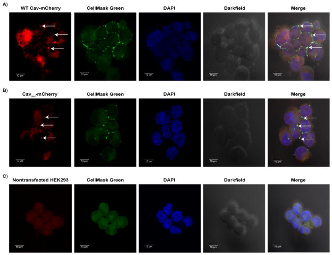
Protein engineering by directed evolution can alter proteins' structures, properties, and functions. However, membrane proteins, despite their importance to living organisms, remain relatively unexplored as targets for protein engineering and directed evolution. This gap in capabilities likely results from the tendency of membrane proteins to aggregate and fail to overexpress in bacteria cells. For example, the membrane protein caveolin-1 has been implicated in many cell signaling pathways and diseases, yet the full-length protein is too aggregation-prone for detailed mutagenesis, directed evolution, and biophysical characterization. Using a phage-displayed library of full-length caveolin-1 variants, directed evolution with alternating subtractive and functional selections isolated a full-length, soluble variant, termed cavsol, for expression in E. coli. Cavsol folds correctly and binds to its known protein ligands HIV gp41, the catalytic domain of cAMP-dependent protein kinase A, and the polymerase I and transcript release factor. As expected, cavsol does not bind off-target proteins. Cellular studies show that cavsol retains the parent protein's ability to localize at the cellular membrane. Unlike truncated versions of caveolin, cavsol forms large, oligomeric complexes consisting of approximately >50 monomeric units without requiring additional cellular components. Cavsol's secondary structure is a mixture of α-helices and β-strands. Isothermal titration calorimetry experiments reveal that cavsol binds to gp41 and PKA with low micromolar binding affinity (KD). In addition to the insights into caveolin structure and function, the approach applied here could be generalized to other membrane proteins.
Keywords: Biophysics; Caveolae; Caveolin; Membrane proteins; Oligomerization; Phage display.
Copyright © 2018 Elsevier B.V. All rights reserved.
Figures


Similar articles
-
Solubilization of a membrane protein by combinatorial supercharging.ACS Chem Biol. 2011 Apr 15;6(4):301-7. doi: 10.1021/cb1001729. Epub 2011 Jan 14. ACS Chem Biol. 2011. PMID: 21192634 Free PMC article.
-
In vitro evolution of ligands to the membrane protein caveolin.J Am Chem Soc. 2011 Jun 29;133(25):9855-62. doi: 10.1021/ja201792q. Epub 2011 Jun 7. J Am Chem Soc. 2011. PMID: 21615158 Free PMC article.
-
Phage display of functional, full-length human and viral membrane proteins.Bioorg Med Chem Lett. 2008 Nov 15;18(22):5937-40. doi: 10.1016/j.bmcl.2008.07.051. Epub 2008 Jul 17. Bioorg Med Chem Lett. 2008. PMID: 18667306 Free PMC article.
-
Recent progress in the topology, structure, and oligomerization of caveolin: a building block of caveolae.Curr Top Membr. 2015;75:305-36. doi: 10.1016/bs.ctm.2015.03.007. Epub 2015 Apr 11. Curr Top Membr. 2015. PMID: 26015287 Review.
-
Caveolin-1: role in cell signaling.Adv Exp Med Biol. 2012;729:29-50. doi: 10.1007/978-1-4614-1222-9_3. Adv Exp Med Biol. 2012. PMID: 22411312 Review.
Cited by
-
Reconstitution of Caveolin-1 into Artificial Lipid Membrane: Characterization by Transmission Electron Microscopy and Solid-State Nuclear Magnetic Resonance.Molecules. 2021 Oct 14;26(20):6201. doi: 10.3390/molecules26206201. Molecules. 2021. PMID: 34684779 Free PMC article.
References
-
- Engel A, Gaub HE. Structure and mechanics of membrane proteins. Annu Rev Biochem. 2008;77:127–148. - PubMed
-
- Yildirim MA, Goh KI, Cusick ME, Barabasi AL, Vidal M. Drug-target network. Nat Biotechnol. 2007;25:1119–1126. - PubMed
-
- Glenney JR. The sequence of human caveolin reveals identity with VIP21, a component of transport vesicles. FEBS Lett. 1992;314:45–48. - PubMed
Publication types
MeSH terms
Substances
Grants and funding
LinkOut - more resources
Full Text Sources
Other Literature Sources
Research Materials
Miscellaneous
