Insulin transport into the brain
- PMID: 29847142
- PMCID: PMC6139500
- DOI: 10.1152/ajpcell.00240.2017
Insulin transport into the brain
Abstract
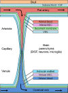
While there is a growing consensus that insulin has diverse and important regulatory actions on the brain, seemingly important aspects of brain insulin physiology are poorly understood. Examples include: what is the insulin concentration within brain interstitial fluid under normal physiologic conditions; whether insulin is made in the brain and acts locally; does insulin from the circulation cross the blood-brain barrier or the blood-CSF barrier in a fashion that facilitates its signaling in brain; is insulin degraded within the brain; do privileged areas with a "leaky" blood-brain barrier serve as signaling nodes for transmitting peripheral insulin signaling; does insulin action in the brain include regulation of amyloid peptides; whether insulin resistance is a cause or consequence of processes involved in cognitive decline. Heretofore, nearly all of the studies examining brain insulin physiology have employed techniques and methodologies that do not appreciate the complex fluid compartmentation and flow throughout the brain. This review attempts to provide a status report on historical and recent work that begins to address some of these issues. It is undertaken in an effort to suggest a framework for studies going forward. Such studies are inevitably influenced by recent physiologic and genetic studies of insulin accessing and acting in brain, discoveries relating to brain fluid dynamics and the interplay of cerebrospinal fluid, brain interstitial fluid, and brain lymphatics, and advances in clinical neuroimaging that underscore the dynamic role of neurovascular coupling.
Keywords: blood CSF barrier; blood-brain barrier; endothelium; insulin; insulin resistance.
Figures


Similar articles
-
Evidence for altered transport of insulin across the blood-brain barrier in insulin-resistant humans.Acta Diabetol. 2014 Aug;51(4):679-81. doi: 10.1007/s00592-013-0546-y. Epub 2013 Dec 27. Acta Diabetol. 2014. PMID: 24370925
-
The insulin receptor is expressed and functional in cultured blood-brain barrier endothelial cells but does not mediate insulin entry from blood to brain.Am J Physiol Endocrinol Metab. 2018 Oct 1;315(4):E531-E542. doi: 10.1152/ajpendo.00350.2016. Epub 2018 Mar 27. Am J Physiol Endocrinol Metab. 2018. PMID: 29584446
-
The cerebrospinal fluid and barriers - anatomic and physiologic considerations.Handb Clin Neurol. 2017;146:21-32. doi: 10.1016/B978-0-12-804279-3.00002-2. Handb Clin Neurol. 2017. PMID: 29110772 Review.
-
Blood-brain barrier and blood-cerebrospinal fluid barrier in normal and pathological conditions.Brain Tumor Pathol. 2016 Apr;33(2):89-96. doi: 10.1007/s10014-016-0255-7. Epub 2016 Feb 26. Brain Tumor Pathol. 2016. PMID: 26920424 Review.
-
Cerebrospinal Fluid, Brain Electrolytes Balance, and the Unsuspected Intrinsic Property of Melanin to Dissociate the Water Molecule.CNS Neurol Disord Drug Targets. 2018;17(10):743-756. doi: 10.2174/1871527317666180904093430. CNS Neurol Disord Drug Targets. 2018. PMID: 30179148 Review.
Cited by
-
Microvascular Dysfunction in Diabetes Mellitus and Cardiometabolic Disease.Endocr Rev. 2021 Jan 28;42(1):29-55. doi: 10.1210/endrev/bnaa025. Endocr Rev. 2021. PMID: 33125468 Free PMC article. Review.
-
Hyperinsulinemia-induced microglial mitochondrial dynamic and metabolic alterations lead to neuroinflammation in vivo and in vitro.Front Neurosci. 2022 Nov 16;16:1036872. doi: 10.3389/fnins.2022.1036872. eCollection 2022. Front Neurosci. 2022. PMID: 36466168 Free PMC article.
-
Goals in Nutrition Science 2020-2025.Front Nutr. 2021 Feb 9;7:606378. doi: 10.3389/fnut.2020.606378. eCollection 2020. Front Nutr. 2021. PMID: 33665201 Free PMC article. Review.
-
Molecular Study of the Protective Effect of a Low-Carbohydrate, High-Fat Diet against Brain Insulin Resistance in an Animal Model of Metabolic Syndrome.Brain Sci. 2023 Sep 28;13(10):1383. doi: 10.3390/brainsci13101383. Brain Sci. 2023. PMID: 37891752 Free PMC article.
-
Facilitation of Insulin Effects by Ranolazine in Astrocytes in Primary Culture.Int J Mol Sci. 2022 Oct 9;23(19):11969. doi: 10.3390/ijms231911969. Int J Mol Sci. 2022. PMID: 36233271 Free PMC article.
References
-
- Ambach G, Palkovits M, Szentágothai J. Blood supply of the rat hypothalamus. IV. Retrochiasmatic area, median eminence, arcuate nucleus. Acta Morphol Acad Sci Hung 24: 93–119, 1976. - PubMed
-
- Arase K, Fisler JS, Shargill NS, York DA, Bray GA. Intracerebroventricular infusions of 3-OHB and insulin in a rat model of dietary obesity. Am J Physiol Regul Integr Comp Physiol 255: R974–R981, 1988. - PubMed
-
- Arnold SE, Lucki I, Brookshire BR, Carlson GC, Browne CA, Kazi H, Bang S, Choi BR, Chen Y, McMullen MF, Kim SF. High fat diet produces brain insulin resistance, synaptodendritic abnormalities and altered behavior in mice. Neurobiol Dis 67: 79–87, 2014. doi:10.1016/j.nbd.2014.03.011. - DOI - PMC - PubMed
Publication types
MeSH terms
Substances
Grants and funding
LinkOut - more resources
Full Text Sources
Other Literature Sources
Medical

