. 2018 Apr 27:9:912.
doi: 10.3389/fimmu.2018.00912. eCollection 2018.

Discrimination Between Human Leukocyte Antigen Class I-Bound and Co-Purified HIV-Derived Peptides in Immunopeptidomics Workflows

Affiliations

Discrimination Between Human Leukocyte Antigen Class I-Bound and Co-Purified HIV-Derived Peptides in Immunopeptidomics Workflows

Thomas Partridge et al. Front Immunol. .

Abstract

Elucidation of novel peptides presented by human leukocyte antigen (HLA) class I alleles by immunopeptidomics constitutes a powerful approach that can inform the rational design of CD8+ T cell inducing vaccines to control infection with pathogens such as human immunodeficiency virus type 1 (HIV-1) or to combat tumors. Recent advances in the sensitivity of liquid chromatography tandem mass spectrometry instrumentation have facilitated the discovery of thousands of natural HLA-restricted peptides in a single measurement. However, the extent of contamination of class I-bound peptides identified using HLA immunoprecipitation (IP)-based immunopeptidomics approaches with peptides from other sources has not previously been evaluated in depth. Here, we investigated the specificity of the IP-based immunopeptidomics methodology using HLA class I- or II-deficient cell lines and membrane protein-specific antibody IPs. We demonstrate that the 721.221 B lymphoblastoid cell line, widely regarded to be HLA class Ia-deficient, actually expresses and presents peptides on HLA-C*01:02. Using this cell line and the C8166 (HLA class I- and II-expressing) cell line, we show that some HLA class II-bound peptides were co-purified non-specifically during HLA class I and membrane protein IPs. Furthermore, IPs of "irrelevant" membrane proteins from HIV-1-infected HLA class I- and/or II-expressing cells revealed that unusually long HIV-1-derived peptides previously reported by us and other immunopeptidomics studies as potentially novel CD8+ T cell epitopes were non-specifically co-isolated, and so constitute a source of contamination in HLA class I IPs. For example, a 16-mer (FLGKIWPSYKGRPGNF), which was detected in all samples studied represents the full p1 segment of the abundant intracellular or virion-associated proteolytically-processed HIV-1 Gag protein. This result is of importance, as these long co-purified HIV-1 Gag peptides may not elicit CD8+ T cell responses when incorporated into candidate vaccines. These results have wider implications for HLA epitope discovery from abundant or membrane-associated antigens by immunopeptidomics in the context of infectious diseases, cancer, and autoimmunity.

Keywords: HIV; antigen presentation; epitope; human leukocyte antigen; immunopeptidomics; major histocompatibility complex; mass spectrometry.


Figures

Figure 1
Figure 1
Immunopeptidomics workflow. (A) C8166, T2, or CD4.221 cell lines were mock-infected or infected with human immunodeficiency virus type 1 (HIV-1) IIIB, then cell lysates were incubated with the pan human leukocyte antigen class I-specific antibody W6/32, CD4-specific antibody OKT4, or the influenza A NP-specific antibody HB-65 for immunoprecipitation. Peptides were purified by reversed-phase high performance liquid chromatography (RP-HPLC), then LC-MS/MS data were acquired on a Fusion Lumos instrument. (B) Peptide identifications were made in PEAKS 8.0 software using a 5% false discovery rate cutoff defined using parallel decoy database searches. Peptides with a length of less than seven amino acids were excluded from all analyses.
Figure 2
Figure 2
Human leukocyte antigen (HLA) immunoprecipitation allows reproducible identification of HLA class I-bound peptides. HLA class I complexes were immunoprecipitated from either uninfected or human immunodeficiency virus type 1 (HIV-1) IIIB-infected C8166 cells (HLA type: A*01:01, B*08:01, B*44:02, C*05:01, C*07:01) using the W6/32 antibody and then sequenced by LC-MS/MS. (A) Summary of the number of unique peptides identified in each biological or technical replicate for uninfected (black bars) or HIV-1 IIIB-infected (gray bars) cells. Samples labeled 1, 2, or 3 are biological replicates and samples labeled 1a and 1b are technical replicates. (B) Charge state of identified peptides from uninfected replicate #1. (C) Left: area-proportional Venn diagrams displaying the numerical overlap in peptide identifications between biological or technical replicates. Right: the measured area under the curve (intensity) for peptides shared between biological and technical replicates is shown. Where intensity values were missing for one or both replicates (uninfected biological replicates n = 69, HIV-1 IIIB-infected replicates n = 49), these peptides were excluded from the analysis. Rho values and p values for the Spearman’s rank correlation are shown.
Figure 3
Figure 3
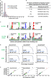
Characteristics of human leukocyte antigen (HLA) class I-bound peptides identified by LC-MS/MS. HLA class I complexes were immunoprecipitated from uninfected C8166 cells (HLA type: A*01:01, B*08:01, B*44:02, C*05:01, C*07:01) using the W6/32 antibody, then peptides were sequenced by LC-MS/MS. (A) Length distribution of peptides identified in the uninfected C8166 replicate #1 at false discovery rates (FDRs) of 0.1, 1, and 5%. (B) All 8–13-mer peptides identified at the indicated FDRs were tested in silico for binding to the HLA class I alleles expressed by C8166 cells using NetMHC4.0. Peptides were classified as “binders” when they were predicted to bind with a higher affinity than 98% of 400,000 randomly generated peptides (i.e., 2% rank or lower). “5% unique” indicates those peptides identified at 5% FDR but not at 1% FDR. (C) All 8–13-mers identified at 5% FDR or those identified only at 5% FDR (not at 1% FDR) in the uninfected replicate #1 sample were clustered using the online GibbsCluster algorithm. Each cluster identified by GibbsCluster is represented by a sequence logo, which corresponds to the indicated HLA class I allele expressed by C8166 cells. The number (n) of peptides in each cluster is also shown. In the sequence logo, amino acids are represented by their single letter code. The more frequently an amino acid occurs at a position within peptides, the larger the letter is displayed. (D) Binding affinity to HLA-A*01:01, HLA-B*08:01, or HLA-B*44:02 was predicted for all 8–13-mers identified at 5% FDR in the uninfected replicate #1 sample (top row) or for all 8–13-mers listed as restricted by each allele in Immune Epitope Database (IEDB) (bottom row). The number of peptides (n) is indicated along with the affinity threshold at which 95% of peptides bound or the proportion of peptides, which bound with higher affinity than 500 nM (a limit set in previous iterations of the NetMHC algorithm). (E) All 8–13-mer peptides identified at the indicated FDR were tested in silico for binding to the HLA class I alleles expressed by C8166 cells using NetMHC4.0. The amino acid frequency for peptides predicted to bind to one of HLA-A*01:01, HLA-B*08:01, or HLA-B*44:02 in the uninfected C8166 replicate #1 was plotted against the amino acid frequency in peptides assigned to each allele in IEDB.
Figure 4
Figure 4
CD4.221 cells express and present peptides on human leukocyte antigen (HLA)-C*01:02. CD4.221 cells were infected with human immunodeficiency virus type 1 IIIB, then lysed and W6/32-conjugated resin was used for immunoprecipitation (IP). Peptides identified were compared to those eluted from W6/32 IPs of 721.221 cells reported by Abelin et al. (36) and peptides reported as HLA-C*01:02-restricted in Immune Epitope Database (IEDB). (A) Length distributions of peptides eluted from CD4.221 cells or 721.221 cells. (B) Sequence logos of 9-mer peptides eluted from CD4.221 cells, 721.221 cells, or 9-mers reported as C*01:02-restricted in IEDB. (C) The protein sequence of the HLA-C*01:02 gene amplified and sequenced from genomic CD4.221 DNA is shown. Highlighted in red is the leader sequence identified in the W6/32 IP from CD4.221 lysates. Highlighted in blue are peptides which were identified in a proteomic analysis of CD4.221 cell lysate and which do not correspond to sequences from HLA-E or HLA-F sequence (which are also expressed by CD4.221 cells).
Figure 5
Figure 5
Some non-specific pull-down of human leukocyte antigen (HLA) molecules occurs in membrane protein IPs. (A) Flow cytometry plots showing HLA-DR expression was measured by cell surface staining with L243 (blue lines) of C8166, CD4.221, peripheral blood mononuclear cells, or T2 cells. Doublets and dead cells were removed from analyses. Unstained controls are indicated by the gray filled histograms. Plots are gated on live single cells. (B) CD4.221, T2, or C8166 cells were infected with human immunodeficiency virus type 1 IIIB for 3 days, then cells were lysed and IP was performed with W6/32-, HB-65- (C8166 only), or OKT4-conjugated resin. Purified peptides were sequenced by LC-MS/MS. The length distributions of peptides eluted from each cell lysate with the indicated antibody are shown. Inset: same data scaled on a reduced y-axis. Data shown are representative of two separate experiments. (C) Sequence logos of 9-mers identified from the same samples as in (B).
Figure 6
Figure 6
Human immunodeficiency virus type 1 (HIV-1) peptides are non-specifically co-purified from HIV-1-infected cells by human leukocyte antigen class I immunoprecipitation (IP). C8166, CD4.221, and T2 cells were infected with HIV-1 IIIB, then lysed and W6/32-, HB-65- (C8166 only), or OKT4-conjugated resin was used for IP. Purified peptides were sequenced by LC-MS/MS. (A) Schematic summary of HIV-1 peptides identified in each sample tested in this study. Numbers above and below the schematic indicate the start and end nucleotide locations of the nine conventional genes encoded by HIV-1 reference strain HXB2. Peptides (or nested sets of peptides) identified in each sample (indicated on the right of each rectangular box) are illustrated as a gray stripe. Diagram adapted from the Los Alamos National Laboratory HIV-1 QuickAlign tool. (B) Length distributions of HIV-1 peptides identified in each sample.

Similar articles

Cited by

References

    1. Borrow P, Lewicki H, Hahn BH, Shaw GM, Oldstone MB. Virus-specific CD8+ cytotoxic T-lymphocyte activity associated with control of viremia in primary human immunodeficiency virus type 1 infection. J Virol (1994) 68(9):6103–10. - PMC - PubMed
    1. Koup RA, Safrit JT, Cao Y, Andrews CA, McLeod G, Borkowsky W, et al. Temporal association of cellular immune responses with the initial control of viremia in primary human immunodeficiency virus type 1 syndrome. J Virol (1994) 68(7):4650–5. - PMC - PubMed
    1. Jones NA, Wei X, Flower DR, Wong M, Michor F, Saag MS, et al. Determinants of human immunodeficiency virus type 1 escape from the primary CD8+ cytotoxic T lymphocyte response. J Exp Med (2004) 200(10):1243–56.10.1084/jem.20040511 - DOI - PMC - PubMed
    1. Goonetilleke N, Liu MKP, Salazar-Gonzalez JF, Ferrari G, Giorgi E, Ganusov VV, et al. The first T cell response to transmitted/founder virus contributes to the control of acute viremia in HIV-1 infection. J Exp Med (2009) 206(6):1253–72.10.1084/jem.20090365 - DOI - PMC - PubMed
    1. Borrow P, Lewicki H, Wei X, Horwitz MS, Peffer N, Meyers H, et al. Antiviral pressure exerted by HIV-l-specific cytotoxic T lymphocytes (CTLs) during primary infection demonstrated by rapid selection of CTL escape virus. Nat Med (1997) 3(2):205–11.10.1038/nm0297-205 - DOI - PubMed

Publication types

MeSH terms

Substances