Mutation of the S and 3c genes in genomes of feline coronaviruses
- PMID: 29769478
- PMCID: PMC6068308
- DOI: 10.1292/jvms.17-0704
Mutation of the S and 3c genes in genomes of feline coronaviruses
Abstract
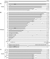
Feline coronavirus (FCoV) is classified into two biotypes based on its pathogenicity in cats: a feline enteric coronavirus of low pathogenicity and a highly virulent feline infectious peritonitis virus. It has been suspected that FCoV alters its biotype via mutations in the viral genome. The S and 3c genes of FCoV have been considered the candidates for viral pathogenicity conversion. In the present study, FCoVs were analyzed for the frequency and location of mutations in the S and 3c genes from faecal samples of cats in an animal shelter and the faeces, effusions, and tissues of cats that were referred to veterinary hospitals. Our results indicated that approximately 95% FCoVs in faeces did not carry mutations in the two genes. However, 80% FCoVs in effusion samples exhibited mutations in the S and 3c genes with remainder displaying a mutation in the S or 3c gene. It was also suggested that mutational analysis of the 3c gene could be useful for studying the horizontal transmission of FCoVs in multi-cat environments.
Keywords: 3c gene; S gene; feline coronavirus; multi-cat environment; mutation.
Figures


Similar articles
-
Limitations of using feline coronavirus spike protein gene mutations to diagnose feline infectious peritonitis.Vet Res. 2017 Oct 5;48(1):60. doi: 10.1186/s13567-017-0467-9. Vet Res. 2017. PMID: 28982390 Free PMC article.
-
Feline Coronavirus 3c Protein: A Candidate for a Virulence Marker?Biomed Res Int. 2016;2016:8560691. doi: 10.1155/2016/8560691. Epub 2016 May 3. Biomed Res Int. 2016. PMID: 27243037 Free PMC article.
-
Feline Coronaviruses: Pathogenesis of Feline Infectious Peritonitis.Adv Virus Res. 2016;96:193-218. doi: 10.1016/bs.aivir.2016.08.002. Epub 2016 Aug 31. Adv Virus Res. 2016. PMID: 27712624 Free PMC article. Review.
-
Feline coronavirus with and without spike gene mutations detected by real-time RT-PCRs in cats with feline infectious peritonitis.J Feline Med Surg. 2020 Aug;22(8):791-799. doi: 10.1177/1098612X19886671. Epub 2019 Nov 15. J Feline Med Surg. 2020. PMID: 31729897 Free PMC article.
-
A Tale of Two Viruses: The Distinct Spike Glycoproteins of Feline Coronaviruses.Viruses. 2020 Jan 10;12(1):83. doi: 10.3390/v12010083. Viruses. 2020. PMID: 31936749 Free PMC article. Review.
Cited by
-
FCoV Viral Sequences of Systemically Infected Healthy Cats Lack Gene Mutations Previously Linked to the Development of FIP.Pathogens. 2020 Jul 24;9(8):603. doi: 10.3390/pathogens9080603. Pathogens. 2020. PMID: 32722056 Free PMC article.
-
Animal coronaviruses and SARS-CoV-2.Transbound Emerg Dis. 2021 May;68(3):1097-1110. doi: 10.1111/tbed.13791. Epub 2020 Sep 9. Transbound Emerg Dis. 2021. PMID: 32799433 Free PMC article. Review.
-
Phylogenetic Analysis of Alphacoronaviruses Based on 3c and M Gene Sequences Isolated from Cats with FIP in Romania.Microorganisms. 2024 Jul 30;12(8):1557. doi: 10.3390/microorganisms12081557. Microorganisms. 2024. PMID: 39203398 Free PMC article.
References
MeSH terms
LinkOut - more resources
Full Text Sources
Other Literature Sources
Miscellaneous

