. 2018 Jul 17;92(15):e00607-18.
doi: 10.1128/JVI.00607-18. Print 2018 Aug 1.

Bag2 Is a Component of a Cytosolic Extraction Machinery That Promotes Membrane Penetration of a Nonenveloped Virus

Affiliations

Bag2 Is a Component of a Cytosolic Extraction Machinery That Promotes Membrane Penetration of a Nonenveloped Virus

Allison Dupzyk et al. J Virol. .

Abstract

During entry, the nonenveloped polyomavirus (PyV) simian virus 40 (SV40) traffics from the cell surface to the endoplasmic reticulum (ER), where it penetrates the ER membrane to reach the cytosol; the virus is then transported into the nucleus to cause infection. Although a coherent understanding of SV40's host entry is emerging, how the virus is ejected from the ER into the cytosol remains mysterious. Our previous analyses revealed that the cytosolic Hsc70-SGTA-Hsp105 complex binds to SV40 and extracts it from the ER into the cytosol. We now report that the nucleotide exchange factor (NEF) Bag2 stimulates SV40 release from Hsc70, thereby enabling successful virus arrival at the cytosol, which leads to infection. Hsp105, another NEF of Hsc70, displays a function overlapping that of Bag2, underscoring the importance of this release reaction. Our findings identify a new component of an extraction machinery essential during membrane penetration of a nonenveloped virus and provide further mechanistic insights into this process.IMPORTANCE How a nonenveloped virus penetrates a biological membrane to cause infection is a mystery. For the nonenveloped polyomavirus SV40, transport across the ER membrane to reach the cytosol is an essential virus infection step. Here, we identify a novel component of a cytosolic Hsc70-dependent chaperone complex called Bag2 that extracts SV40 from the ER into the cytosol. Bag2 does this by triggering SV40 release from Hsc70, thus ensuring that the virus reaches the cytosol en route for productive infection.

Keywords: endoplasmic reticulum; membrane transport; nonenveloped virus; protein chaperone; simian virus 40.


Figures

FIG 1
FIG 1
Bag2 binds to ER membrane J proteins. (A) Total numbers of peptides corresponding to B12, C18, and Bag2 identified by mass spectrometry using 3×FLAG-B12- or 3×FLAG-C18-immunoprecipitated material from HEK 293T cells. (B) HEK 293T cells were transfected with either GFP-S, B12-S, B14-S, or C18-S. The cells were lysed, and the resulting cell extracts were subjected to affinity purification and SDS-PAGE, followed by immunoblotting using the indicated antibodies. Input represents 8% of the total sample used for affinity purification. (C) HEK 293T cell extracts were incubated with an anti-B12 or a control IgG antibody. The immunoprecipitated (IP) materials were subjected to SDS-PAGE, followed by immunoblotting using the indicated antibodies. (D) As for panel C, except that HEK 293T cells were transfected with GFP-FLAG, B12-FLAG, or QPD B12-FLAG, and the cell extracts were incubated with FLAG antibody-conjugated agarose beads. The asterisk indicates an unidentified protein that cross-reacted with the Hsc70 antibody. (E) As for panel C, except that HEK 293T cells were transfected with GFP-FLAG, WT Bag2*-FLAG, or I160A Bag2*-FLAG, and the cell extracts were incubated with FLAG antibody-conjugated agarose beads.
FIG 2
FIG 2
Bag2 promotes SV40 infection. (A) siRNA knockdown of Bag2 and Hsp105. Cell extracts derived from CV-1 cells transfected with the indicated siRNA(s) were subjected to SDS-PAGE and immunoblotting with the indicated antibodies. The asterisk indicates an unidentified protein that cross-reacted with the actin antibody. (B) CV-1 cells transfected with the indicated siRNAs were infected with SV40; 24 hpi, the cells were permeabilized, fixed, and stained for large T antigen. At least 300 cells were counted per condition over three biological replicates. The graph represents means and SD. Student's two-tailed t test was used to determine statistical significance. (C) As for panel B, except cells were transfected with the indicated constructs 24 h prior to infection with SV40. The cells were then fixed, permeabilized, and stained for large T antigen and FLAG. Only cells expressing the FLAG construct were counted. At least 100 cells were counted per condition over three biological replicates. The graph represents means and SD. Student's two-tailed t test was used to determine statistical significance.
FIG 3
FIG 3
Bag2 is important for ER-to-cytosol transport of SV40. (A) CV-1 cells transfected with the indicated siRNAs were infected with SV40 and subjected to semipermeabilization. The resulting cytosol-, membrane-, and ER-localized fractions were subjected to SDS-PAGE and immunoblotting with the indicated antibodies. The amount loaded for the cytosol was 50% of the total cytosol fraction, whereas the amount loaded for the membrane was 20% of the total membrane fraction. Hsp90 and PDI acted as both loading and fractionation controls. (B) The VP1 band intensity in the cytosol fraction from panel A was quantified with Image J software (National Institutes of Health), normalized relative to the Hsp90 loading control bands, and graphed as a percentage of the VP1 band intensity in the scrambled-siRNA-treated sample. The graph represents the means and SD from at least 3 biological replicates. Student's two-tailed t test was used to determine statistical significance.
FIG 4
FIG 4
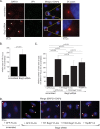
Depletion of Bag2 traps SV40 in the ER-to-cytosol membrane penetration site. (A) CV-1 cells were transfected with the indicated siRNAs for 48 h, followed by 16 h of SV40 infection. The cells were fixed, permeabilized, stained using anti-BAP31 and anti-VP1 antibodies, and imaged by fluorescence microscopy. The enlarged images (3× zoom) correspond to the boxed areas in the merged images. The arrowheads indicate BAP31 foci. (B) The size of the VP1 focus was quantified based on the measured area (in pixels) using Image J software. The graph represents the means and SD of at least 30 cells counted in at least three biological replicates. Student's two-tailed t test was used to determine statistical significance. (C) As for panel A, except cells were transfected with the indicated constructs 24 h prior to infection with SV40. The cells were then fixed, permeabilized, and stained using anti-BAP31, anti-FLAG, or anti-S antibodies. Only cells expressing the indicated FLAG- or S-tagged constructs were counted. The data plotted are the numbers of cells with at least one BAP31 focus, graphed as a percentage of the scrambled siRNA-treated sample. The graph represents the means and SD of at least 100 cells counted in at least three biological replicates. Student's two-tailed t test was used to determine statistical significance. (D) Representative images of the data in panel C. The arrowheads indicate BAP31 foci. The asterisks indicate cells expressing the indicated constructs.
FIG 5
FIG 5
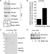
Bag2 releases SV40 from Hsc70. (A) COS-7 cells were treated with the indicated siRNAs for 24 h, followed by transfection of the Hsc70-S construct. After 24 h, the cells were infected with SV40 for 16 h, and the resulting cell extracts were subjected to affinity purification, SDS-PAGE, and immunoblotting with the indicated antibodies. (B) The VP1 band intensity from panel A was quantified with Image J software (National Institutes of Health), normalized relative to the affinity-purified Hsc70-S level, and graphed as a percentage of the VP1 band intensity in the scrambled-siRNA-treated sample. The graph represents the means and SD from three biological replicates. Student's two-tailed t test was used to determine statistical significance. (C) Coomassie staining of commercially available Hsc70 and Bag2, along with purified SV40. Note that the level of VP2 in the SV40 preparation is below the threshold of detection. (D) After SV40 was incubated with Hsc70 (in the presence of 1 mM DTT), precipitation of SV40 VP1 coprecipitated Hsc70. The SV40-Hsc70 complex was incubated with a control buffer containing only ATP, or the indicated amount of Bag2 with ATP, followed by reimmunoprecipitation of SV40 VP1. The reimmunoprecipitated material was subjected to SDS-PAGE followed by silver staining.
FIG 6
FIG 6
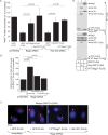
Bag2 and Hsp105 play overlapping roles. (A) CV-1 cells were transfected with the indicated siRNAs for 24 h, followed by DNA transfection of the indicated FLAG constructs. After 24 additional hours, the cells were infected with SV40 for 24 h, fixed, permeabilized, and stained for FLAG and large T antigen. Only cells expressing the indicated FLAG constructs were counted. At least 100 cells were counted per condition over three biological replicates. The graph represents means and SD. Student's two-tailed t test was used to determine statistical significance. (B) As for panel A, except cells were infected with SV40 for 16 h prior to being fixed, permeabilized, and stained for FLAG and BAP31. Only cells expressing the indicated FLAG constructs were counted. The graph represents the means and SD of at least 100 cells counted in at least three biological replicates. (C) Representative images of the data in panel B. The arrowheads indicate BAP31 foci. The asterisks indicate cells expressing the indicated constructs. (D) HEK 293T cells were transfected with the indicated FLAG-tagged constructs and lysed, and the resulting cell extract was subjected to immunoprecipitation, SDS-PAGE, and immunoblotting with the indicated antibodies.
FIG 7
FIG 7
The Hsc70-SGTA-Bag2 complex docks on an ER membrane J protein and promotes SV40 cytosol entry. In our model, Hsc70-containing protein complexes dock on an ER membrane J protein, such as B12. In association with its cochaperone, SGTA, an Hsc70 molecule recruits Bag2 or Hsp105, forming either the Hsc70-SGTA-Bag2 or Hsc70-SGTA-Hsp105 cytosolic extraction complex. During the final step of SV40's ER-to-cytosol membrane transport, a single membrane-embedded viral particle recruits many copies of either the Hsc70-SGTA-Bag2 or Hsc70-SGTA-Hsp105 protein complex. Iterative cycles of SV40 binding to and release from Hsc70 in turn provide the energy to extract the virus into the cytosol. (Inset) Importantly, Bag2 functions to trigger SV40 release from Hsc70 in this cycle. Bag2 does this by promoting ADP release from Hsc70, generating ATP-Hsc70, which displays low affinity for its substrate, such as SV40. “Focus” refers to the SV40 cytosol entry site in the ER membrane, where the cytosolic extraction machinery is recruited to the ER membrane J protein.

Similar articles

Cited by

References

    1. Poranen MM, Daugelavicius R, Bamford DH. 2002. Common principles in viral entry. Annu Rev Microbiol 56:521–538. doi:10.1146/annurev.micro.56.012302.160643. - DOI - PubMed
    1. Tsai B. 2007. Penetration of nonenveloped viruses into the cytoplasm. Annu Rev Cell Dev Biol 23:23–43. doi:10.1146/annurev.cellbio.23.090506.123454. - DOI - PubMed
    1. Walczak CP, Ravindran MS, Inoue T, Tsai B. 2014. A cytosolic chaperone complexes with dynamic membrane J-proteins and mobilizes a nonenveloped virus out of the endoplasmic reticulum. PLoS Pathog 10:e1004007. doi:10.1371/journal.ppat.1004007. - DOI - PMC - PubMed
    1. Ravindran MS, Bagchi P, Inoue T, Tsai B. 2015. A non-enveloped virus hijacks host disaggregation machinery to translocate across the endoplasmic reticulum membrane. PLoS Pathog 11:e1005086. doi:10.1371/journal.ppat.1005086. - DOI - PMC - PubMed
    1. Dalianis T, Hirsch HH. 2013. Human polyomaviruses in disease and cancer. Virology 437:63–72. doi:10.1016/j.virol.2012.12.015. - DOI - PubMed

Publication types

MeSH terms

LinkOut - more resources