Comparative Study
. 2018 Sep 1;114(11):1499-1511.
doi: 10.1093/cvr/cvy110.

PDE4 and mAKAPβ are nodal organizers of β2-ARs nuclear PKA signalling in cardiac myocytes

Affiliations
Comparative Study

PDE4 and mAKAPβ are nodal organizers of β2-ARs nuclear PKA signalling in cardiac myocytes

Ibrahim Bedioune et al. Cardiovasc Res. .

Abstract

Aims: β1- and β2-adrenergic receptors (β-ARs) produce different acute contractile effects on the heart partly because they impact on different cytosolic pools of cAMP-dependent protein kinase (PKA). They also exert different effects on gene expression but the underlying mechanisms remain unknown. The aim of this study was to understand the mechanisms by which β1- and β2-ARs regulate nuclear PKA activity in cardiomyocytes.

Methods and results: We used cytoplasmic and nuclear targeted biosensors to examine cAMP signals and PKA activity in adult rat ventricular myocytes upon selective β1- or β2-ARs stimulation. Both β1- and β2-AR stimulation increased cAMP and activated PKA in the cytoplasm. Although the two receptors also increased cAMP in the nucleus, only β1-ARs increased nuclear PKA activity and up-regulated the PKA target gene and pro-apoptotic factor, inducible cAMP early repressor (ICER). Inhibition of phosphodiesterase (PDE)4, but not Gi, PDE3, GRK2 nor caveolae disruption disclosed nuclear PKA activation and ICER induction by β2-ARs. Both nuclear and cytoplasmic PKI prevented nuclear PKA activation and ICER induction by β1-ARs, indicating that PKA activation outside the nucleus is required for subsequent nuclear PKA activation and ICER mRNA expression. Cytoplasmic PKI also blocked ICER induction by β2-AR stimulation (with concomitant PDE4 inhibition). However, in this case nuclear PKI decreased ICER up-regulation by only 30%, indicating that other mechanisms are involved. Down-regulation of mAKAPβ partially inhibited nuclear PKA activation upon β1-AR stimulation, and drastically decreased nuclear PKA activation upon β2-AR stimulation in the presence of PDE4 inhibition.

Conclusions: β1- and β2-ARs differentially regulate nuclear PKA activity and ICER expression in cardiomyocytes. PDE4 insulates a mAKAPβ-targeted PKA pool at the nuclear envelope that prevents nuclear PKA activation upon β2-AR stimulation.


Figures
Figure 1
Figure 1
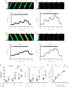
Stimulation of β1- and β2-ARs induce differential activation of cytoplasmic and nuclear PKA activity in ARVMs. A–D Representative time course of cytoplasmic and nuclear PKA activities reported by the normalized yellow fluorescent protein (YFP)/cyan fluorescent protein (CFP) ratio in ARVMs transduced with Ad.AKAR3-NES (A and C) or Ad.AKAR3-NLS (B and D) for 24 h at a multiplicity of infection (MOI) of 1000 active viral particles/cell. β1-AR stimulation was achieved using a combination of 10 nM Iso and 10 nM ICI 118, 551 (ICI) (A and B); β2-AR stimulation using 100 nM Iso in combination with 100 nM CGP 20712A (CGP) (C and D). Pseudo-colour images of the YFP/CFP ratio were recorded at the times indicated by the letters on the graphs. Scale bars represent 20 µm. (E and F) Mean variation (±S.E.M.) of the YFP/CFP ratio in ARVMs expressing either AKAR3-NES or AKAR3-NLS upon β1-AR stimulation using 1, 3 and 10 nM Iso in combination with 10 nM ICI (E) or β2-AR stimulation using 10, 30, and 100 nM Iso in combination with 100 nM CGP (F). Number of cells/animals is indicated in brackets. Statistical significance is indicated as ***P < 0.001 vs. ICI+Iso 1 nM or CGP+Iso 10 nM in the cytoplasm, $$P < 0.01 vs. ICI+Iso 1 nM in the nucleus, ###P < 0.001 by nested ANOVA with Tukey’s post hoc test. (G) Nuclear PKA activation (% increase in YFP/CFP ratio in ARVMs expressing AKAR3-NLS) is plotted as a function of cytoplasmic PKA activation (% increase in YFP/CFP ratio in ARVMs expressing AKAR3-NES) for β1- and β2-AR stimulations. Values (±S.E.M.) of (E and F) were used for this graph.
Figure 2
Figure 2
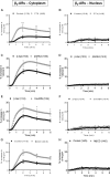
Both β1- and β2-AR increase cytoplasmic and nuclear cAMP levels in ARVMs. Representative time course of the normalized CFP/YFP ratio upon selective β1-AR stimulation with Iso (1, 3, and 10 nM) in combination with 10 nM ICI (A and B) or β2-AR stimulation with Iso (10, 30 and 100 nM) in combination with 100 nM CGP (C and D) in ARVMs transduced with Ad.Epac-SH187(A and C) or Ad.Epac-SH187-3NLS (B and D) for 24 h at a MOI of 1000 active viral particles/cell. Pseudo-colour images of the CFP/YFP ratio were recorded at the times indicated by the letters on the graphs. Scale bars represent 20 µm. (E and F) Mean variation (±S.E.M.) of the CFP/YFP ratio in ARVMs expressing either Epac-SH187 or Epac-SH187-3NLS upon β1-AR stimulation (E) or β2-AR stimulation (F). Number of cells/animals is indicated in brackets. Statistical significance is indicated as * P < 0.05; ** P < 0.01; *** P < 0.001 vs. ICI+Iso 1 nM or CGP+Iso 10 nM in the cytoplasm, $$P < 0.01, $$$P < 0.001 vs. ICI+Iso 1 nM or CGP+Iso 10 nM in the nucleus, #P< 0.05, ###P < 0.001, £££P < 0.001 by nested ANOVA with Tukey’s post-hoc test. (G) Nuclear cAMP elevation (% increase in CFP/YFP ratio in ARVMs expressing Epac-SH187-3NLS) is plotted as a function of cytoplasmic cAMP elevation (% increase in CFP/YFP ratio in ARVMs expressing Epac-SH187) in response to either β1- or β2-AR stimulation. Values (±S.E.M.) of (E and F) were used for this graph.
Figure 3
Figure 3
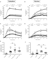
Gi proteins, caveolae and GRK2 regulate cytoplasmic but not nuclear PKA activation in response to β2-ARs stimulation. Average time course of the YFP/CFP ratio upon β2-AR stimulation in ARVMs expressing AKAR3-NES (A, C, E, and G) or AKAR3-NLS (B, D, F, and H). (A and B) ARVMs treated or not with PTX (1.5 µg/ml, 2 h) were exposed to 10 nM Iso plus 100 nM CGP to stimulate β2-ARs. In all other protocols, β2-ARs were stimulated with 30 nM Iso plus 100 nM CGP. (C and D) ARVMs were treated or not with 2 mM MβCD for 1 h. (E) ARVMs were co-transduced with Ad.AKAR3-NES (MOI 200) and Ad.β-Galactosidase (β-Gal, MOI 2000) or Ad.AKAR3-NES and an adenovirus encoding a dominant-negative Cav3 mutant (Ad. Cav3DN, MOI 2000) for 48 h. (F) ARVMs were co-transduced with Ad.AKAR3-NLS and Ad. β-Gal or Ad.AKAR3-NLS and Ad.Cav3DN for 48 h. (G) ARVMs were co-transduced with Ad.AKAR3-NES (MOI 200) and Ad.β-Gal (MOI 1000) or Ad.AKAR3-NES (MOI 200) and Ad.βARK-ct (MOI 1000) for 48 h. (H) ARVMs were co-transduced with Ad.AKAR3-NLS (MOI 200) and Ad.β-Gal (MOI 1000) or Ad.AKAR3-NLS (MOI 200) and Ad.βARK-ct (MOI 1000) for 48 h. In each panel, the number of cells/animals is indicated in brackets for the different experimental conditions. Statistical significance is indicated as *P < 0.05; **P , 0.01; ***P < 0.001 by nested ANOVA with Tukey’s post-hoc test.
Figure 4
Figure 4
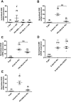
PDE4 is predominant for regulation of β1- and β2-AR induced cytoplasmic and nuclear PKA activation. (A and B) Average variation of the YFP/CFP ratio upon β1-AR stimulation using 1 nM Iso plus 10 nM ICI alone or in the presence of 1 µM cilostamide (Cil), a PDE3 inhibitor, or 10 µM Ro-201724 (Ro) a PDE4 inhibitor in ARVMs transduced with Ad.AKAR3-NES (A) or Ad.AKAR3-NLS (B) at MOI 1000 for 24 h. (C and D) Average variation of the YFP/CFP ratio upon β2-AR stimulation using 30 nM Iso plus 100 nM CGP alone or in the presence of 1 µM Cil or 10 µM Ro in ARVMs transduced with Ad.AKAR3-NES (C) or Ad.AKAR3-NLS (D) at MOI 1000 for 24 h. Number of cells/animals is indicated in brackets. Statistical significance is indicated as *P < 0.05; **P < 0.01; ***P < 0.001; $$P < 0.01 vs. β1- or β2-AR by nested ANOVA with Tukey’s post-hoc test. (E and F) ICER mRNA expression in ARVMs in primary culture for 24 h and stimulated or not by β1-ARs (100 nM Iso plus 10 nM ICI during 2 h) or β2-ARs (100 nM Iso plus 100 nM CGP during 2 h) alone or in combination with 10 µM Ro or 1 µM Cil. Number of animals is indicated in brackets. Statistical significance is indicated as ***P < 0.001 vs. control; $$P < 0.01, $$$P < 0.001 by Kruskal-Wallis test with Dunn’s post-hoc test (E) or one-way ANOVA with Tukey’s post-hoc test (F).
Figure 5
Figure 5
β1-ARs and β2-ARs differentially regulate the PKA target gene, ICER in cardiomyocytes. (A) ICER mRNA expression in ARVMs transduced during 24 h with Ad.β-Gal (MOI 250) with or without β1-AR stimulation (100 nM Iso plus 10 nM ICI during 2 h), Ad. PKI-NES (MOI 250) with β1-AR stimulation (100 nM Iso plus 10 nM ICI during 2 h) and Ad.PKI-3NLS (MOI 250) with β1-AR stimulation (100 nM Iso plus 10 nM ICI during 2 h). (B) Effects of β1-AR stimulation (100 nM Iso plus 10 nM ICI during 2 h) with concomitant PDE4 inhibition (Ro, 10 µM) on ICER mRNA expression in ARVMs transduced during 24 h with Ad.β-Gal (MOI 250) or Ad.PKI-3NLS (MOI 250). (C) Effects of β2-AR stimulation (100 nM Iso plus 100 nM CGP during 2 h) with concomitant PDE4 inhibition (Ro, 10 µM) on ICER mRNA expression in ARVMs transduced during 24 h with Ad.β-Gal (MOI 250) or Ad.PKI-3NLS (MOI 250). (D) Effects of β2-AR stimulation (100 nM Iso plus 100 nM CGP during 2 h) with concomitant PDE4 inhibition (Ro, 10 µM) on ICER mRNA expression in ARVMs transduced during 24 h with Ad.β-Gal (MOI 250) and pre-treated or not with 10 µM CE3F4 during 15 min at 37°C. (E) Effects of β2-AR stimulation (100 nM Iso plus 100 nM CGP during 2 h) with concomitant PDE4 inhibition (Ro, 10 µM) on ICER mRNA expression in ARVMs transduced during 24 h with Ad.β-Gal (MOI 250) or Ad.PKI-NES (MOI 250). Number of rats is indicated in the respective panels. Statistical significance is indicated as **P < 0.01; ***P < 0.001 vs. β-Gal; $$$P < 0.001 by Kruskal-Wallis test with Dunn’s post-hoc test.
Figure 6
Figure 6
The scaffold protein mAKAPβ controls β2-AR induced nuclear PKA activity when PDE4 is inhibited. (A) Immunocytochemical detection of mAKAPβ in ARVMs 72 h after sequential infection with adenoviruses encoding either a scrambled shRNA (Ad.Control shRNA MOI 2000) or a shRNA against mAKAPβ (Ad-mAKAPβ shRNA MOI 2000) for 48 h followed by infection with Ad.AKAR3-NLS (MOI 1000) for 24 h. Scale bars represent 20 µm. (B) Quantification of mAKAPβ fluorescence in ARVMs co-transduced with Ad.AKAR3-NLS and Ad.Control shRNA or Ad-mAKAPβ shRNA at 72 h. (C) Mean variation of the YFP/CFP ratio upon β1-AR stimulation with 3 nM Iso plus 10 nM ICI in ARVMs co-transduced with Ad.AKAR3-NLS and Ad.Control shRNA or Ad.AKAR3-NLS and Ad.mAKAPβ shRNA. (D) Mean variation of the YFP/CFP ratio upon β1-AR stimulation with 1 nM Iso plus 10 nM ICI in the presence of 10 µM Ro 201724 (Ro) to block PDE4. ARVMs were co-transduced with Ad.AKAR3-NLS and Ad.Control shRNA or with Ad.AKAR3-NLS and Ad.mAKAPβ shRNA. Mean variation of the YFP/CFP ratio upon β2-AR stimulation (using 30 nM Iso plus 100 nM CGP) alone (E) or in the presence of 10 µM Ro (F) in ARVMs co-transduced with Ad.AKAR3-NLS and Ad.Control shRNA or with Ad.AKAR3-NLS Ad-mAKAPβ shRNA. Number of cells/rats is indicated in brackets. Statistical significance is indicated as *P < 0.05; **P < 0.01 by nested ANOVA with Tukey’s post-hoc test.
Figure 7
Figure 7
Proposed model for β1- and β2-AR regulation of cytoplasmic and nuclear PKA activity and ICER expression in adult cardiac myocytes. Stimulation of β1-ARs generate cAMP signals (in red) diffusing in the cytoplasm and the nucleus. Upon β1-ARs stimulation, PDE4 (in green) regulates cAMP levels to control PKA activity in the cytoplasm. A fraction of catalytic subunits (C) of PKA dissociate from regulatory subunits (R) and translocate inside the nucleus to increase nuclear PKA activity. Elevation of nuclear PKA activity allows induction of ICER transcription, presumably through CREB phosphorylation, which may be direct or indirect (dotted arrow between C and CREB). Stimulation of β2-ARs also generates cAMP elevation in the cytoplasm and the nucleus resulting in activation of cytoplasmic PKA. Cytoplasmic PKA activation upon β2-AR stimulation is restricted by caveolin3, Gi, GRK2, PDE3, and PDE4. In addition, PDE4 prevents activation by β2-ARs of a specific pool of PKA tethered by mAKAPβ at the perinuclear membrane (illustrated by the dotted line surrounding the mAKAPβ-PKA-PDE4 complex) which controls access of C subunits to the nucleus (dotted arrow) and nuclear PKA activation by β2-ARs. When PDE4 is inhibited, nuclear PKA activation contributes to ICER up-regulation by β2-ARs, although other mechanisms depending on cytoplasmic PKA activity are involved.

Similar articles

Cited by

References

    1. Xiao RP, Lakatta EG.. Beta 1-adrenoceptor stimulation and beta 2-adrenoceptor stimulation differ in their effects on contraction, cytosolic Ca2+, and Ca2+ current in single rat ventricular cells. Circ Res 1993;73:286–300. - PubMed
    1. Milano CA, Allen LF, Rockman HA, Dolber PC, McMinn TR, Chien KR, Johnson TD, Bond RA, Lefkowitz RJ.. Enhanced myocardial function in transgenic mice overexpressing the beta 2-adrenergic receptor. Science 1994;264:582–586. - PubMed
    1. Engelhardt S, Hein L, Wiesmann F, Lohse MJ.. Progressive hypertrophy and heart failure in beta1-adrenergic receptor transgenic mice. Proc Natl Acad Sci U S A 1999;96:7059–7064. - PMC - PubMed
    1. Communal C, Singh K, Sawyer DB, Colucci WS.. Opposing effects of beta(1)- and beta(2)-adrenergic receptors on cardiac myocyte apoptosis: role of a pertussis toxin-sensitive G protein. Circulation 1999;100:2210–2212. - PubMed
    1. Xiao RP. Beta-adrenergic signaling in the heart: dual coupling of the beta2-adrenergic receptor to G(s) and G(i) proteins. Sci STKE 2001;2001:re15. - PubMed

Publication types

MeSH terms

Substances