Comparative Study
. 2018 May 2;14(1):149.
doi: 10.1186/s12917-018-1469-1.

Comparison of the virulence of three H3N2 canine influenza virus isolates from Korea and China in mouse and Guinea pig models

Affiliations
Comparative Study

Comparison of the virulence of three H3N2 canine influenza virus isolates from Korea and China in mouse and Guinea pig models

Xing Xie et al. BMC Vet Res. .

Abstract

Background: Avian-origin H3N2 canine influenza virus (CIV) has been the most common subtype in Korea and China since 2007. Here, we compared the pathogenicity and transmissibility of three H3N2 CIV strains [Chinese CIV (JS/10), Korean CIV (KR/07), and Korean recombinant CIV between the classic H3N2 CIV and the pandemic H1N1 virus (MV/12)] in BALB/c mouse and guinea pig models. The pandemic H1N1 (CA/09) strain served as the control.

Results: BALB/c mice infected with H1N1 had high mortality and obvious body weight loss, whereas no overt disease symptoms were observed in mice inoculated with H3N2 CIV strains. The viral titers were higher in the group MV/12 than those in groups JS/10 and KR/07, while the mice infected with JS/10 showed higher viral titers in all tissues (except for the lung) than the mice infected with KR/07. The data obtained in guinea pigs also demonstrated that group MV/12 presented the highest loads in most of the tissues, followed by group JS/10 and KR/07. Also, direct contact transmissions of all the three CIV strains could be observed in guinea pigs, and for the inoculated and the contact groups, the viral titer of group MV/12 and KR/07 was higher than that of group JS/10 in nasal swabs. These findings indicated that the matrix (M) gene obtained from the pandemic H1N1 may enhance viral replication of classic H3N2 CIV; JS/10 has stronger viral replication ability in tissues as compared to KR/07, whereas KR/07 infected guinea pigs have more viral shedding than JS/10 infected guinea pigs.

Conclusions: There exists a discrepancy in pathobiology among CIV isolates. Reverse genetics regarding the genomes of CIV isolates will be helpful to further explain the virus characteristics.

Keywords: BALB/c mice; Guinea pigs; H3N2 canine influenza virus; Pathogenicity; Transmissibility.


Conflict of interest statement

Ethics approval

This study is not involved human participants. Veterinarians took the samples for analysis purposes and/or to check the health status of the mice and guinea pig population. Before conducting the study, approval for conducting the animal experiments was obtained from the Animal Ethics Committee of Korea University.

Competing interests

The authors declare that they have no competing interests.

Publisher’s Note

Springer Nature remains neutral with regard to jurisdictional claims in published maps and institutional affiliations.

Figures
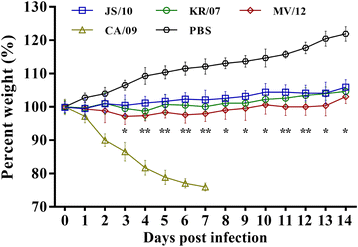
Fig. 1
Fig. 1
Body weight changes in mice inoculated with four influenza virus strains. Four experimental groups of 6-week-old BALB/c mice were challenged with 106 EID50/mL of the JS/10, KR/07, MV/12 and CA/09 strains. Mice inoculated with same volume of PBS served as the negative control. Mice were monitored for body weight loss throughout the observation period for 14 days. Each error bar indicates the standard deviation. The results are expressed in terms of percent body weight. *, P < 0.05, or **, P < 0.01, indicates significantly different weight compared between group JS/10 and MV/12
Fig. 2
Fig. 2
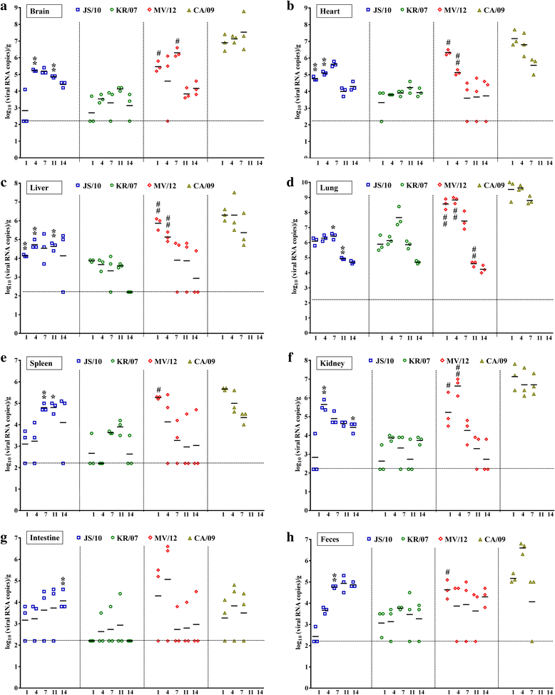
Viral loads in collected tissues and fecal samples from mice at five different time points after infection with four virus strains. Mice were inoculated with 106 EID50/mL of the JS/10, KR/07, MV/12 and CA/09 strains. In each virus group, the brain (a), heart (b), liver (c), lung (d), spleen (e), kidney (f), intestine (g) and feces (h) were collected from the mice to determine the viral loads using real-time PCR at 1, 4, 7, 11 and 14 days post-challenge. *, P < 0.05, or **, P < 0.01, indicates significantly different virus titers compared between group JS/10 and KR/07. #, P < 0.05, or ##, P < 0.01, indicates significantly different virus titers compared between group MV/12 and KR/07. For viral loads in different organs mentioned above, the results are expressed as log10 (viral RNA copies)/g. The horizontal line means the detection limit of this assay (158 copies of RNA per g)
Fig. 3
Fig. 3
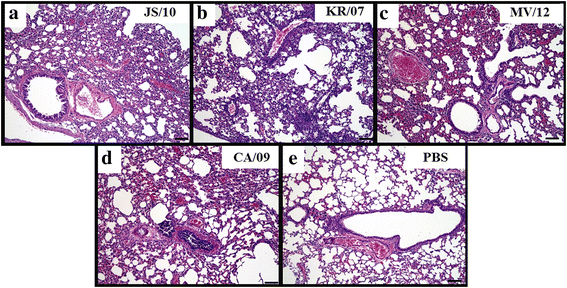
Histopathological lesions in lung samples from mice infected with the four virus strains at 7 dpi. Histopathological findings in the lungs of mice at 7 days post-inoculation with 106 EID50/mL of the JS/10, KR/07, MV/12 and CA/09 strains. All inoculated groups demonstrated histopathological pneumonic lesions. (A) – (E) are representative microscopic images of the histopathological pneumonic lesions from each group (X100). (A) JS/10 and (B) KR/07 resulted in mild lymphocyte infiltration and congestion. (C) MV/12 and (D) CA/09 resulted in mild lymphocyte infiltration and moderate congestion and hemorrhaging. (E) Lung tissue in the normal state
Fig. 4
Fig. 4
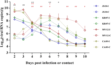
Nasal swab shedding of guinea pigs infected with the four virus strains in both the inoculation and contact groups. Guinea pigs were inoculated with the JS/10, KR/07, MV/12 and CA/09 virus strains. After 24 h on 1 dpi, additional naive guinea pigs were placed into each virus group as the contact group. Nasal swabs were collected every day for determination of the viral loads using real-time PCR and the results are expressed as log10 (viral RNA copies)/g. The solid line represents for the viral inoculation group (-I) and the dotted line represents for the virus contact group (-C) of four viruses. Each error bar indicates the standard deviation. *, P < 0.05, or **, P < 0.01, indicates significantly different virus titers in nasal swabs compared between group JS/10 and KR/07. * in black, represents significant difference in inoculation group, while * in red demonstrates significant difference in contact group. #, P < 0.05, or ##, P < 0.01, indicates a significant difference in virus titers for group MV/12 compared with group KR/07. # in black, represents significant difference in inoculation group, while # in red demonstrates significant difference in contact group. All significant differences are shown above the figure
Fig. 5
Fig. 5
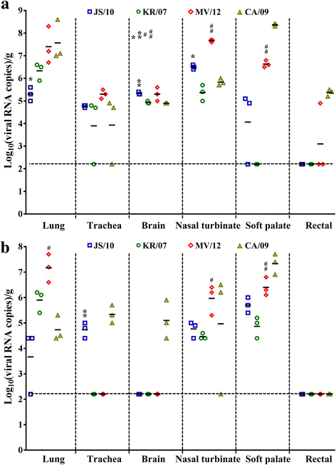
Viral loads in tissues collected from guinea pigs infected with the four virus strains at 3 dpi for the inoculated group and 5 dpe for the contact group. Guinea pigs were inoculated with 106 EID50/mL of the JS/10, KR/07, MV/12 and CA/09 strains. Organs including the lung, trachea, brain, nasal turbinate, soft palate and rectal were collected for the determination of the viral loads using real-time PCR at 3 dpi and 5 dpe, for the inoculated group (a) and contact group (b) for each virus, respectively. The results are expressed as log10 (viral RNA copies)/g. *, P < 0.05, or **, P < 0.01, indicates significantly different virus titers compared between JS/10 and KR/07 virus group. #, P < 0.05, or ##, P < 0.01, indicates a significant difference in virus load for the group MV/12 compared with group KR/07. For viral loads in different organs mentioned above, the results are expressed in terms of mean virus titer logEID50. The horizontal line means the detection limit of this assay (158 copies of RNA per g)
Fig. 6
Fig. 6
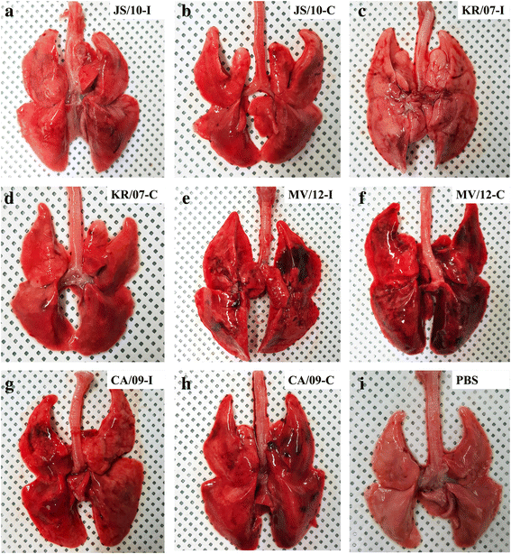
Gross lesions of lung samples from guinea pigs infected with the four virus strains in both the inoculation and contact groups. Guinea pigs were inoculated with 106 EID50/mL of the JS/10, KR/07, MV/12 and CA/09 strains. After 24 h on 1 dpi, additional naïve guinea pigs were placed into each virus group as the contact group. Pictures were taken of the gross lesions of the viral inoculation groups at 3 dpi for JS/10 (a), KR/07 (c), MV/12 (e) and CA/09 (G) and of the virus contact groups at 6 dpi (5 dpe) for JS/10 (b), KR/07 (d), MV/12 (f) and CA/09 (H). Macroscopic images of guinea pig lungs in the PBS negative control (I) were also taken
Fig. 7
Fig. 7
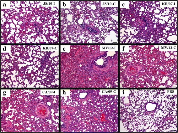
Histopathological lesions in guinea pig lung samples after infection with the four virus strains at 3 dpi for the inoculation group and 5 dpe for the contact group. Guinea pigs were inoculated with 106 EID50/mL of the JS/10, KR/07, MV/12 and CA/09 strains. Each microscopic image represents histopathological pneumonic lesions in the viral inoculation group (-I) at 3 dpi for JS/10 (a), KR/07 (c), MV/12 (e) and CA/09 (g) and in the virus contact group (-C) at 6 dpi (5 dpe) for JS/10 (b), KR/07 (d), MV/12 (f) and CA/09 (h) (X100). The lesion scores of each image was as following: (a) LI: 1 and CH: 2; (b) LI: 1 and CH: 1; (c) LI: 1 and CH: 1;(d) LI: 1 and CH: 1; (e) LI: 3 and CH:3; (f) LI: 2 and CH: 3; (g) LI: 2 and CH: 2;(h) LI: 2 and CH: 1; (i) LI: 0 and CH: 0

Similar articles

Cited by

References

    1. Ali A, Daniels JB, Zhang Y, Rodriguez-Palacios A, Hayes-Ozello K, Mathes L, Lee CW. Pandemic and seasonal human influenza virus infections in domestic cats: prevalence, association with respiratory disease, and seasonality patterns. J Clin Microbiol. 2011;49:4101–4105. doi: 10.1128/JCM.05415-11. - DOI - PMC - PubMed
    1. Crawford PC, Dubovi EJ, Castleman WL, Stephenson I, Gibbs EP, Chen L, Smith C, Hill RC, Ferro P, Pompey J, Bright RA, Medina MJ, Johnson CM, Olsen CW, Cox NJ, Klimov AI, Katz JM, Donis RO. Transmission of equine influenza virus to dogs. Science. 2005;310:482–485. doi: 10.1126/science.1117950. - DOI - PubMed
    1. Song D, Kang B, Lee C, Jung K, Ha G, Kang D, Park S, Park B, Oh J. Transmission of avian influenza virus (H3N2) to dogs. Emerg Infect Dis. 2008;14:741–746. doi: 10.3201/eid1405.071471. - DOI - PMC - PubMed
    1. Song D, Lee C, Kang B, Jung K, Oh T, Kim H, Park B, Oh J. Experimental infection of dogs with avian-origin canine influenza a virus (H3N2) Emerg Infect Dis. 2009;15:56–58. doi: 10.3201/eid1501.080755. - DOI - PMC - PubMed
    1. Lyoo KS, Na W, Yeom M, Jeong DG, Kim CU, Kim JK, Song D. Virulence of a novel reassortant canine H3N2 influenza virus in ferret, dog and mouse models. Arch Virol. 2016;161:1915–1923. doi: 10.1007/s00705-016-2868-x. - DOI - PubMed

Publication types

LinkOut - more resources