. 2018 May 29;131(10):jcs210781.
doi: 10.1242/jcs.210781.

Multiple roles of epithelial heparan sulfate in stomach morphogenesis

Affiliations

Multiple roles of epithelial heparan sulfate in stomach morphogenesis

Meina Huang et al. J Cell Sci. .

Abstract

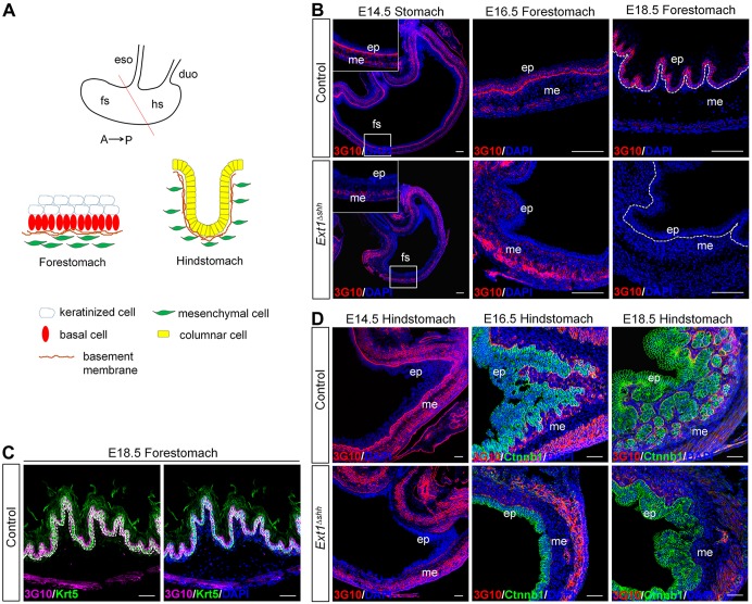
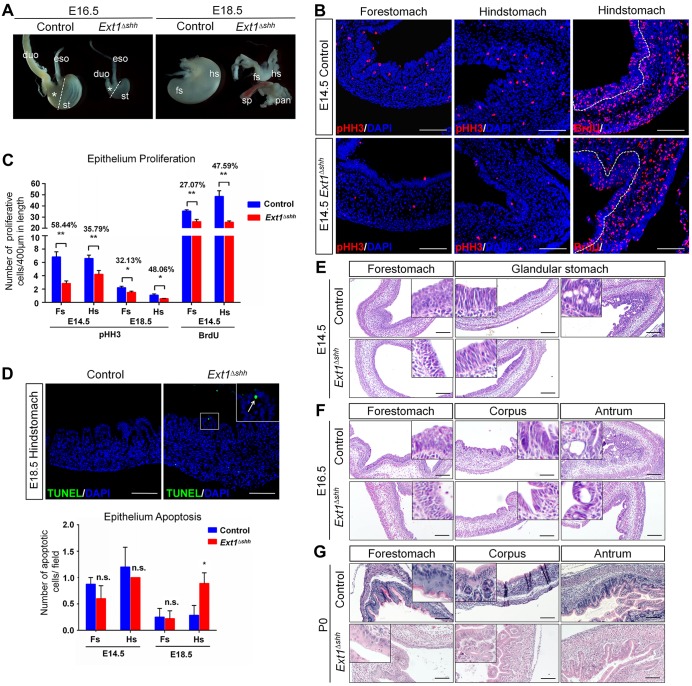
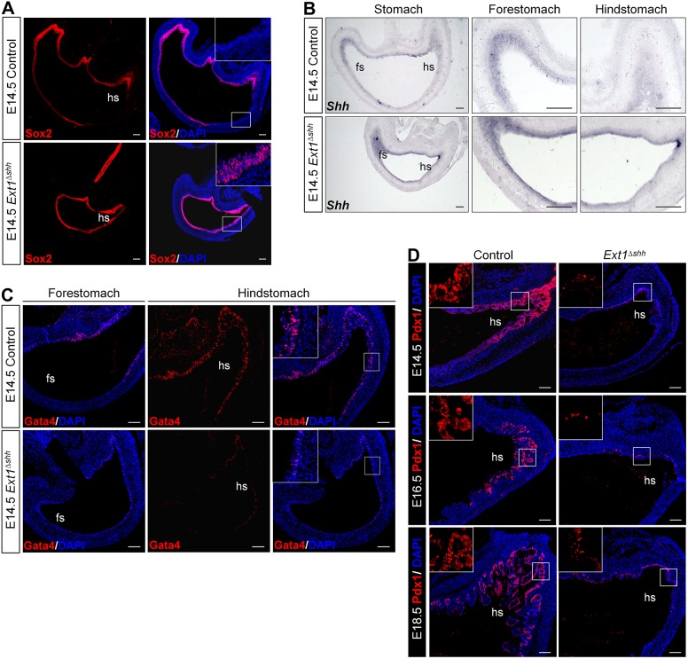
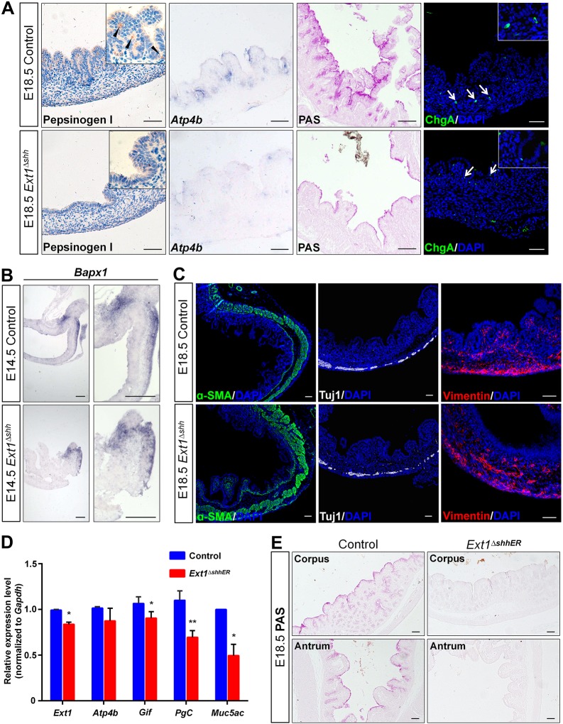
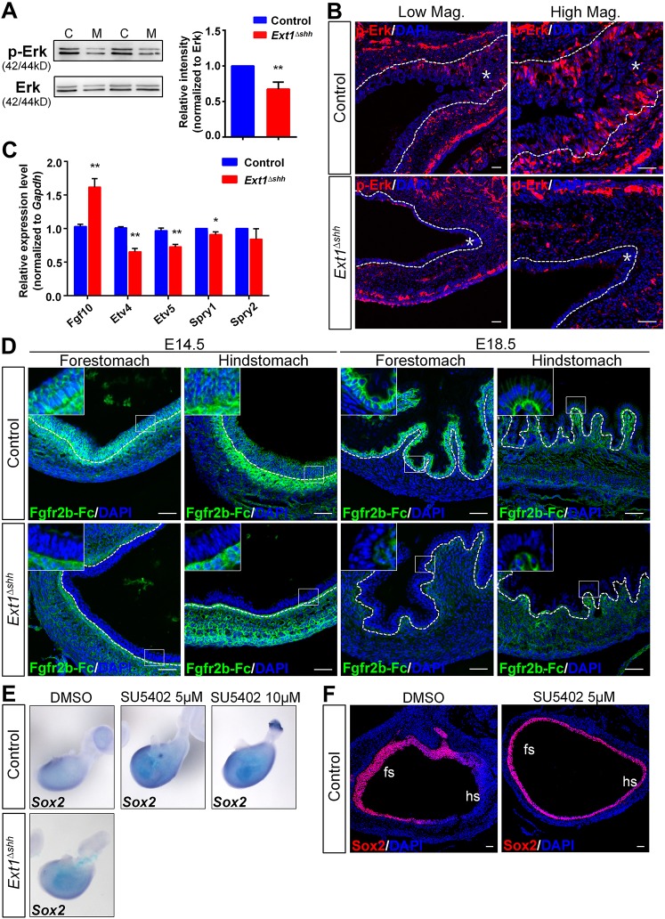
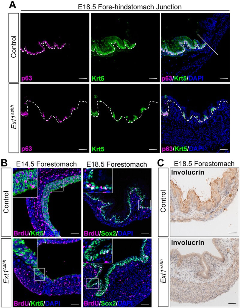
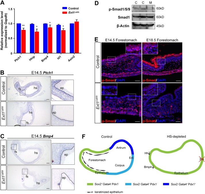
Heparan sulfate proteoglycans (HSPGs) have been shown to regulate various developmental processes. However, the function of heparan sulfate (HS) during the development of mammalian stomach has not been characterized yet. Here, we investigate the role of epithelial HS in embryonic stomach by examining mice deficient in the glycosyltransferase gene Ext1 We show that HS exhibits a specific and dynamic expression pattern in mouse embryonic stomach. Depletion of the epithelial HS leads to stomach hypoplasia, with phenotypic differences in the gastric mucosa between the forestomach and hindstomach. In the posterior stomach, HS depletion disrupts glandular stomach patterning and cytodifferentiation via attenuation of Fgf signaling activity. Inhibition of Fgf signaling in vitro recapitulates the patterning defect. Ligand and carbohydrate engagement assay (LACE) reveals a diminished assembly of Fgf10 and Fgfr2b in the mutant. In the anterior stomach, loss of epithelial HS leads to stratification and differentiation defects of the multilayered squamous epithelium, along with reduced Hh and Bmp signaling activity. Our data demonstrate that epithelial HS plays multiple roles in regulating mammalian stomach morphogenesis in a regional-specific manner.

Keywords: Ext1; Fgf; Glandular development; Heparan sulfate; Hh-Bmp; Stratification.


Conflict of interest statement

Competing interestsThe authors declare no competing or financial interests.

Figures

Fig. 1.
Fig. 1.
Dynamic expression of HS in gastric epithelium during embryonic development. (A) Schematic representation of the anatomy of stomach epithelium. The mouse embryonic stomach is subdivided into the anterior/non-glandular (forestomach) and the posterior/glandular (hindstomach) compartment, overlaid by multilayered keratinized squamous epithelia and complex glandular epithelia, respectively. A straight line distinguishes the forestomach and hindstomach. Distinct cell lineages are indicated. (B,C) HS is recognized by anti-3G10 antibody in control and mutant forestomach. An abundance of 3G10 signals are observed in the basement membrane and gastric epithelium in the forestomach. There is a switch of HS location from basement membrane to the cell surface from E14.5 to E18.5 (B, top panel). After epithelial deletion of Ext1, 3G10 staining is abolished both in the basement membrane and epithelium (B, bottom panel). At E18.5, expression of HS is highly enriched on the cell surface of Krt5-labeled basal cells (C). Magnified views of the boxed area are presented in the insets. Dashed lines separate the epithelium and mesenchyme. (D) HS is expressed in the basement membrane around forming glands in the hindstomach, which is consistent with glandular morphogenesis (top panel). HS is absent in the mutant (bottom panel). Ctnnb1 (green) staining denotes the epithelial cells. eso, esophagus; duo, duodenum; fs, forestomach; hs, hindstomach; ep, epithelium; me, mesenchyme. Scale bars: 100 μm (B), 50 μm (C and D).
Fig. 2.
Fig. 2.
Depletion of epithelial HS causes stomach growth defects and impaired gastric morphogenesis. (A) Gross images of stomachs at indicated embryonic stages from control and Ext1Δshh mutant mice. Mutant embryos exhibit smaller stomachs and thinner intestines. Asterisks represent the glandular stomachs. Dashed lines separate the forestomach and hindstomach. Note that abnormal and rudimentary ‘buds’ are observed from some E18.5 mutant embryos. eso, esophagus; duo, duodenum; st, stomach; fs, forestomach; hs, hindstomach; sp, spleen; pan, pancreas. (B,C) Analysis of gastric epithelium proliferation by pHH3 and BrdU immunostaining. The mutant stomach shows a decreased proliferation in both the forestomach and hindstomach at E14.5. Quantification is achieved by counting the number of pHH3- and BrdU-positive cells relative to the length of the epithelium. The rates of reduction between control and mutant are shown. Nuclei are visualized with DAPI. Dashed lines outline the epithelium. n=5 per group. (D) Increased apoptosis at E18.5 is observed in mutant glandular stomach, demonstrated by TUNEL assay. The inset represents magnified image of the boxed area. The arrow indicates an apoptotic cell in the mutant epithelium. n=3 per group. (E-G) Hematoxylin and Eosin stained sections of mutant stomachs showing histological defects during gastric epithelium morphogenesis. *P<0.05, **P<0.01; n.s., no significant difference. Error bars indicate s.e.m. Scale bars: 100 μm.
Fig. 3.
Fig. 3.
Epithelial HS is required for A-P patterning and glandular specification during the development of stomach. (A) Immunofluorescence staining of Sox2 in E14.5 stomach. HS deletion results in strong expression of Sox2 in the glandular stomach epithelium. (B) In situ hybridization (ISH) using Shh riboprobe on E14.5 paraffin sections. Expression of Shh is spread throughout the entire gastric epithelium in the mutant. Images at high magnification are shown on the right. (C) Immunofluorescence staining with antibody against Gata4. Nuclear stained Gata4 is decreased in mutant glandular stomach, compared with the strong and wide Gata4 pattern in the control. (D) Immunofluorescence staining with antibody against Pdx1 reveals a dramatic reduction in the pre-antral region of the mutant stomach during embryonic development. Insets represent magnified images of the boxed area with no DAPI stain. hs, hindstomach; fs, forestomach. Scale bars: 100 μm (A,B), 50 μm (C,D).
Fig. 4.
Fig. 4.
Epithelial HS is indispensable for differentiation of epithelial cell lineages in the glandular stomach. (A) Differentiation of the glandular epithelium at E18.5. Immunohistochemistry and ISH analysis indicate loss of chief cells (pepsinogen I), parietal cells (Atp4b) and fewer mucus cell (PAS) in the corpus after depletion of HS, whereas ChgA-positive endocrine cells are present. Arrowheads denote chief cells, and arrows indicate endocrine cells. Magnified views are presented in the insets. (B) ISH on paraffin sections using Bapx1 riboprobe. Expression of Bapx1 in the distal gastric mesenchyme is comparable between the control and mutant. (C) Differentiation of the gastric mesenchyme is examined by immunostaining of α-SMA for smooth muscle cells, Tuj1 for enteric neurons and vimentin for mesenchyme cells. Major mesenchymal cell types emerge normally at E18.5 in HS-deficient mice. (D) Ext1 and gastric-specific markers examined by qPCR in Ext1 inducible knockout mice. Several cell lineages are impaired, suggesting that HS is essential for the differentiation of glandular lineages. *P<0.05, **P<0.01. Error bars indicate s.e.m. (n=3). (E) PAS staining confirms the reduction in mucus production. Scale bars: 50 μm (A,C,E), 100 μm (B).
Fig. 5.
Fig. 5.
Epithelial HS modulates Fgf signaling activity in embryonic stomach. (A) Representative image of western blot of p-Erk shows a reduction in its protein level. Graph shows the quantification of band intensity from three independent western blots. C, control; M, mutant (n=3). (B) p-Erk signals are absent in mutant glandular epithelium. Asterisks indicate epithelium. (C) Analysis of Fgf signaling targets. qPCR shows decreased expression of Etv4, Etv5 and Spry1 in mutant stomach in spite of elevated Fgf10 mRNA level (n=5 per group). (D) LACE staining of Fgf10 and Fgfr2b in forestomach and hindstomach. Strong signals near the basement membrane, as well as in the epithelial cells both in the forestomach and hindstomach are detected in the control, which is rarely observed in the mutant. Magnified views of the boxed areas are shown in the insets. (E,F) Fgfr inhibitor (SU5402) treatment leads to enhanced Sox2 expression in a concentration-dependent manner shown by ISH and immunostaining. Organ culture experiments were performed independently three times (n=6 under each control condition; n=2 for mutant). fs, forestomach; hs, hindstomach. The dashed lines outline the gastric epithelium. **P<0.05 and *P<0.01. Error bars indicate s.e.m. Scale bars: 50 μm.
Fig. 6.
Fig. 6.
Epithelial HS regulates forestomach stratification and differentiation. (A) Immunostaining for basal cell markers on forestomach sections. The number of p63+ and Krt5+ basal cells declined and their distribution is sporadic rather than continuous in the mutant epithelium. The straight line separates the forestomach and glandular stomach. White dashed lines outline the epithelium. (B) Analysis of the proliferation in forestomach epithelium. Co-staining for BrdU and Krt5 indicates that remaining basal cells in mutant have proliferation capacity. However, those abnormal Sox2-stained cells in the supralayer are negative for BrdU staining. Magnified views of the boxed area are presented in the insets. (C) Immunohistochemistry analysis of keratinization marker involucrin reveals that the formation of squamous layers is disrupted in the mutant. Scale bars: 50 μm.
Fig. 7.
Fig. 7.
Epithelial HS depletion attenuates Hh-Bmp signaling activity in the stomach. (A) Expression of Hh, Bmp and Wnt signaling targets. Both Hh targets (Ptch1, Hhip1, Bmp4) and Bmp target (Id1) are downregulated after HS ablation at E14.5. **P<0.05, *P<0.01. Error bars indicate s.e.m. (n=4 per group). (B) ISH for Ptch1 transcripts. Mutant displays decreased Ptch1 signal strength in the mesenchyme, compared with the control. (C) ISH for Bmp4 confirms its decreased expression in mutant mesenchyme. (D,E) Phosphorylation of Smad1/5/9 is reduced in mutant revealed by western blotting and immunostaining. C, control; M, mutant (n=3). White dashed lines outline the epithelium. Magnified views of the boxed areas are presented in the insets (B,C,E). Scale bars: 100 μm (B,C), 50 μm (E). (F) Model for the functions of epithelial HS on stomach patterning and specification. In wild-type glandular stomach, epithelial HS promotes Fgf-Erk signaling activation in the overlying epithelium to regulate proper patterning and glandular cytodifferention. In the forestomach, epithelial HS is required for epithelial stratification and modulation of Hh-Bmp signaling. In HS-deficient mice, gastric epithelium is disorganized in both forestomach and hindstomach.

Similar articles

Cited by

References

    1. Allen B. L. and Rapraeger A. C. (2003). Spatial and temporal expression of heparan sulfate in mouse development regulates FGF and FGF receptor assembly. J. Cell Biol. 163, 637-648. 10.1083/jcb.200307053 - DOI - PMC - PubMed
    1. Aubin J., Dery U., Lemieux M., Chailler P. and Jeannotte L. (2002). Stomach regional specification requires Hoxa5-driven mesenchymal-epithelial signaling. Development 129, 4075-4087. - PubMed
    1. Balasubramanian R. and Zhang X. (2016). Mechanisms of FGF gradient formation during embryogenesis. Semin. Cell Dev. Biol. 53, 94-100. 10.1016/j.semcdb.2015.10.004 - DOI - PMC - PubMed
    1. Bishop J. R., Schuksz M. and Esko J. D. (2007). Heparan sulphate proteoglycans fine-tune mammalian physiology. Nature 446, 1030-1037. 10.1038/nature05817 - DOI - PubMed
    1. Bitgood M. J. and McMahon A. P. (1995). Hedgehog and Bmp genes are coexpressed at many diverse sites of cell-cell interaction in the mouse embryo. Dev. Biol. 172, 126-138. 10.1006/dbio.1995.0010 - DOI - PubMed

Publication types

Substances