. 2018 Apr 26;14(4):e1006991.
doi: 10.1371/journal.ppat.1006991. eCollection 2018 Apr.

Cross-sectional analysis of CD8 T cell immunity to human herpesvirus 6B

Affiliations

Cross-sectional analysis of CD8 T cell immunity to human herpesvirus 6B

Larissa K Martin et al. PLoS Pathog. .

Abstract

Human herpesvirus 6 (HHV-6) is prevalent in healthy persons, causes disease in immunosuppressed carriers, and may be involved in autoimmune disease. Cytotoxic CD8 T cells are probably important for effective control of infection. However, the HHV-6-specific CD8 T cell repertoire is largely uncharacterized. Therefore, we undertook a virus-wide analysis of CD8 T cell responses to HHV-6. We used a simple anchor motif-based algorithm (SAMBA) to identify 299 epitope candidates potentially presented by the HLA class I molecule B*08:01. Candidates were found in 77 of 98 unique HHV-6B proteins. From peptide-expanded T cell lines, we obtained CD8 T cell clones against 20 candidates. We tested whether T cell clones recognized HHV-6-infected cells. This was the case for 16 epitopes derived from 12 proteins from all phases of the viral replication cycle. Epitopes were enriched in certain amino acids flanking the peptide. Ex vivo analysis of eight healthy donors with HLA-peptide multimers showed that the strongest responses were directed against an epitope from IE-2, with a median frequency of 0.09% of CD8 T cells. Reconstitution of T cells specific for this and other HHV-6 epitopes was also observed after allogeneic hematopoietic stem cell transplantation. We conclude that HHV-6 induces CD8 T cell responses against multiple antigens of diverse functional classes. Most antigens against which CD8 T cells can be raised are presented by infected cells. Ex vivo multimer staining can directly identify HHV-6-specific T cells. These results will advance development of immune monitoring, adoptive T cell therapy, and vaccines.


Conflict of interest statement

The authors have declared that no competing interests exist.

Figures

Fig 1
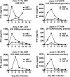
Fig 1. HHV-6B peptide-specific polyclonal T cell lines and CD8 T cell clones.
(A) Peptide-specific T cells in PBMCs from four healthy HLA-B*08:01-positive donors (a-d) were detected in an IFN-γ ELISPOT assay. PBMCs were stimulated with pools of 146 octameric or 153 nonameric HHV-6B/HLA-B*08:01 candidate peptides, 29 Epstein-Barr virus peptides (positive control), or no peptides. Mean+SD of 3 replicates is shown. (B) T cell cultures from donor 1 were stimulated with the complete octamer or nonamer peptide pools for 42 or 56 days as indicated, and then tested in IFN-γ ELISA or ELISPOT assays for reactivity to non-overlapping peptide subpools. HLA-B*08:01-positive activated B cells (mini-LCLs) were used to present peptides. Mean+range of 2 replicates is shown. (C) CD8 T cell clones were screened for reactivity to HHV-6B peptide pools in IFN-γ ELISA, as shown in this example for 12 T cell clones. Autologous CD40-activated B cells were used to present peptides. (D) To identify each T cell clone's target within the peptide libraries, T cells were stimulated with "crossed" peptide subpools. One positive signal each for horizontal and vertical subpools identifies the target peptide, as shown here for one T cell clone, which turned out to be specific for the QTR peptide from U41.
Fig 2
Fig 2. Overview of HLA-B*08:01-restricted peptides and epitopes recognized by CD8 T cells.
Each entry corresponds to one or more CD8 T cell clones that recognized the HHV-6B peptide as indicated. Peptides were derived from HHV-6B strain Z29. Amino acid positions that differ between HHV-6B and HHV-6A are underlined. Kinetic classification of antigens follows 1Oster et al. [43], 2Tsao et al. [44], or 3Taniguchi et al. [45]. Recognition of infected cells was analyzed using HHV-6B strain HST and HHV-6A strain U1102. Restriction through HLA-B*08:01 was verified by analyzing reactivity to sets of HLA-matched and mismatched targets ("match"), by HLA/peptide multimer staining ("mult."), or by analyzing recognition of HLA-transfected targets ("trfec."); "nt" means not tested. "–S" indicates that lack of recognition of infected cells may be explained by differences between HHV-6B strains Z29 and HST. In strain HST, the TSK peptide has the sequence TSKTRQTV (the same as in U1102), and the LPR peptide is probably not expressed due to an upstream frameshift mutation [46].
Fig 3
Fig 3. Verification of HLA-B*08:01 restriction of HHV-6B peptides.
CD8 T cell clones were tested for specific recognition of 293T kidney cells transiently transfected with HLA-B*08:01 and loaded with the specific cognate peptide. HLA-B*08:01-matched and mismatched LCLs were also tested. IFN-γ was quantified by ELISA. Mean and range of two replicates from one of two experiments is shown. Peptide specificities are indicated by the first three letters of the amino acid sequence (see Fig 2), the gene name, and in parentheses the protein name or function.
Fig 4
Fig 4. HHV-6-infected cells present HLA-B*08:01-restricted epitopes to CD8 T cells.
(A, B) Analysis of IFN-γ secretion in response to infected cells. PHA-activated primary HLA-B*08:01-positive CD4 T cells were infected with HHV-6B strain HST (panel A) or HHV-6A strain U1102 (panel B), or remained uninfected. After six days, infected cells or controls were cocultivated with CD8 T cell clones of diverse specificities. After overnight coincubation, IFN-γ secretion was measured by ELISA. Mean and range of two replicates is shown. (C–E) Cytotoxic activity in response to infected cells. A T cell clone specific for the DFK epitope from U86 (C,D) or a polyclonal DFK-specific T cell line (E), both from donor 5, were tested against primary CD4 T cells infected with HHV-6B strain HST for six days or parallel controls in a calcein release assay. (C, E) HLA-B8-positive targets. (D) HLA-B8-negative targets.
Fig 5
Fig 5. Time course of presentation of HLA-B*08:01-restricted HHV-6B epitopes to CD8 T cells.
PHA-activated primary HLA-B*08:01-positive CD4 T cells were infected with HHV-6B strain HST, and cocultivated overnight from the indicated time after infection with CD8 T cell clones of diverse specificities. IFN-γ secretion was measured by ELISA. Mean and range of two replicates from one of three experiments is shown.
Fig 6
Fig 6. Antigen presentation by cells infected with HHV-6B and HHV-6A.
HLA-B*08:01-matched PHA-activated CD4 T cells were infected with HHV-6B strain HST (panel A) or HHV-6A strain U1102 (panel B). As a further control, the viral replication inhibitor ganciclovir (GCV) was added where indicated. At the indicated time after infection, infected cells were cocultivated with T cell clones of diverse specificities. Data are shown as mean and range of duplicates from one of two experiments.
Fig 7
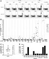
Fig 7. Frequencies of HLA-B*08:01-restricted HHV-6-specific T cells in peripheral blood.
PBMCs from eight healthy adult HLA-B*08:01-positive donors were stained with thirteen HLA-B*08:01/peptide multimers (dextramers) carrying HHV-6 peptides as indicated. An HLA-B*08:01 dextramer carrying the EBV epitope RAK served as a control. (A) Examples of dextramer staining for two donors and six dextramers. (B) Frequencies of multimer-staining cells in eight healthy donors. Donors were known to be HHV-6-seropositive, except donor 9, whose serostatus was not known. Only four donors could be stained with RAK; donor 3 was not stained with TNK and MAR. A median of 7×10⁵ cells was stained for FACS analysis. (C) Number per donor of HHV-6B/6A multimer-staining populations that exceeded 0.01% of CD8 T cells. (D) For each HHV-6 epitope, the proportion of donors is shown who had multimer-staining populations that exceeded 0.01% of CD8 T cells.
Fig 8
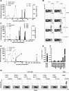
Fig 8. Reconstitution of HHV-6B-specific T cells in patients after allo-HSCT.
(A) HHV-6 DNA and HHV-6-specific CD8 T cells in patient 1. Two peripheral blood samples (day 57, day 68) were available for staining with three HLA-B*08:01/peptide multimers, DFK and QTR from HHV-6B and RAK from EBV BZLF1. (B) Dot plots for multimer stainings of patient 1. (C) Detection of HHV-6 DNA and of HHV-6-specific CD8 T cells in patient 2. Two peripheral blood samples (day 182, day 266) were stained with HLA-B*08:01/peptide multimers carrying HHV-6B peptides DFK, SPR, EGR, and RSK. (D) Detection of HHV-6 DNA and time of HLA/peptide multimer analysis in patient 3. Peripheral blood samples were available for multimer staining from an early and a late time point (day 56, day 1221). (E) Multimer staining for detection of HHV-6-specific T cells (DFK) and EBV-specific T cells in patient 3 at day 56. (F) Multimer staining for HHV-6-specific T cells (14 epitopes as indicated) and EBV-specific T cells (RAK) in patient 3 at day 1221. (G) Exemplary dot plots of multimer stainings of patient 3 at day 1221.
Fig 9
Fig 9. Amino acid composition of HLA-B*08:01-restricted HHV-6B epitopes and their flanking sequences.
(A) For 16 confirmed epitopes presented by infected cells, N-terminal flanking amino acid sequences (N8' to N1'), epitope sequences (N1 to N7, C2, C1), and C-terminal flanking sequences (C1' to C8') were aligned. Predefined anchors are shaded in grey. The box indicates that the amino acids at position N7 = C2) of octameric peptides were considered twice; they were included in the calculations shown in panel B as instances of the N7 position and of the C2 position. (B) Amino acid frequency in each position, categorized into different chemical classes of amino acids. AGP, small or not otherwise categorized; LIVM, aliphatic; FYW, aromatic; DE, acidic; CSTNQ, uncharged polar; RKH, basic.

Similar articles

Cited by

References

    1. Baillargeon J, Piper J, Leach CT (2000) Epidemiology of human herpesvirus 6 (HHV-6) infection in pregnant and nonpregnant women. J Clin Virol 16: 149–157. - PubMed
    1. De Bolle L, Naesens L, De Clercq E (2005) Update on human herpesvirus 6 biology, clinical features, and therapy. Clin Microbiol Rev 18: 217–245. doi: 10.1128/CMR.18.1.217-245.2005 - DOI - PMC - PubMed
    1. Yamanishi K, Okuno T, Shiraki K, Takahashi M, Kondo T, Asano Y et al. (1988) Identification of human herpesvirus-6 as a causal agent for exanthem subitum. Lancet 1: 1065–1067. - PubMed
    1. Zerr DM, Meier AS, Selke SS, Frenkel LM, Huang ML, Wald A et al. (2005) A population-based study of primary human herpesvirus 6 infection. N Engl J Med 352: 768–776. doi: 10.1056/NEJMoa042207 - DOI - PubMed
    1. Agut H, Bonnafous P, Gautheret-Dejean A (2015) Laboratory and clinical aspects of human herpesvirus 6 infections. Clin Microbiol Rev 28: 313–335. doi: 10.1128/CMR.00122-14 - DOI - PMC - PubMed

Publication types

MeSH terms

Grants and funding

Funding: This work was supported by Deutsche Forschungsgemeinschaft (SFB-Transregio 36, project A4, to AM; www.dfg.de). The funders had no role in study design, data collection and analysis, decision to publish, or preparation of the manuscript.