Necroptosis increases with age and is reduced by dietary restriction
- PMID: 29696779
- PMCID: PMC6052392
- DOI: 10.1111/acel.12770
Necroptosis increases with age and is reduced by dietary restriction
Abstract
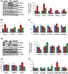
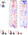
Necroptosis is a newly identified programmed cell death pathway that is highly proinflammatory due to the release of cellular components that promote inflammation. To determine whether necroptosis might play a role in inflammaging, we studied the effect of age and dietary restriction (DR) on necroptosis in the epididymal white adipose tissue (eWAT), a major source of proinflammatory cytokines. Phosphorylated MLKL and RIPK3, markers of necroptosis, were increased 2.7- and 1.9-fold, respectively, in eWAT of old mice compared to adult mice, and DR reduced P-MLKL and P-RIPK3 to levels similar to adult mice. An increase in the expression of RIPK1 (1.6-fold) and MLKL (2.7-fold), not RIPK3, was also observed in eWAT of old mice, which was reduced by DR in old mice. The increase in necroptosis was paralleled by an increase in 14 inflammatory cytokines, including the pro-inflammatory cytokines IL-6 (3.9-fold), TNF-α (4.7-fold), and IL-1β (5.1-fold)], and 11 chemokines in old mice. DR attenuated the expression of IL-6, TNF-α, and IL-1β as well as 85% of the other cytokines/chemokines induced with age. In contrast, inguinal WAT (iWAT), which is less inflammatory, did not show any significant increase with age in the levels of P-MLKL and MLKL or inflammatory cytokines/chemokines. Because the changes in biomarkers of necroptosis in eWAT with age and DR paralleled the changes in the expression of pro-inflammatory cytokines, our data support the possibility that necroptosis might play a role in increased chronic inflammation observed with age.
Keywords: adipose tissue; aging; dietary restriction; inflammaging; inflammation; necroptosis.
© 2018 The Authors. Aging Cell published by the Anatomical Society and John Wiley & Sons Ltd.
Figures


Similar articles
-
RIPK3-MLKL-mediated necroinflammation contributes to AKI progression to CKD.Cell Death Dis. 2018 Aug 29;9(9):878. doi: 10.1038/s41419-018-0936-8. Cell Death Dis. 2018. PMID: 30158627 Free PMC article.
-
Necroptosis increases with age in the brain and contributes to age-related neuroinflammation.Geroscience. 2021 Oct;43(5):2345-2361. doi: 10.1007/s11357-021-00448-5. Epub 2021 Sep 13. Geroscience. 2021. PMID: 34515928 Free PMC article.
-
RIPK3 deficiency or catalytically inactive RIPK1 provides greater benefit than MLKL deficiency in mouse models of inflammation and tissue injury.Cell Death Differ. 2016 Sep 1;23(9):1565-76. doi: 10.1038/cdd.2016.46. Epub 2016 May 13. Cell Death Differ. 2016. PMID: 27177019 Free PMC article.
-
The diverse role of RIP kinases in necroptosis and inflammation.Nat Immunol. 2015 Jul;16(7):689-97. doi: 10.1038/ni.3206. Nat Immunol. 2015. PMID: 26086143 Review.
-
The Inflammatory Signal Adaptor RIPK3: Functions Beyond Necroptosis.Int Rev Cell Mol Biol. 2017;328:253-275. doi: 10.1016/bs.ircmb.2016.08.007. Epub 2016 Sep 22. Int Rev Cell Mol Biol. 2017. PMID: 28069136 Free PMC article. Review.
Cited by
-
MLKL overexpression leads to Ca2+ and metabolic dyshomeostasis in a neuronal cell model.Cell Calcium. 2024 May;119:102854. doi: 10.1016/j.ceca.2024.102854. Epub 2024 Feb 6. Cell Calcium. 2024. PMID: 38430790
-
Necroptosis in Pneumonia: Therapeutic Strategies and Future Perspectives.Viruses. 2024 Jan 7;16(1):94. doi: 10.3390/v16010094. Viruses. 2024. PMID: 38257794 Free PMC article. Review.
-
Characterization of novel mouse models to study the role of necroptosis in aging and age-related diseases.Geroscience. 2023 Dec;45(6):3241-3256. doi: 10.1007/s11357-023-00955-7. Epub 2023 Oct 4. Geroscience. 2023. PMID: 37792157 Free PMC article.
-
The ionome and proteome landscape of aging in laying hens and relation to egg white quality.Sci China Life Sci. 2023 Sep;66(9):2020-2040. doi: 10.1007/s11427-023-2413-4. Epub 2023 Jul 27. Sci China Life Sci. 2023. PMID: 37526911
-
Establishing a genomic radiation-age association for space exploration supplements lung disease differentiation.Front Public Health. 2023 May 11;11:1161124. doi: 10.3389/fpubh.2023.1161124. eCollection 2023. Front Public Health. 2023. PMID: 37250098 Free PMC article.
References
Publication types
MeSH terms
Grants and funding
LinkOut - more resources
Full Text Sources
Other Literature Sources
Miscellaneous

