. 2018 Sep;127(3):375-386.
doi: 10.1007/s00412-018-0670-0. Epub 2018 Apr 14.

P190RhoGAP prevents mitotic spindle fragmentation and is required to activate Aurora A kinase at acentriolar poles

Affiliations

P190RhoGAP prevents mitotic spindle fragmentation and is required to activate Aurora A kinase at acentriolar poles

Arkadi Manukyan et al. Chromosoma. 2018 Sep.

Abstract

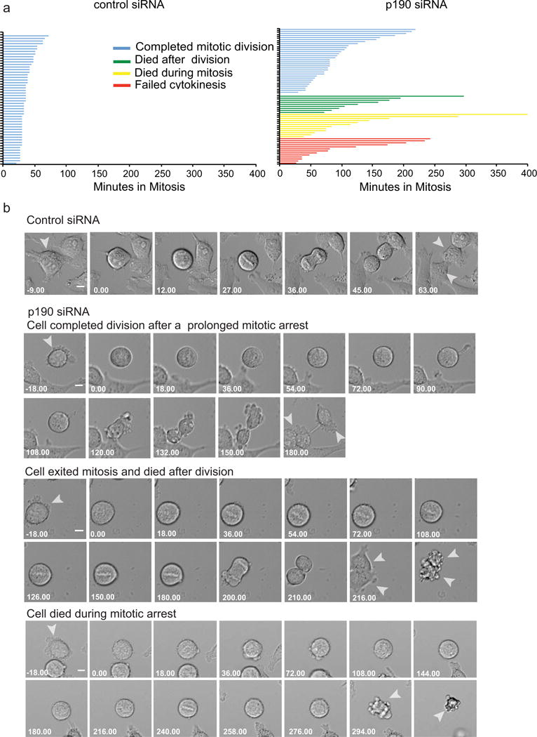
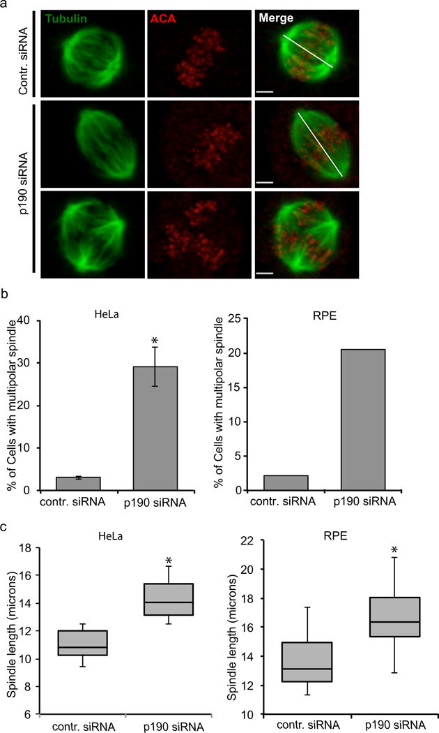
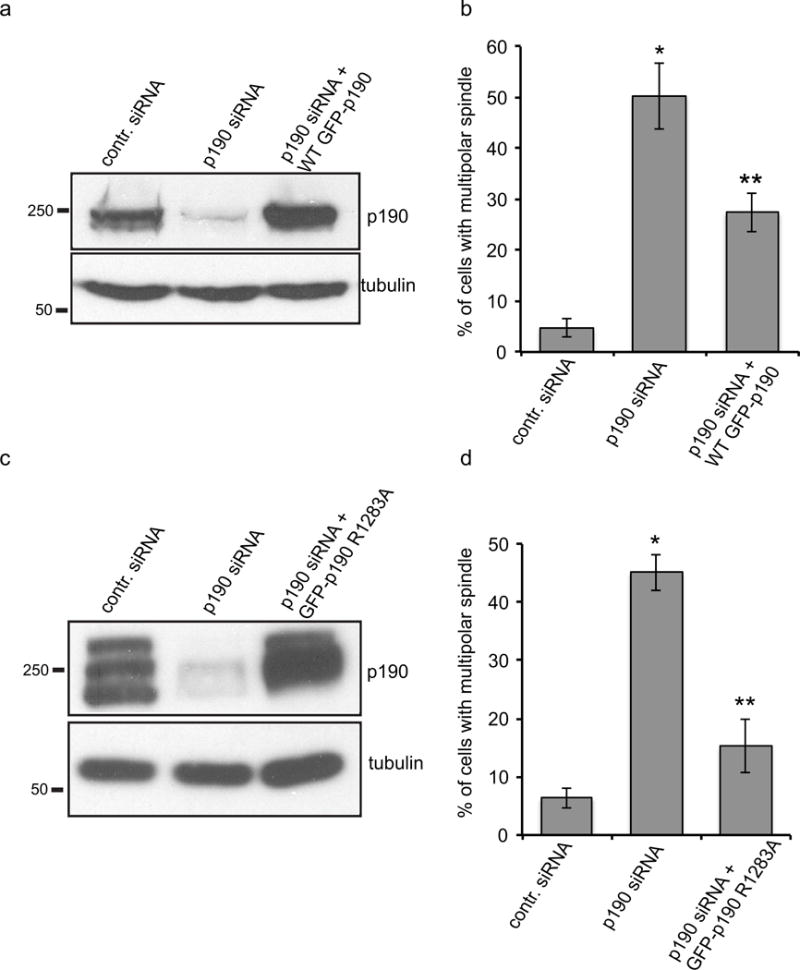
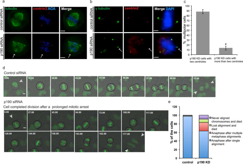
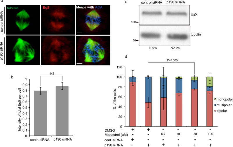
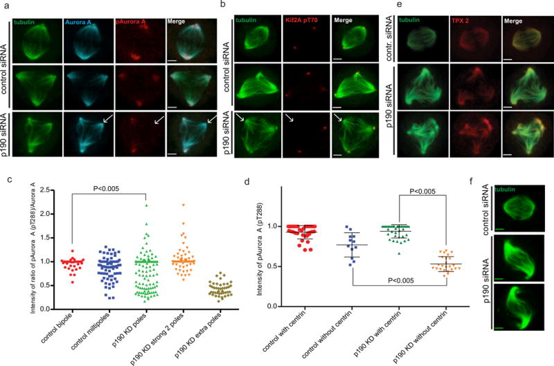
Assembly of the mitotic spindle is essential for proper chromosome segregation during mitosis. Maintenance of spindle poles requires precise regulation of kinesin- and dynein-generated forces, and improper regulation of these forces disrupts pole integrity leading to pole fragmentation. The formation and function of the mitotic spindle are regulated by many proteins, including Aurora A kinase and the motor proteins Kif2a and Eg5. Here, we characterize a surprising role for the RhoA GTPase-activating protein, p190RhoGAP, in regulating the mitotic spindle. We show that cells depleted of p190RhoGAP arrest for long periods in mitosis during which cells go through multiple transitions between having bipolar and multipolar spindles. Most of the p190RhoGAP-depleted cells finally achieve a stable bipolar attachment and proceed through anaphase. The multipolar spindle phenotype can be rescued by low doses of an Eg5 inhibitor. Moreover, we show that p190RhoGAP-depleted multipolar cells localize Aurora A to all the poles, but the kinase is only activated at the two centriolar poles. Overall, our data identify an unappreciated connection between p190RhoGAP and the proteins that control spindle poles including Aurora A kinase and Eg5 that is required to prevent or correct spindle pole fragmentation.

Keywords: Aurora A; Centrosome; Eg5; Mitotic spindle; p190RhoGAP.


Conflict of interest statement

Conflict of Interest

All of the authors declare that he/she has no conflict of interest.

Figures

Fig. 1
Fig. 1. Mitotic fates of HeLa cells depleted of p190
a. Cell fates of HeLa cells as measured by time-lapse imaging after transfection with either control or p190 siRNA. The length of each line represents the time a single cell spent in mitosis, calculated as the time between cell rounding until it re-entered interphase. The color of the line represents the four cell fates triggered by p190 depletion as indicated in the key. b. Representative time-lapse images of the cell fates quantified in the Top panel represent control-siRNA-treated cells. The next three panels show p190- siRNA-treated cells that represent the three cell fates described in the text. Failure to cytokinesis was not shown since it was characterized in our previous paper [Manukyan A et al., 2015]. Arrows indicate single cell divisions after treatment with either siRNA. Time is shown in min. Scale bars-20 μm.
Fig. 2
Fig. 2. p190RhoGAP regulates bipolar spindle formation and spindle length
a. Confocal images of control- and p190-siRNA-treated HeLa cells co-stained with alpha tubulin (green) and anti-centromere antibody (ACA) (red). Top panel images represent control-siRNA-treated cell; middle panel images represent p190-depleted cells with longer bipolar spindles; bottom panel images represent p190-depleted cells with multipolar spindles. White lines show how spindle length was measured for control- and p190-siRNA-treated cells. Images shown are representative of n>50. Scale bars represent 2.1 μm. b. Quantification of cells with multipolar spindles stained with tubulin and anti-centromere antibody (n ≥ 200). Results are mean ± s.d from three independent experiments. *P< 0.05 (T-test) as compared to control siRNA-treated cells. c. p190 depletion causes increase of spindle length in HeLa and RPE cells. Spindle lengths were measured in p190-depleted HeLa and RPE cells stained with tubulin and anticentromere antibody. Box-and-whisker plots show median, 25th and 75th percentiles (box) and 5th and 95th percentiles (whiskers) (n ≥ 50). *P< 0.05 (T-test) as compared to control-siRNA-treated cells.
Fig. 3
Fig. 3. p190RhoGAP regulates bipolar spindle formation independent of its GAP activity
a. Immunoblot to compare endogenous p190 levels to those induced after knockdown of the endogenous p190 levels and expression of the transgene by doxycycline. b. Quantification of multipolar spindle cells after depletion of endogenous p190 and replacement with doxycycline inducible exogenous wild type (n≥200). Results are mean ±s.d from three independent experiments. *P<0.005 (T-test) for both p190-siRNA-treated cells compared to control, and p190-siRNA-treated cells compared to cells rescued by wild-type p190. c. Immunoblot to compare endogenous p190 levels to those induced after knockdown of the endogenous p190 levels and expression of the p190 Gap mutant (R1283A) transgene by doxycycline. d. Quantification of multipolar spindle cells after depletion of endogenous p190 and replacement with the doxycycline-inducible GAP mutant of p190. 48 hrs post-transfection and doxycycline treatment, cells were fixed and stained with tubulin and ACA and scored blindly for multipolar spindle cells (n≥200). Results are mean ± s.d from three independent experiments. *P<0.005 for both p190-siRNA-treated cells compared to control, and p190-siRNA-treated cells compared to cells rescued by p190 GAP mutant (p190 R1283A) compared to control.
Fig. 4
Fig. 4. p190 depletion results in pericentriolar, but not centriolar, fragmentation
a. Confocal images of control- and p190-depleted cells co-stained with α-tubulin and a centriole marker (centrin2). Arrows indicate extra spindle poles. Images shown are representative of n>20. Scale bars represent 4 μm b. Confocal images of control- and p190-depleted cells co-stained with centrin2 and pericentriolar material component (PCM) γ-tubulin. Arrow indicates the pole that did not stain with centrin2, suggesting that multipolarity generated by loss of p190 is the result of PCM fragmentation. Images shown are representative of n>20. Scale bars represent 4 μm. c. Percentage of the cells with two centrioles are graphed versus percentage of the cells that contained more than two centrioles (n≥50, means of 2 experiments). d. Representative time-lapse images of the control and p190 depleted cells with GFP tagged H2B chromatin marker. Top panel represent control-siRNA-treated cells. Bottom panels show p190-siRNA-treated cells. Arrows indicate single cell divisions after treatment with either siRNA. Time is shown in min. Scale bars 20 μm. e. Quantification of time-lapse images of control and p190 siRNA treated HeLa cells according to chromosome alignment (p=1.8e−06, Fisher’s exact test).
Fig. 5
Fig. 5. p190 controls Eg5activity
a. Eg5 localized properly in p190-depleted cells. Confocal images of control- and p190-siRNA treated cells that were fixed and stained with α-tubulin, ACA and anti-Eg5 antibody. Images shown are representative of n>20 bars represent 4 μm. b. Quantification of Eg5 intensity at poles in HeLa cells (p =NS). c. Immunoblot of endogenous Eg5 in control and p190 KD cells. Eg5:tubulin ratio were quantified and amount were reported by percentage. d. Multipolar spindles induced by p190 depletion are rescued by inhibition of Eg5 activity. p190-depleted and double thymidine-blocked cells were released from thymidine for 10 hours and treated with DMSO or 6.7μM, 10μM, 20μM or 100μM monastrol for one and a half hrs. Cells were fixed and stained with tubulin and ACA antibodies, and monopolar, bipolar and multipolar cells were quantified. Results are mean ± s.d from three independent experiments. The rescue of bipolar spindles by 20 uM monastrol is statistically significant (*P<0.005).
Fig. 6
Fig. 6. p190 is required to activate Aurora A kinase on acentriolar poles
a. Aurora A is properly localized to all poles but it is not active at all poles. Confocal images of control- and p190-depleted cells fixed and co-stained with α-tubulin, Aurora A and T-loop phosphorylation for Aurora A (pT288) antibodies. First two panels represent examples of bipolar and multipolar HeLa cells treated with control siRNA; the third panel represents a p190-depleted multipolar cell. Arrows indicate an extra spindle pole in a p190-depleted cell. Images shown are representative of n>20. Scale bars represent 3 μm. b. Aurora A activity as measured with a phosphoantibody against an Aurora site on Kif2a is missing from poles that lack centrosomes. Arrows indicate extra spindle poles in p190-depleted cells. Images shown are representative of n>20. Scale bars represent 3 μm. c. Quantification of Aurora A activation in spindle poles measured as a ratio of T-loop divided by total Aurora A. Each dot in the plot represents a paired measurement of T-loop and Aurora A at a single centrosome. Ratio’s are not statistically different between control bipoles and multipoles but they are both different from p190-depleted poles P<0.005 (T test). To demonstrate that the bimodal distribution of Aurora activation is generated by differences between poles in the same cell we replotted the data in the third lane. The degree of Aurora A activation in the highest two poles is different than the levels in the additional pole in the same cell. d. p190 activates Aurora A at acentriolar poles. Control- and p190-depleted cells were fixed and co-stained with α-tubulin, centrin 2 and phospho-Aurora A pT288 antibodies. Intensity of phospho –T288 Aurora A was measured in each pole with or without a centriole in both control- and p190-depleted cells and plotted as a dot plot P<0.005 (ANOVA). e. Tpx2 levels at poles are not affected by p190 knockdown. Images shown are representative of n>20. Scale bars represent 3 μm. f. Confocal images of tubulin stained control and p190-depleted cells with wavy” bipolar spindles. P<0.005

Similar articles

Cited by

References

    1. Asteriti IA, Giubettini M, Lavia P, Guarguaglini G. Aurora-A inactivation causes mitotic spindle pole fragmentation by unbalancing microtubule-generated forces. Mol Cancer. 2011;10:131. - PMC - PubMed
    1. Balchand SK, Mann BJ, Titus J, Ross JL, Wadsworth P. TPX2 Inhibits Eg5 by Interactions with Both Motor and Microtubule. J Biol Chem. 2015;290(28):17367–79. - PMC - PubMed
    1. Bayliss R, Sardon T, Vernos I, Conti E. Structural basis of Aurora-A activation by TPX2 at the mitotic spindle. Mol Cell. 2003;12(4):851–62. - PubMed
    1. Brouns MR, Matheson SF, Hu KQ, Delalle I, Caviness VS, Silver J, Bronson RT, Settleman J. The adhesion signaling molecule p190 RhoGAP is required for morphogenic processes in normal development. Development. 2000;127:4891–903. - PubMed
    1. Brouns MR, Matheson SF, Settleman J. P190 RhoGAP is the principal Src substrate in brain and regulates axon outgrowth, guidance and fasciculation. Nat Cell Biol. 2001;3(4):361–7. - PubMed

Publication types

LinkOut - more resources