. 2018 Apr 13;50(4):1-12.
doi: 10.1038/s12276-018-0055-8.

Vascular endothelial growth factor mediates the therapeutic efficacy of mesenchymal stem cell-derived extracellular vesicles against neonatal hyperoxic lung injury

Affiliations

Vascular endothelial growth factor mediates the therapeutic efficacy of mesenchymal stem cell-derived extracellular vesicles against neonatal hyperoxic lung injury

So Yoon Ahn et al. Exp Mol Med. .

Abstract

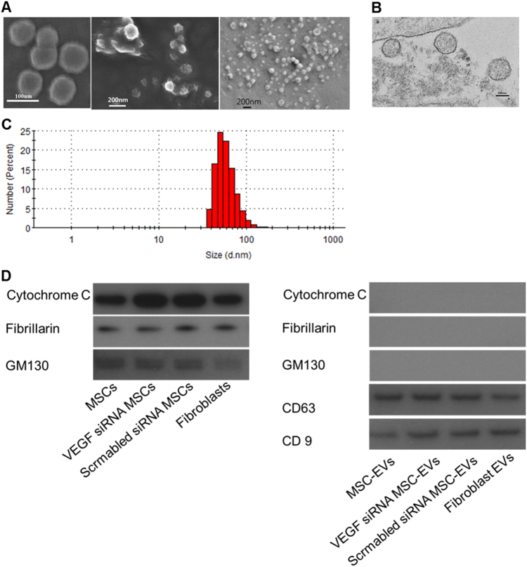
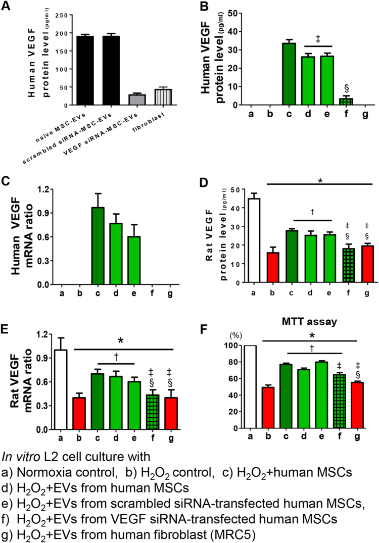
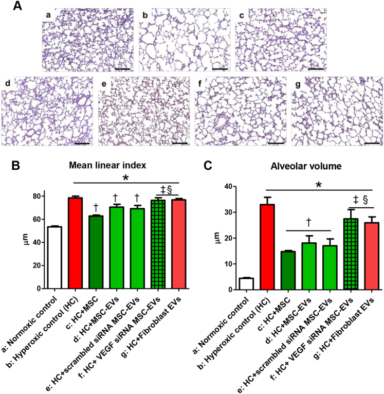
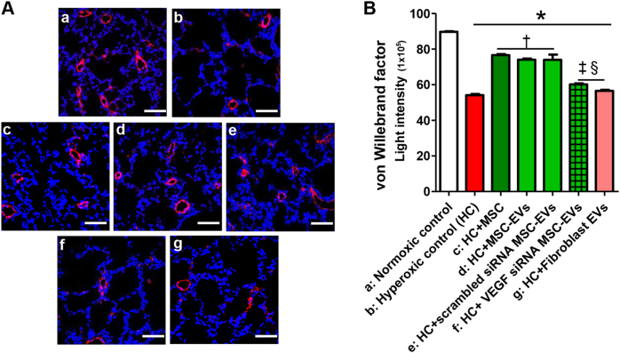
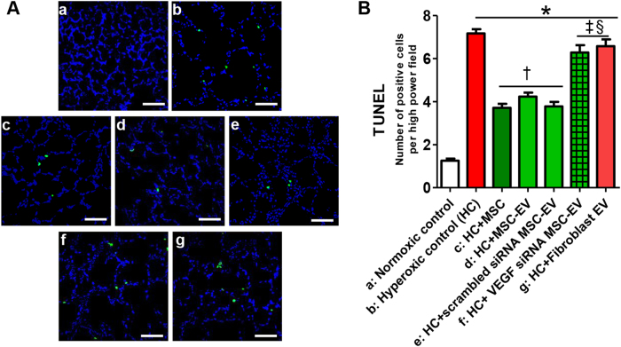
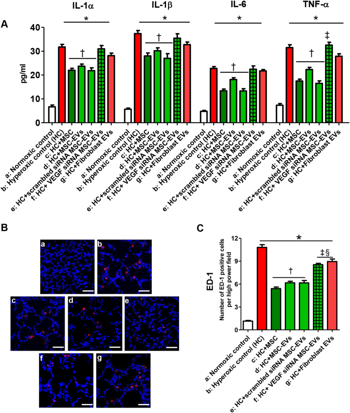
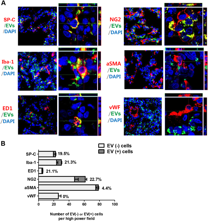
We previously reported the role of vascular endothelial growth factor (VEGF) secreted by mesenchymal stem cells (MSCs) in protecting against neonatal hyperoxic lung injuries. Recently, the paracrine protective effect of MSCs was reported to be primarily mediated by extracellular vesicle (EV) secretion. However, the therapeutic efficacy of MSC-derived EVs and the role of the VEGF contained within EVs in neonatal hyperoxic lung injury have not been elucidated. The aim of the study was to determine whether MSC-derived EVs attenuate neonatal hyperoxic lung injury and, if so, whether this protection is mediated via the transfer of VEGF. We compared the therapeutic efficacy of MSCs, MSC-derived EVs with or without VEGF knockdown, and fibroblast-derived EVs in vitro with a rat lung epithelial cell line challenged with H2O2 and in vivo with newborn Sprague-Dawley rats exposed to hyperoxia (90%) for 14 days. MSCs (1 × 10⁵ cells) or EVs (20 µg) were administered intratracheally on postnatal day 5. The MSCs and MSC-derived EVs, but not the EVs derived from VEGF-knockdown MSCs or fibroblasts, attenuated the in vitro H2O2-induced L2 cell death and the in vivo hyperoxic lung injuries, such as impaired alveolarization and angiogenesis, increased cell death, and activated macrophages and proinflammatory cytokines. PKH67-stained EVs were internalized into vascular pericytes (22.7%), macrophages (21.3%), type 2 epithelial cells (19.5%), and fibroblasts (4.4%) but not into vascular endothelial cells. MSC-derived EVs are as effective as parental MSCs for attenuating neonatal hyperoxic lung injuries, and this protection was mediated primarily by the transfer of VEGF.


Conflict of interest statement

Samsung Medical Center and MEDIPOST Co., Ltd. own issued or filed patents for “Method of treating lung diseases using cells separated or proliferated from umbilical cord blood” in the names of the inventors Y.S.C. and W.S.P. The authors declare that they have no conflict of interest.

Figures

Fig. 1
Fig. 1. Confirmation of extracellular vesicles (EVs).
a Scanning electron microscopy images of the EVs derived from mesenchymal stem cells (MSCs). b Transmission electron microscopy images of EVs derived from mesenchymal stem cells (MSCs). c Particle size distribution (number-weighted size distribution) of MSC-derived EVs. d Western blot assays of MSCs and MSC-derived EVs; cytochrome C (mitochondria marker), fibrillarin (nucleus marker), GM130 (Golgi apparatus marker), CD63, and CD9 (exosome marker)
Fig. 2
Fig. 2. Induction of VEGF expression in rat lung epithelial L2 cells by the VEGF from MSC-derived extracellular vesicles (EVs) rescues oxidative injury in vitro.
a The VEGF levels were measured in the EVs derived from naive mesenchymal stem cells (MSCs), the EVs from scramble siRNA-transfected MSCs, the EVs from VEGF siRNA-transfected MSCs, and the EVs from fibroblasts. The EVs from VEGF siRNA-transfected MSCs or fibroblasts presented with substantially decreased VEGF levels compared to the levels in EVs from naive MSCs or scramble siRNA-transfected MSCs. Rat lung epithelial (L2) cells were treated with H2O2 for 1 h to induce oxidative stress. L2 cells were co-treated with naive MSCs, EVs from naive MSCs, EVs from scramble siRNA-transfected MSCs, EVs from VEGF siRNA-transfected MSCs, or EVs from fibroblasts. In the culture medium, the expression of human VEGF protein (b), human VEGF mRNA (c), rat VEGF protein (d), and rat VEGF mRNA (e) was measured. f The cell survival rate in each group was evaluated by 3-(4,5-dimethylthiazol-2-yl)-2,5-diphenyltetrazolium bromide (MTT) assays. The in vitro L2 cell culture groups are as follows: a, normoxic control; b, H2O2 control; c, H2O2 + human MSCs; d, H2O2 + EVs from human MSCs; e, H2O2 + EVs from scramble siRNA-transfected human MSCs; f, H2O2 + EVs from VEGF siRNA-transfected human MSCs; and g, H2O2 + EVs from human fibroblast (MRC5). Data are presented as the means ± SEM. *P < 0.05 compared to the normoxic control, P < 0.05 compared to the H2O2 control, P < 0.05 compared to H2O2 + human MSCs, §P < 0.05 compared to H2O2 + EVs from human MSCs
Fig. 3
Fig. 3. Extracellular vesicles (EVs) released from MSCs attenuated hyperoxia-induced impaired alveolarization in the lung tissue partly by transferring VEGF.
a Representative photomicrographs of hematoxylin and eosin staining in the lungs of P14 rats from each group (scale bar = 25 μm). The degree of alveolarization was assessed by the (b) mean linear intercept and (c) mean alveolar volume. The EV injection dose per rat was 20 µg. The experimental in vivo groups are as follows: a, normoxic control; b, hyperoxic control; c, hyperoxia + MSCs; d, hyperoxia + MSC-derived EVs; e, hyperoxia + scramble siRNA-transfected human MSC-derived EVs; f, in vivo hyperoxia + VEGF siRNA-transfected human MSCs EVs; and g, in vivo hyperoxia human fibroblast (MRC5)-derived EVs. Data are presented as the means ± SEM. *P < 0.05 compared to the normoxic control, P < 0.05 compared to the hyperoxic control, P < 0.05 compared to hyperoxia + human MSCs, §P < 0.05 compared to hyperoxia + EVs derived from human MSCs
Fig. 4
Fig. 4. Extracellular vesicles (EVs) released from MSCs improved the hyperoxia-induced impaired pulmonary vascularization partly via transferring VEGF. Pulmonary angiogenesis was determined by staining for vWF in histological sections of P14 rat lungs.
a Representative immunofluorescence photomicrographs of vWF staining in the lungs of P14 rats in each group. Nuclei were labeled with 4′,6-diamidino-2-phenylindole (DAPI, blue), and vWF was labeled with the fluorescent marker 5(6)-carboxyfluorescein diacetate N-succinimidyl ester (CFSE, red) (×200, scale bar = 50 μm). b The mean light intensity of vWF immunofluorescence per high power field (HPF) in each group. The EV injection dose was 20 µg per rat. The experimental in vivo groups are as follows: a, normoxic control; b, hyperoxic control; c, hyperoxia + MSCs; d, hyperoxia + MSC-derived EVs; e, hyperoxia + scramble siRNA-transfected human MSC-derived EVs; f, in vivo hyperoxia + VEGF siRNA-transfected human MSCs-derived EVs; and g, in vivo hyperoxia human fibroblast (MRC5)-derived EVs. Data are presented as the means ± SEM. *P < 0.05 compared to the normoxic control, P < 0.05 compared to the hyperoxic control, P < 0.05 compared to hyperoxia + human MSCs, §P < 0.05 compared to hyperoxia + EVs derived from human MSCs
Fig. 5
Fig. 5. Extracellular vesicles (EVs) released from MSCs improved hyperoxia-induced cell death partly via VEGF.
a Representative optical microscopy photomicrographs of TUNEL-stained lung histological sections from P14 rats in each group. The nuclei are labeled with DAPI (blue), and the TUNEL-positive cells are labeled with FITC (green) (×200, scale bar = 50 μm). b The average number of TUNEL-positive cells per high-power field (HPF) in each group. The EV injection dose was 20 µg per rat. The experimental in vivo groups are as follows: a, normoxic control; b, hyperoxic control; c, hyperoxia + MSCs; d, hyperoxia + MSC-derived EVs; e, hyperoxia + scramble siRNA-transfected human MSC-derived EVs; f, in vivo hyperoxia + VEGF siRNA-transfected human MSCs-derived EVs; and g, in vivo hyperoxia human fibroblast (MRC5)-derived EVs. Data are presented as the means ± SEM. *P < 0.05 compared to the normoxic control, P < 0.05 compared to the hyperoxic control, P < 0.05 compared to hyperoxia + human MSCs, §P < 0.05 compared to hyperoxia + EVs derived from human MSCs
Fig. 6
Fig. 6. Extracellular vesicles (EVs) released from MSCs attenuated the hyperoxia-induced increase in the inflammatory response in lung tissue partly via transferring VEGF.
a The expression of interleukin (IL)-1α, IL-1β, IL-6, and TNF-α in P14 rat lungs from each group was measured with ELISA. b Representative immunofluorescence photomicrographs of ED-1 staining, which indicates active macrophages in the lungs of the P14 rats from each group. ED-1 positive alveolar macrophages were labeled with CFSE (red), and nuclei were labeled with DAPI (blue) (×200, scale bar = 50 μm). c The average number of ED-1 positive cells per HPF in each group. The EV injection dosage was 20 µg per rat. The experimental in vivo groups are as follows: a, normoxic control; b, hyperoxic control; c, hyperoxia + MSCs; d, hyperoxia + MSC-derived EVs; e, hyperoxia + scramble siRNA-transfected human MSC-derived EVs; f, in vivo hyperoxia + VEGF siRNA-transfected human MSCs-derived EVs; and g, in vivo hyperoxia human fibroblast (MRC5)-derived EVs. Data are presented as the means ± SEM. *P < 0.05 compared to the normoxic control, P < 0.05 compared to the hyperoxic control, P < 0.05 compared to hyperoxia + human MSCs, §P < 0.05 compared to hyperoxia + EVs derived from human MSCs
Fig. 7
Fig. 7. Distribution of the intratracheally delivered extracellular vesicles (EVs) according to the pulmonary tissue cell type. MSC-derived EVs displayed a cell-type dependent distribution in the pulmonary tissue.
a Representative immunofluorescence photomicrographs of donor EVs and host pulmonary cell staining in the lungs of P14 rats from each group. The nuclei are labeled with DAPI (blue), and the type 2 alveolar cells, total macrophages, activated macrophages, vascular smooth muscle cells, vascular endothelial cells, and vascular pericytes were each immunohistochemically stained with pro-surfactant protein C (SP-C), Iba-1, ED1, α-smooth muscle actin, von Willebrand factor (vWF), and NG2 (red), respectively. The extracellular vesicles (EVs) were prestained with PKH67 dye (green). The three-dimensional images of the co-localized donor EVs and recipient cells were obtained using confocal z-stack images. b The number of each type of host pulmonary cell was counted and evaluated according to EVs co-localization. The rate of donor EVs incorporation into each type of host pulmonary cell is presented as the percentage of double-labeled pulmonary cells with EVs among the non-EVs-merged pulmonary cells. EVs: Extracellular vesicles

Similar articles

Cited by

References

    1. Bhandari A, Panitch HB. Pulmonary outcomes in bronchopulmonary dysplasia. Semin. Perinatol. 2006;30:219–226. doi: 10.1053/j.semperi.2006.05.009. - DOI - PubMed
    1. Bland RD. Neonatal chronic lung disease in the post-surfactant era. Biol. Neonate. 2005;88:181–191. doi: 10.1159/000087581. - DOI - PubMed
    1. Chang YS, et al. Human umbilical cord blood-derived mesenchymal stem cells attenuate hyperoxia-induced lung injury in neonatal rats. Cell Transplant. 2009;18:869–886. doi: 10.3727/096368909X471189. - DOI - PubMed
    1. Chang YS, et al. Intratracheal transplantation of human umbilical cord blood-derived mesenchymal stem cells dose-dependently attenuates hyperoxia-induced lung injury in neonatal rats. Cell Transplant. 2011;20:1843–1854. doi: 10.3727/096368910X520056. - DOI - PubMed
    1. Ahn SY, et al. Long-term (postnatal day 70) outcome and safety of intratracheal transplantation of human umbilical cord blood-derived mesenchymal stem cells in neonatal hyperoxic lung injury. Yonsei Med. J. 2013;54:416–424. doi: 10.3349/ymj.2013.54.2.416. - DOI - PMC - PubMed

Publication types

MeSH terms