Review
. 2018 Apr 10;6(2):43.
doi: 10.3390/biomedicines6020043.

The Many Roles of Ubiquitin in NF-κB Signaling

Affiliations
Review

The Many Roles of Ubiquitin in NF-κB Signaling

Gilles Courtois et al. Biomedicines. .

Abstract

The nuclear factor κB (NF-κB) signaling pathway ubiquitously controls cell growth and survival in basic conditions as well as rapid resetting of cellular functions following environment changes or pathogenic insults. Moreover, its deregulation is frequently observed during cell transformation, chronic inflammation or autoimmunity. Understanding how it is properly regulated therefore is a prerequisite to managing these adverse situations. Over the last years evidence has accumulated showing that ubiquitination is a key process in NF-κB activation and its resolution. Here, we examine the various functions of ubiquitin in NF-κB signaling and more specifically, how it controls signal transduction at the molecular level and impacts in vivo on NF-κB regulated cellular processes.

Keywords: nuclear factor κB; signal transduction; ubiquitin; ubiquitination/deubiquitination.


Conflict of interest statement

The authors declare no conflict of interest.

Figures

Figure 1
Figure 1
The NF-κB proteins and their inhibitors. (A) Members of the Rel/NF-κB family. The five NF-κB subunits are presented with their functional domains. RHD = Rel homology domain; TAD = transcription activation domain; LZ = leucine zipper; GR = glycine-rich domain; DD = death domain. The bold arrows indicate the C-terminus of p50 and p52 after processing of p105 and p100, respectively. (B) Members of the IκB family. The three IκB inhibitors are presented with their functional domains. PEST = proline/glutamic acid/serine/threonine-rich sequence.
Figure 2
Figure 2
The canonical and non-canonical pathways of NF-κB activation. On the left is presented the canonical pathway which involves phosphorylation of IκBs by IKK to induce their degradation. On the right is presented the non-canonical pathway which is dependent on NIK stabilization and IKK1 activation. In each case specific NF-κB dimers are induced that regulates different classes of genes participating in various biological processes. Steps that are controlled by ubiquitination processes are indicated by “Ub”. See text for details.
Figure 3
Figure 3
The subunits of IKK and TAK1 complexes. (A) IKK complex. The three subunits of this complex are presented with their functional domains. KD = kinase domain; ULD = ubiquitin-like domain; SDD = scaffold/dimerization domain; N = NEMO binding domain; Hl1/Hl2 = Helix 1 and 2; CC1/CC2 = coiled coil 1 and 2; LZ = leucine zipper; ZF = zinc finger; (B) TAK1 complex. The three subunits of this complex are presented with their functional domains. KD = kinase domain; T2BD = TAB2/TAB3-binding domain; φPhD = pseudo-phosphatase domain; p38 = p38-interacting domain; TkBD = TAK1-binding domain; CUE = coupling of ubiquitin conjugation to ER degradation domain; ZF = novel zinc finger (Npl4 class).
Figure 4
Figure 4
The ubiquitination process. (A) The enzymatic machinery. The three components (E1/E2/E3) involved in substrate (S) polyubiquitination and major steps of the ubiquitination process are shown; (B) key amino acids of ubiquitin. Indicated are Met1 and the seven internal Lys that can be used to form polyubiquitin chains through peptide (Met) or isopeptide (Lys) bonds involving Gly76. Main biological functions of these chains are indicated. See text for details.
Figure 5
Figure 5
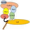
The degradation machinery of IκBs. The SCF E3 ligase complex that induces degradative ubiquitination of IκBs is depicted. K48-linked polyubiquitination is indicated with brown hexagons. See text for details.
Figure 6
Figure 6
Regulated processing/degradation of p105. KPC1-dependent constitutive processing of p105 to generate p50, which can be slightly augmented upon IKK2 activation, is shown at the top. Complete proteolysis or limited processing to release active NF-κB dimers (p50/p50 or p50/RelA) upon cell activation is shown at the bottom. See text for details.
Figure 7
Figure 7
TNF-R1 signaling pathway. Components and mechanisms ensuring signal transduction in this pathway are depicted on the left, with black arrows indicating ubiquitination processes and grey arrows phosphorylation. M1-, K11- and K63-linked polyubiquitination is indicated with yellow, pink and green hexagons, respectively. Components and mechanisms participating in signal shut-off are presented on the right. An induced proteolysis of RIPK1, in addition to its deubiquitination, is indicated although its relevance in NF-κB signaling is uncertain. K48-linked polyubiquitination is indicated with brown hexagons. See text for details.
Figure 8
Figure 8
IL-1βR1 signaling pathway. Components and mechanisms ensuring signal transduction in this pathway are depicted, with black arrows indicating ubiquitination processes and grey arrows phosphorylation. M1- and K63-linked polyubiquitination is indicated with yellow and green hexagons, respectively. See text for details.
Figure 9
Figure 9
Nod1/2 signaling pathway. Components and mechanisms ensuring signal transduction in this pathway are depicted, with black arrows indicating ubiquitination processes and grey arrows phosphorylation. M1- and K63-linked polyubiquitination is indicated with yellow and green hexagons, respectively. Auto-activating Tyrosine phosphorylation of RIPK2 is indicated with a red Y. See text for details.
Figure 10
Figure 10
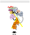
RIG-I/MAVS signaling pathway. Components and mechanisms ensuring signal transduction in this pathway are depicted, with black arrows indicating ubiquitination processes. K63-linked polyubiquitination is indicated with green hexagons. Components shown to be (formally or putatively (question mark)) required in this pathway but whose exact relationship is not defined are shown connected by broken lines. Red filaments represent activating double strand RNA. Activation occurs at the surface of the mitochondria. E3 ligases TRIM4 and MEX3C may also participate in RIG-I activating ubiquitination. See text for details.
Figure 11
Figure 11
cGAS/STING signaling pathway. Components and mechanisms ensuring signal transduction in this pathway are depicted, with black arrows indicating ubiquitination processes and grey arrows phosphorylation. K63-linked polyubiquitination is indicated with green hexagons. Blue filaments represent activating double strand DNA. Activation occurs at the surface of the endoplasmic reticulum. See text for details.
Figure 12
Figure 12
TCR signaling pathway. Components and mechanisms ensuring signal transduction in this pathway are depicted, with black arrows indicating ubiquitination processes and grey arrows phosphorylation. M1- and K63-linked polyubiquitination is indicated with yellow and green hexagons, respectively. Events occurring upstream of PKC activation are not shown and indicated by broken arrows. See text for details.
Figure 13
Figure 13
Genotoxic stress signaling pathway. Components and mechanisms ensuring signal transduction in the two proposed pathways are depicted, with black arrows indicating ubiquitination processes and grey arrows phosphorylation. M1- and K63-linked polyubiquitination is indicated with yellow and green hexagons, respectively. Blue squares labeled S indicate sumoylation whereas red hexagons indicate monoubiquitination. The participation of RIPK1 in the cytoplasmic events is indicated but with a question mark since how it relates to these two pathways, or another one, is unclear. See text for details.

Similar articles

Cited by

References

    1. Karin M., Ben-Neriah Y. Phosphorylation meets ubiquitination: The control of NF-κB activity. Annu. Rev. Immunol. 2000;18:621–663. doi: 10.1146/annurev.immunol.18.1.621. - DOI - PubMed
    1. Aggarwal B.B., Takada Y., Shishodia S., Gutierrez A.M., Oommen O.V., Ichikawa H., Baba Y., Kumar A. Nuclear transcription factor NF-κB: Role in biology and medicine. Indian J. Exp. Biol. 2004;42:341–353. - PubMed
    1. Hinz M., Scheidereit C. The IκB kinase complex in NF-κB regulation and beyond. EMBO Rep. 2014;15:46–61. doi: 10.1002/embr.201337983. - DOI - PMC - PubMed
    1. Dai L., Aye Thu C., Liu X.Y., Xi J., Cheung P.C. TAK1, more than just innate immunity. IUBMB Life. 2012;64:825–834. doi: 10.1002/iub.1078. - DOI - PubMed
    1. Sun S.C. The noncanonical NF-κB pathway. Immunol. Rev. 2012;246:125–140. doi: 10.1111/j.1600-065X.2011.01088.x. - DOI - PMC - PubMed

LinkOut - more resources