. 2018 May 18;293(20):7645-7658.
doi: 10.1074/jbc.RA118.002200. Epub 2018 Apr 3.

Proline hydroxylation in collagen supports integrin binding by two distinct mechanisms

Affiliations

Proline hydroxylation in collagen supports integrin binding by two distinct mechanisms

Kalle H Sipilä et al. J Biol Chem. .

Abstract

Collagens are the most abundant extracellular matrix proteins in vertebrates and have a characteristic triple-helix structure. Hydroxylation of proline residues is critical for helix stability, and diminished prolyl hydroxylase activity causes wide-spread defects in connective tissues. Still, the role of proline hydroxylation in the binding of collagen receptors such as integrins is unclear. Here, we isolated skin collagen from genetically modified mice having reduced prolyl 4-hydroxylase activity. At room temperature, the reduced proline hydroxylation did not affect interactions with the recombinant integrin α2I domain, but at 37 °C, collagen hydroxylation correlated with the avidity of α2I domain binding. Of note, LC-MS/MS analysis of isolated skin collagens revealed no major changes in the hydroxyproline content of the main integrin-binding sites. Thus, the disrupted α2I domain binding at physiological temperatures was most likely due to structural destabilization of the collagenous helix. Integrin α2I binding to the triple-helical GFPGER motif was slightly weaker than to GFOGER (O = hydroxyproline). This phenomenon was more prominent when α1 integrin was tested. Integrin α1β1 expressed on CHO cells and recombinant α1I domain showed remarkably slower binding velocity and weaker avidity to GFPGER when compared with GFOGER. Structural modeling revealed the critical interaction between Arg-218 in α1I and the hydroxyproline residue in the integrin-binding motif. The role of Arg-218 was further validated by testing a variant R218D α1I domain in solid-phase binding assays. Thus, our results show that the lack of proline hydroxylation in collagen can affect integrin binding by a direct mechanism and via structural destabilization of the triple helix.

Keywords: cell adhesion; collagen; connective tissue; hydroxyproline; integrin; post-translational modification (PTM).


Conflict of interest statement

J. M. owns equity in FibroGen Inc., which develops hypoxia-inducible factor P4H inhibitors as potential therapeutics. This company supports hypoxia response research in the J. M. group

Figures

Figure 1.
Figure 1.
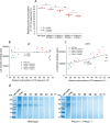
At 37 °C, but not at room temperature, the binding of integrin α2I domain is weaker to collagen isolated from P4ha1+/−;P4ha2−/− mice than to collagen from WT mice. A, binding of α2I domain to collagen isolated from 6 P4ha1+/−;P4ha2−/− and 5 WT mice at room temperature (RT). Two independent experiments are shown. B, binding of α2I domain to collagen isolated from 6 P4ha1+/−;P4ha2−/− and 5 WT mice at 37 °C. Two independent experiments are shown. C, binding of α2I domain to triple-helical GFOGER and GFPGER peptides at room temperature. Two independent experiments are shown. D, binding of α2I domain to triple-helical GFOGER and GFPGER peptides at 37 °C. Two independent experiments are shown. p values from Student's t test and mean ± S.D. are shown. *, p < 0.01; **, p < 0.001. Integrin αI domain concentrations used in the experiments were 200 (A and B) or 400 nm (C and D).
Figure 2.
Figure 2.
Integrin binding correlates with the proline hydroxylation levels at 37 °C but not at room temperature. A, relative hydroxylation of the -X-P-G- sequence in skin collagen of C-P4H mutant mice calculated by dividing the number of unique tandem mass spectra matching to a peptide containing -X-O-G- sequences (but no -X-P-G- sequences) with the number of unique spectra matching to the peptides containing -X-O-G or -X-P-G- sequences. The data points represent the average of two LC–MS/MS runs of collagen preparations from individual mice. p values from Tukey HSD test and mean ± S.D. are shown. B and C, Pearson correlation analyses between integrin binding (α2I domain) and relative hydroxylation of collagen (isolated separately from 4 P4ha1+/+;P4ha2+/−, 5 P4ha1+/−;P4ha2+/−, 7 P4ha1+/+;P4ha2−/−, 6 P4ha1+/−;P4ha2−/−, and 5 WT mice) at room temperature (RT) (B) or 37 °C (C). Integrin αI domain concentration used in the experiments was 200 nm. D, analysis of thermal stability of fibrillar (mainly type I) collagen isolated from the skin of the WT and P4ha1+/−;P4ha2−/− mice by trypsin/chymotrypsin digestion. The collagen samples were treated with a mixture of trypsin and chymotrypsin at temperatures between 36 and 41 °C and analyzed by 8% SDS-PAGE under reducing conditions followed by Coomassie Blue staining, and representative images are shown. An undigested sample without trypsin/chymotrypsin is shown as a control. Molecular weight markers are shown on the left of the gels and the arrows show the positions of the α1(I) and α2(I) collagen chains.
Figure 3.
Figure 3.
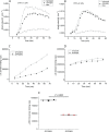
The hydroxylation of infrequent integrin-binding sites in collagen is changed due to genetic ablation of C-P4Hs. A and B, relative hydroxylation, calculated by dividing the number of unique spectra matching to a peptide containing a hydroxylated motif with the number of unique spectra matching a peptide containing hydroxylated or nonhydroxylated motifs, of unaffected sites (A) and affected sites (B). C, relative abundance of sequences containing the first triplet of the hydroxylated integrin binding sequence calculated as number of spectra matching to a peptide containing the hydroxylated triplet divided by number of all spectra matching to collagen peptides in the sample. The data points represent the average of two LC–MS/MS runs of collagen preparations from individual mice. Mean ± S.D. are shown.
Figure 4.
Figure 4.
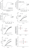
The lack of hydroxyproline in the integrin-binding motif GFOGER reduces the avidity of integrins α1 and α11 but only slightly the binding of integrin α2. A, CD-spectra for GFOGER and GFPGER peptides. The data were collected between 260 and 190 nm 22 °C using a 0.1-cm path length quartz cuvette at 0.1 mg/ml of protein concentration using triplicates. Melting temperatures for the peptides are shown. B and C, binding of α1I (B) and α2I (C) domains to GFPGER or GFOGER in a solid-phase binding assay (BSA as a background control). D, E, and G, spread of CHO cells transfected with α1 (D), α2 (E), or α11 (G) integrin on GFOGER or GFPGER analyzed by impedance-based xCELLigence technology (BSA as a background control). Mean ± S.D. is shown. Each sample was measured with three parallel wells. The difference in peptide binding was confirmed in 4 (α2I) or 3 (α1I) independent experiments. Estimates for the dissociation constants were obtained using an equation: measured binding = maximal binding/(1 + Kd/[αI]). F, difference of cell adhesion between GFPGER and GFOGER in 2 h in the case of CHO-α1 and CHO-α2. p values from one-sample t test and mean ± S.D. of three independent experiments are shown. H, difference of cell adhesion between GFPGER and GFOGER in 2 h in the case of CHO-α11. p values from one-sample t test and mean ± S.D. of 7 independent experiments are shown.
Figure 5.
Figure 5.
Lack of hydroxylation affects the binding of integrin α1β1 both in nonactivated and preactivated conformation. A and B, spreading velocity of CHO-α1 (A) or CHO-α2 (B) cells on GFOGER and GFPGER as a function of time measured by xCELLigence technology. The experiments have been repeated three times with similar results. C and D, binding of recombinant α1I domain (C) or α2I domain (D), to GFPGER and GFOGER in a solid-phase binding assay as a function of time. The integrin αI domain concentration used in the experiments was 200 nm. The experiments have been repeated twice with similar results. E, binding of recombinant α1 E317I domain (α1 domain containing preactivation mutation) to GFPGER and GFOGER in a solid-phase binding assay. p values from Student's t test and mean ± S.D. are shown. The integrin αI domain concentration used in the experiment was 400 nm. The experiment has been repeated twice with similar results.
Figure 6.
Figure 6.
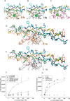
Interaction of the GFOGER and GFPGER peptides with the αI domains. Despite the differences in the peptides and their binding modes to αI domain, the interactions with the αI domain including the key interaction between the metal ion and the Glu in peptide are similar in (A) the α2I-GFOGER (PDB ID 1DZI), (B) the modeled α1I-GFOGER, and (C) the α1I-GLOGEN (PDB ID 2M32) complexes. The leading, middle, and trailing strands are colored in cyan, green, and orange, respectively, and the hydroxyprolines within each strand in a darker color. The hydrogen bonds between the αI domain and the hydroxyprolines are shown as a dashed line and colored according to the color of the connected strands. In the GFOGER–α2I complex (A), one of the hydroxyprolines interacts with the main chain of His-528 and the other one with the main chain oxygens of Ile-156 and Asn-154. Furthermore, the side chain of His-528 interacts with the main chain oxygen of Arg in the peptide and the side chain nitrogen of Asn-154 with the main oxygen of the hydroxyproline in the peptide. These interactions are conserved in the modeled α1I–GFOGER complex (B) and in the X-ray structure of the GLOGEN–α1I complex (C). The salt bridge between the middle strand Arg of the peptide and Asp-219 in α2I (D; circled in black) is not conserved, as the corresponding residue Arg-218 in α1I cannot form a similar interaction (E; circled in black). Based on the α1I–GFOGER model (E), the middle strand Arg of GFOGER stacks with Arg-218 and forms a hydrogen bond with Gln-219. Glu-188 stabilizes the orientation of Arg-218 in α1I by a salt bridge (E) and, similarly, Asn-189 in α2I positions Asp-219 by a hydrogen bond (D). The waters and hydrogen bonds involving hydroxyprolines in the αI domain–peptide complexes are shown in red and the rest of in gray. One of the hydroxyprolines in the trailing strand (circled in red) makes a connection to the middle strand via a water-mediated hydrogen bond and the same hydroxyproline is also involved in the conserved interactions with the αI domain. F, binding of α1I domain and α1I R218D domain to GFPGER or GFOGER in a solid-phase binding assay (BSA as a background control). Each sample was measured with three parallel wells. Estimates for the dissociation constants were obtained using equation: measured binding = maximal binding/(1 + Kd/[αI]). G, binding of the α1I domain and α1I R218D domain to collagen I in a solid-phase binding assay (BSA as a background control). Each sample was measured with three parallel wells. Integrin αI domain concentration used in the experiments was 400 nm. Estimates for the dissociation constants were obtained using equation: measured binding = maximal binding/(1 + Kd/[αI]). The experiments in F and G have been repeated twice with similar results.

Similar articles

Cited by

References

    1. Myllyharju J., and Kivirikko K. I. (2004) Collagens, modifying enzymes and their mutations in humans, flies and worms. Trends Genet. 20, 33–43 10.1016/j.tig.2003.11.004 - DOI - PubMed
    1. Rosenbloom J., Harsch M., and Jimenez S. (1973) Hydroxyproline content determines the denaturation temperature of chick tendon collagen. Arch. Biochem. Biophys. 158, 478–484 10.1016/0003-9861(73)90539-0 - DOI - PubMed
    1. Kivirikko K. I., Myllylä R., and Pihlajaniemi T. (1992) Prolyl hydroxylase, protein disulfide isomerase, and other structurally related proteins. in Post-Translational Modifications of Proteins (Harding J. J., and Crabbe M. J. C., eds) pp. 1–51, CRC Press, Boca Raton, FL
    1. Grosso G., Bei R., Mistretta A., Marventano S., Calabrese G., Masuelli L., Giganti M. G., Modesti A., Galvano F., and Gazzolo D. (2013) Effects of vitamin C on health: a review of evidence. Front. Biosci. 18, 1017–1029 10.2741/4160 - DOI - PubMed
    1. Helaakoski T., Annunen P., Vuori K., MacNeil I. A., Pihlajaniemi T., and Kivirikko K. I. (1995) Cloning, baculovirus expression, and characterization of a second mouse prolyl 4-hydroxylase α-subunit isoform: formation of an α2β2 tetramer with the protein disulfide-isomerase/β subunit. Proc. Natl. Acad. Sci. U.S.A. 92, 4427–4431 10.1073/pnas.92.10.4427 - DOI - PMC - PubMed

Publication types