Review
. 2018 May;30(22):e1706245.
doi: 10.1002/adma.201706245. Epub 2018 Mar 26.

Neutrophil-Based Drug Delivery Systems

Affiliations
Review

Neutrophil-Based Drug Delivery Systems

Dafeng Chu et al. Adv Mater. 2018 May.

Abstract

White blood cells (WBCs) are a major component of immunity in response to pathogen invasion. Neutrophils are the most abundant WBCs in humans, playing a central role in acute inflammation induced by pathogens. Adhesion to vasculature and tissue infiltration of neutrophils are key processes in acute inflammation. Many inflammatory/autoimmune disorders and cancer therapies have been found to be involved in activation and tissue infiltration of neutrophils. A promising strategy to develop novel targeted drug delivery systems is the targeting and exploitation of activated neutrophils. Herein, a new drug delivery platform based on neutrophils is reviewed. There are two types of drug delivery systems: neutrophils as carriers and neutrophil-membrane-derived nanovesicles. It is discussed how nanoparticles hijack neutrophils in vivo to deliver therapeutics across blood vessel barriers and how neutrophil-membrane-derived nanovesicles target inflamed vasculature. Finally, the potential applications of neutrophil-based drug delivery systems in treating inflammation and cancers are presented.

Keywords: cancer; inflammation; nanovesicles; neutrophils; targeted drug delivery.


Conflict of interest statement

Conflicts of Interest

The authors declare no conflicts of interest.

Figures

Figure 1.
Figure 1.
Adhesion and transmigration of neutrophils during inflammation. Adhesive molecules on both neutrophils and the endothelium are indicated. PSGL-1: P-selectin glycoprotein ligand-1; GlyCAM-1: Glycosylation-dependent cell adhesion molecule-1; LFA-1: Lymphocyte function-associated antigen-1; VLA-4: Integrin α4β1 (Very Late Antigen-4); ICAM-1: Intercellular Adhesion Molecule-1; ICAM-2: Intercellular Adhesion Molecule-2; VCAM-1: Vascular cell adhesion protein-1; Mac-1: Macrophage-1 antigen; PECAM-1: Platelet endothelial cell adhesion molecule-1; JAMs: Junctional adhesion molecules; VE-cadherin: vascular endothelial cadherin.
Figure 2.
Figure 2.
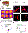
NP targeting of adherent neutrophils. (A) Cy5-loaded and (B) Alex Fluor 647-conjugated albumin NPs (red) were internalized by Gr-1-positive neutrophils (green). Scale bars, 10 µm. (C) Native albumin-conjugated polystyrene NPs (green) were bound to the neutrophil surface (red), and (D) Cy5-conjugated native albumin (red) was not internalized by Gr-1-positive neutrophils (green). Scale bars, 10 µm. (E) Quantitative analysis of percentage of Gr-1-positive neutrophils internalizing three types of NPs and Cy5-labeled albumin. (F) Quantitative analysis of uptake of Cy5-loaded and Cy5-conjugated albumin NPs was carried out by measuring fluorescence intensity per neutrophil. The results are shown as the mean ± s.e.m. (n = 13–20 vessels with three mice per group.) ND, not detected. Copyright 2014, Springer Nature.[11]
Figure 3.
Figure 3.
Neutrophil-mediated targeted delivery of nanotherapeutics and neutrophil membrane-derived vesicles for targeted drug delivery.
Figure 4.
Figure 4.
Neutrophil-mediated delivery of NPs to tumors across blood vessel walls. (A) The concept of neutrophil-mediated delivery of NPs to tumor tissues inflamed by photosensitization (PS). Intravital microscopic 3D images of mouse tumors treated with (B) both PS and NPs-CD11b (green), (C) PS and NPs-PEG (green), (D) NPs-CD11b only and (E) PS and NPs-CD11b after neutrophil depletion. The tumor was treated with PS (intravenous (i.v.) injection of pyropheophorbide-a (Ppa) and 660-nm laser). Approximately 0.75 h later, NPs were i.v. administered. LY-6G Abs (pink) was subcutaneously (s.c.) injected around the tumor to stain neutrophils. Cy3-BSA (red) was i.v. administered to visualize the blood vessel. Anti-LY-6G Abs were intraperitoneally (i.p.) injected 24 h before the administration of Ppa to deplete microphils. (F) Thermal graphic imaging of mouse tumors after the tumors were irradiated with a laser for 20 min. (G) Tumor size and (H) survival rate of the tumor-bearing mice. The mice were irradiated with an 808-nm laser after the injection of GNRs-PEG and GNRs-CD11b with or without PS (denoted as PS/PEG, PS/CD11b, PEG, and CD11b, respectively) or irradiated with an 808-nm laser after the treatment of both PS and GNRs-CD11b with neutrophil depletion (denoted as LY-6G/PS/CD11b). For PBS (without PS or laser irradiation at 808 nm), PS (with PS but without laser irradiation at 808 nm), and PS/808 nm laser (with both PS and laser irradiation at 808 nm), only PBS (pH 7.4) was injected. Data represent mean ± SD (n = 6 mice per group). Copyright 2017, Wiley.[13]
Figure 5.
Figure 5.
Neutrophils deliver NPs to tumors induced by antibody dependent cell-mediated cytotoxicity (ADCC). (A) Concept of NPs delivered by neutrophils to tumors mediated by ADCC. Percentage of neutrophils in tumor tissue with i.v. administration of (B) PBS and (C) TA99 at 48 h. The samples were single cell suspensions prepared from mouse tumors 48 h after treatment with PBS or TA99 in PBS. The cell suspension was analyzed by flow cytometry. (D) MFI of NPs in neutrophils in tumor by flow cytometry 48 h after the injection of Cy5-BSA NPs or both TA99 (i.v. injected at 24 h) and Cy5-BSA NPs (i.v. injected at 48 h) (n = 3). Neutrophils were stained with Alexa-488 anti-mouse Gr-1 antibody, and nucleus was stained by DAPI. (E) Tumor size and (F) survival rates of mice bearing melanoma illuminated with a 660-nm laser 48 h after i.v. injections of vehicles, TA99, Ppa-loaded NPs, or both TA99 (i.v. injected at 24 h) and Ppa-loaded NPs. Copyright 2016, Wiley.[14]
Figure 6.
Figure 6.
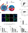
Neutrophil-mediated delivery of NPs to inflamed alveoli by crossing a blood-air barrier. (A) Schematic diagram of neutrophil-mediated delivery of NPs for the treatment of lung inflammation. Flow cytometry of bronchoalveolar lavage fluid (BALF) (B) 2 h and (C) 20 h after i.v. injection of Cy5-BSA NPs. (D) Fluorescence confocal microscopy of neutrophils from BALF 2 and 20 h after i.v. injection of Cy5-BSA NPs (red). Neutrophils were labeled with Gr-1 antibody (green). Nuclei were stained by DAPI (blue). (E) Cy5-BSA NPs in BALF with or without i.p. injection of Gr-1 antibody. Cy5-BSA NPs were i.v. injected 4 h after LPS challenge. Gr-1 antibodies were i.p. injected after LPS challenge. The samples were collected 20 h after the administration of NPs. NPs was administered in mice 4 h after LPS challenge (8 mg/kg). Concentrations of (F) IL-6 and (G) TNF-α in BALF 20 h after i.v. injection of vehicle of 2-[(Aminocarbonyl)amino]-5-(4-fluorophenyl)-3-thiophenecarboxamide (TPCA-1) solution, 5% glucose, TPCA-1 solution or TPCA-1 BSA NPs in mice 4 h after LPS challenge (8 mg/kg). (H) Total colony forming units of P. aeruginosa in BALF after i.v. injection of 5% glucose, vehicle of cefoperazone acid (Cefo-A) solution, Cefo-A solution (25 mg/kg) and Cefo-A BSA NPs (25 mg/kg) in mice 12 h after P. aeruginosa infection. Samples were collected 12 h later. All data represent mean (SD) (3–4 mice per group). Copyright 2015, American Chemical Society.[15]
Figure 7.
Figure 7.
Preparation of cell membrane-derived nanovesicles by nitrogen cavitation. (A) The process to generate the nanovesicles via nitrogen cavitation, centrifugation and extrusion. Differentiated HL-60 cells were collected and disrupted by nitrogen cavitation under a pressure of approximately 350 psi, and membrane-formed vesicles, intracellular molecules, and the nucleus exist in the resulting solution. Afterwards, differential centrifugation was used to purify and obtain the needed vesicles. Copyright 2016, Elsevier.[16] (B) Cryo-TEM of cell membrane-formed vesicles, which are approximately 150 nm in diameter. (C) Proteomics of nanovesicles and their source cells. The total proteins (left) and membrane proteins (right) in nitrogen cavitation-generated EVs (NC-EVs), naturally secreted EVs (NS-EVs) and their parent cell analyzed by mass spectrometry. The shared proteins between EVs and their parent cells suggested that EVs were derived from their parent cells. (D) Quantification analysis of the yield of NC-EVs and NS-EVs indicated the reproductively of NC-EVs. Copyright 2017, Elsevier.[17]
Figure 8.
Figure 8.
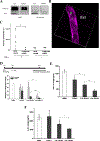
HL-60 cell membrane-formed nanovesicles target and treat inflammation. (A) Western blot of integrin β2 on HL-60 and erythrocyte cells and their nanovesicles. (B) Intravital images of the cremaster venule of a mouse treated with TNF-α and DiO-fluorescently labeled HVs (green). The endothelium was labeled by Alex Fluor-647-labeled anti-CD31 (pink) to indicate the blood vessel. Intravenously (i.v.) injected HV nanovesicles were found adherent to cremaster venules. (C) Quantification of the adherent HVs on cremaster venules based on intravital images. (n = 3). P<0.001. Numbers of neutrophils (D), TNF-α (E) and IL-6 (F) in BALF 10 h after injection of HBSS, erythrocyte vesicles-TPCA-1 (EV-TPCA-1) and HV-TPCA-1 in mice 3 h after LPS challenge. Compared with EV and free drug, HV showed a reduction of infiltrated neutrophils and amount of cytokines, which indicated the therapeutic efficacy of HV nanovesicles. *, **, and *** represent P<0.05, 0.01 and 0.001 in two-way t-test. Copyright 2016, Elsevier.[16]
Figure 9.
Figure 9.
Drug-loaded neutrophil membrane vesicles for anti-inflammation therapy. (A) Schematic illustration of remote loading of piceatannol into cavitation-generated nanovesicles (NC-EVs) via pH gradient. (B) Neutrophil numbers in BALF 12 h after lung LPS challenge at a dose of 10 mg/kg. Vehicle, piceatannol (pic) (2 mg/kg) and pic-NC-EVs (pic at 2 mg/kg) were i.v. injected 2 h after LPS challenge. (C) MPO activity in mouse lung, liver and kidney 8 h after intraperitoneal (i.p.) LPS (22 mg/kg) challenge (LPS-induced sepsis model). Vehicle, piceatannol (3 mg/kg) and pic-NC-EVs (pic at 3 mg/kg) were i.v. injected 2 h after LPS challenge. The result showed that piceatannol-loaded NC-EVs decreased neutrophil tissue infiltration in all the three organs. (D) Survival rates after i.p. LPS (22 mg/kg) challenge (LPS-induced sepsis model). Vehicle, piceatannol (3 mg/kg) and pic-NC-EVs (pic at 3 mg/kg) were i.v. injected 2 h after i.p. LPS challenge. Piceatannol-loaded NC-EVs prevented death in 80% of mice. *, **, and *** represent P<0.05, 0.01 and 0.001 in two-way t-test. Copyright 2017, Elsevier.[17]
Figure 10.
Figure 10.
Scheme of Carfilzomib (CFZ), a second-generation proteasome inhibitor, loaded neutrophil-mimicking NPs (NM-NP-CFZ) that target CTCs and their site of colonization. Copyright 2017, American Chemical Society.[18]
Figure 11.
Figure 11.
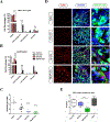
Neutrophil membrane-coated NPs treat cancer metastasis. Cell percentage of viable, early apoptotic, late apoptotic, and necrotic cells among GFP+ 4T1 cells (A) and leukocytes (B) in blood after treatment with free CFZ, NP-CFZ, and NM-NP-CFZ. (C) Quantification of metastasis nodules in lung tissue in the model of NM-NP-CFZ preventing the formation of early metastasis. Metastasis nodules were significantly reduced after treatment with NM-NP-CFZ compared to treatment with saline or free CFZ. (D) Imaging of in vivo apoptosis of successfully colonized GFP+ tumor cells in the lung by deoxynucleotidyl transferase dUTP nick end labeling (TUNEL) assay. 4T1 tumor metastasis models were established 16 days before TUNEL analysis. Apoptosis was significantly improved by NM-NP-CFZ compared to other groups in the previously formed 4T1 lung metastasis sites. Green indicated tumor cells, red indicated TUNEL labeling and nuclei were stained as blue. Scale bar is 100 μm. (E) Quantification of GFP+ nodules per slice in the model of NM-NP-CFZ inhibiting the development of previously formed lung metastasis. All data were represented as the mean ± SD. *** P<0.001, * P< 0.05 and ## P<0.01. Copyright 2017, American Chemical Society.[18]

Similar articles

Cited by

References

    1. Fullerton JN, Gilroy DW, Nat Rev Drug Discov 2016, 15, 551. - PubMed
    1. Summers C, Rankin SM, Condliffe AM, Singh N, Peters AM, Chilvers ER, Trends Immunol 2010, 31, 318. - PMC - PubMed
    1. Wright HL, Moots RJ, Bucknall RC, Edwards SW, Rheumatology (Oxford) 2010, 49, 1618. - PubMed
    1. Kruger P, Saffarzadeh M, Weber AN, Rieber N, Radsak M, von Bernuth H, Benarafa C, Roos D, Skokowa J, Hartl D, PLoS Pathog 2015, 11, e1004651; - PMC - PubMed
    1. Kolaczkowska E, Kubes P, Nat Rev Immunol 2013, 13, 159; - PubMed